Citation:
Yi Liu, Xiaolin Yu, Wenyun Mu, Minsi Meng, Baixue Li, Jie Liu, Haixin Qian, Lin Weng, Tingting Yu, Nan Hu, Xin Chen, Yi Hao. Substrate-independent nano-coating with persistent antibacterial and tooth whitening activities for dental health[J]. Chinese Chemical Letters,
2026, 37(5): 111338.
doi:
10.1016/j.cclet.2025.111338
Substrate-independent nano-coating with persistent antibacterial and tooth whitening activities for dental health
English
Substrate-independent nano-coating with persistent antibacterial and tooth whitening activities for dental health
College of Chemical Engineering, Sichuan University of Science & Engineering, Zigong 643000, China
b.
Institute of Precision Medicine, Zigong Academy of Big Data and Artificial Intelligence in Medical Science, Zigong Fourth People's Hospital, Zigong 643000, China
c.
Department of Chemical Engineering, Shaanxi Key Laboratory of Energy Chemical Process Intensification, Institution of Polymer Science in Chemical Engineering, School of Chemical Engineering and Technology, Xi'an Jiaotong University, Xi'an 710049, China
d.
Sichuan Clinical Research Center for Clinical Laboratory, Zigong Fourth People's Hospital, Zigong 643000, China
e.
Department of Laboratory Medicine, Zigong Fourth People's Hospital, Zigong 643000, China
f.
Department of Prosthodontics, Shanghai Ninth People's Hospital College of Stomatology, Shanghai Jiao Tong University School of Medicine, National Clinical Research Center of Stomatology, Shanghai Key Laboratory of Stomatology and Shanghai Research Institute of Stomatology, Shanghai 200240, China
haoyi5315@163.com (Y. Hao). 1 These authors contributed equally to this work.
Received Date:
11 March 2025 Accepted Date:
16 May 2025 Revised Date:
15 May 2025 Available Online:
15 May 2026
Abstract:
Bacteria and stains on tooth and various dental materials severely harm dental health and beauty and require feasible solutions. In this study, a simple strategy was developed to produce nano-coating on different substrates for persistent antibacterial and whitening. The coating is formed by the lysozyme (Lys), hemoglobin (Hb), and glucose oxidase (GOD) via co-assembly, in which the phase transition of Lys initiated the co-assembly to anchor other two proteins. During therapy, the GOD continuously oxidizes glucose in the oral environment to cut off the nutrition of bacteria meanwhile generating H2O2, which would be further catalyzed by the ferrous ions in Hb to produce reactive oxygen species (ROS) for effective decomposition of surrounding bacteria and stains. Moreover, the Hb can perform persistent release of oxygen, which not only enhances the efficiency of glucose oxidation to produce more ROS but directly suppresses anaerobic bacteria via reversing the local hypoxia environment in the mouth. The experimental results indicated that our strategy is able to form nano-film of proteins both on the surface of dental orthosis and human tooth, which further causes obvious reduction of the bacteria not only on the coated substrate but in the surrounding tissue with up to 100% of the bacteriostatic rate. In addition, both the dental orthosis and human tooth were also rapidly cleaned due to the local ROS generation, leading to a sustained anti-staining property in the long term.
It has been reported that there are over 700 different bacterial strains in the mouth of human, including various pathogenic bacteria [1]. These pathogenic bacteria are able to accumulate on both real tooth and dental materials, which not only produce harmful metabolites such as endotoxins, exotoxins and teichoic acid to directly damage oral tissues but form dental plaque to encourage the colonization of more pathogenic bacteria [2], resulting in dental staining, dental caries, periodontal even systemic diseases such as bacteremia, bacterial endocarditis, pneumonia and diabetes [3]. In addition, the problem could be further promoted by the absorption of organic matter during daily diet, which leads to additional tooth staining as well as the enhanced adhesion of pathogenic bacteria on both dental enamel and various dental materials [4–7]. Therefore, it is necessary to suppress the accumulation of both bacteria and organic matter on tooth and dental materials for oral health.
Although some household products containing fungicides, such as mouthwashes, toothpaste, gels and films have been used to inhibit the growth of pathogenic bacteria in the mouth, these products majorly relied on toxic chlorinated compounds such as cetylpyridinium chloride and chlorhexidine gluconate [8–11], which not only brought a potential threat to human health but performed a limited anti-bacteria efficiency due to its short working duration. In addition, these products paid little attention to the oral microenvironments such as abundant salivary glucose (energy source of bacteria) and hypoxia in dental plaque (survival condition of anaerobe), which are hotbeds for bacteria to live [12], resulting in significant insufficiency to eliminate the pathogenic bacteria. Therefore, developing a safe strategy with sustained anti-bacterial properties and effective manipulation of microenvironments surrounding these bacteria is highly expected for oral health.
Beside the directly damage of bacteria, some teeth whitening technologies were also developed to further ensure the oral health, which was expected to remove the dental stains caused by bacteria plaque and extrinsic organic matter to achieve dental esthetics meanwhile assist bacteria inhibition [13–15]. Currently, these strategies mainly based on the bleaching agents containing hydrogen peroxide (H2O2) or carbamide peroxide [16,17]. In order to achieve effective whitening, dentists often use high concentration of bleaching agents due to their limited oxidation property, which may cause irritation of the soft tissue, tooth sensitivity even enamel damage [18]. Although low-concentration (below 3%) of peroxides would effectively lessen the side effects, their effectiveness in tooth whitening is also significantly reduced as well [19]. Thus, the contradiction between efficiency and safety remains a challenge for current tooth whitening procedures.
Catalytic nanomedicine, an emerging strategy that utilizes enzymatic reactions or biomimetic catalysts to generate therapeutic agents in situ, has shown promise in resolving such biomedical trade-offs [20–25]. By harnessing the catalytic activity of natural enzymes or engineered nanomaterials, these systems can produce reactive oxygen species (ROS) or oxygen on demand, enabling efficient killing of aerobic [26–28] and anaerobic bacteria [29]. For instance, glucose oxidase (GOD)-based nanoreactors have been designed to deplete glucose in tumor microenvironments [30], while metal nanoparticles mimicking peroxidase activity can convert endogenous H2O2 into bactericidal radicals [31–33]. These successes highlight the potential of catalytic approaches to achieve both high efficacy and biocompatibility. However, translating such strategies to oral healthcare remains challenging due to the dynamic nature of the oral cavity-saliva flow constantly disrupting foreign materials, pH fluctuates with food intake, and microbial communities rapidly adapt to environmental changes [34–36]. Recent advances in catalytic coating strategies, such as multifunctional titanium implants integrating photodynamic and gas therapies [37], have demonstrated the feasibility of tailored surface modifications to address complex oral conditions. Simultaneously, new applications of protein-based materials as nanocarriers in the therapeutic field have brought new inspirations for biocompatible surface engineering [38–40]. Inspired by these innovations, hopefully, it will be possible to design a catalytic nano-pharmaceutical coating that will enable substrate-independent adhesion and autonomous catalytic activity on different dental materials.
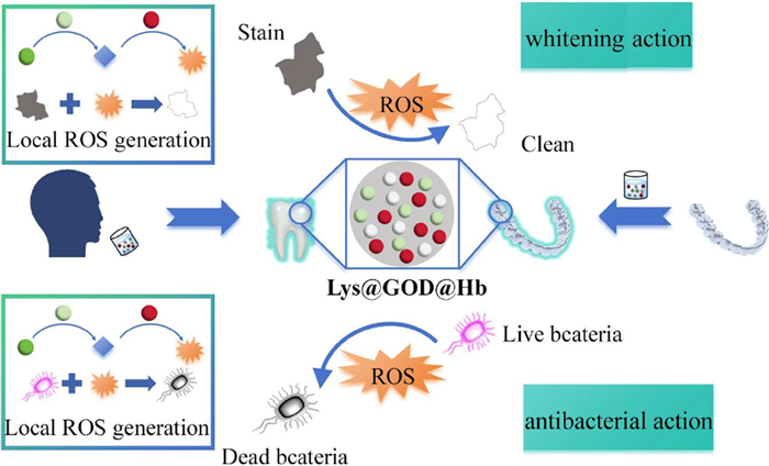
To address the above issues, we plan to introduce catalytic nanomedicine into the mouth of human beings, which would continuously utilize and manipulate the glucose-rich oral microenvironment to generate free radicals to rapidly damage bacteria and degrade various organic stains, hopefully simultaneously achieving effective elimination of pathogenic bacteria and tooth whitening (Scheme 1). Due to the rapid assembly of β-lysozyme (β-Lys) on the surface of various substrates both β-Lys, Hb and GOD would be co-superimposed on both plastic dental orthotics and real tooth to form protein nanofilm only after 5 min soak. The system's molecular foundation begins with the TCEP-induced reduction of Lys's disulfide bonds, triggering a conformational shift from α-helix/random coil to β-sheet-rich amyloid fibrils [41,42]. These amphiphilic fibrils spontaneously assemble into an adhesive nanofilm at interfaces, featuring abundant functional groups that enable robust both plastic dental orthotics and real tooth attachment while serving as an ideal scaffold for functional components [43]. Crucially, this amyloid network serves as a scaffold for co-assembling hemoglobin (Hb, oxygen supplier and ROS catalyst [44]) and GOD (glucose depleting agent and H2O2 generator [45]) through synergistic hydrophobic interactions, electrostatic complementarity, and hydrogen bonding - while preserving their native biological functions. As expected, the nanocoating can achieve the Hb-supported O2 supply [46], GOD-mediated glucose consumption and H2O2 generation [47,48], as well as the Hb-catalyzed conversion from H2O2 to ROS in an oral environment, which not only direct damage surrounding anaerobic and aerobic bacteria by catalytic medicine and hypoxia reversion but cut off their food source (glucose) via starvation therapy to further enhance the antibacterial effect [49]. Moreover, the local production of ROS also effectively oxidized the deposited stains on the plastic dental orthotics and real teeth as well as the free staining agents, resulting in the additional whitening and sustained prevention of staining. Importantly, the application of this multiple-functional coating did not affect the function and aesthetics of dental orthotics and teeth, making our nano-coating consisting of Lys, hemoglobin, and GOD (Lys@GOD@Hb) a promising candidate for oral applications [50,51].
Scheme 1
Scheme 1.
Schematic illustration of the construction, function and application of the composite nano-coating with persistent antibacterial and whitening activities for both human tooth and dental orthosis. (a) The construction of nano-coating on human teeth and dental orthosis. (b) Mechanism of whitening and stain prevention for teeth and orthosis after coating. (c) Mechanism of antibacterial action and bacteria prevention for teeth and orthosis after coating.
As to obtain the Lys@GOD@Hb nanocoating, the pro-solution containing β-Lys, Hb and GOD was prepared by mixing the above proteins with tris(2-carboxyethyl)phosphine hydrochloride in Tris solution. Then the antibacterial effect of this freshly prepared pre-solution against Gram-positive bacteria (Escherichia coli (E. coli)) and Gram-negative bacteria (Staphylococcus aureus (S. aureus)) was investigated by the paper inhibition method. As can be seen from Fig. S1a (Supporting information). The inhibition circles were respectively 13.04 cm2 for S. aureus and 5.87 cm2 for E. coli, indicating the excellent antibacterial performance of this combination. The quantitative data was also presented in Fig. S1b (Supporting information). Cytocompatibility evaluation via indirect contact testing showed > 90% L929 fibroblast viability after 72 h exposure to leachates from Lys@GOD@Hb-coated aligners (Fig. S2 in Supporting information), confirming biosafety for prolonged oral use.
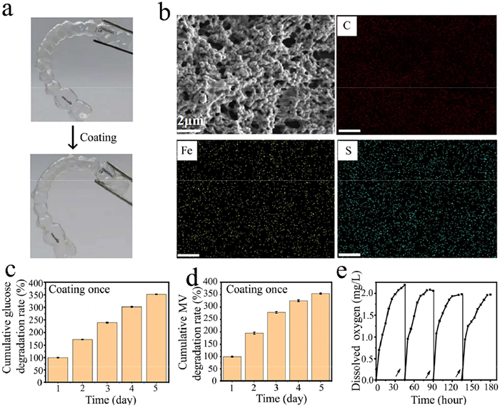
For nanocoating deposition, dental orthotics was immersed in the pre-solution for 5 min at room temperature, followed by ultrapure water rinsing to remove unbound components. The resultant transparent coating preserved device aesthetics and functionality (Fig. 1a). To confirm nanocoating formation, parallel scanning electron microscopy (SEM)/elemental mapping of coated (Fig. 1b) and uncoated (Fig. S3 in Supporting information) orthotics revealed nanoparticle coverage on coated surfaces versus featureless uncoated substrates. Elemental mapping showed uniform S (Lys-derived) and Fe (hemoglobin-derived) distribution exclusively on coated samples. This structural-compositional correlation underpins enhanced antibacterial/whitening performance in subsequent assays. As expected, the resulting Lys@GOD@Hb nanofilm was able to continuously consume glucose and generate ROS in at least 5 days only after one-time coating (Figs. 1c and d), which were detected by the glucose meter and methyl violet (typical ROS indicator [52]), respectively. This indicated the well-maintained function of GOD and Hb even in the nanofilm, which could gradually oxide the glucose, produce H2O2, and convert the H2O2 to ROS [53,54]. Moreover, the hemoglobin-integrated film also exhibited sustained O2 capture-release cyclability (Fig. 1e). By periodically reoxygenating the system to simulate oral cavity conditions, the nanofilm maintained a continuous oxygen supply that fueled glucose oxidation catalysis while simultaneously creating an oxygen-rich microenvironment to suppress anaerobic pathogens.
Figure 1
Figure 1.
(a) Photographs of plastic dental orthotics before and after nano-coating. (b) SEM analysis and elemental distribution of C, Fe and S on the plastic dental orthotics after nano-coating. Scale bar: 2 µm. (c) Glucose consumption of the plastic dental orthotics after nanocoating was detected by the glucose meter. (d) ROS generation of the plastic dental orthotics after nanocoating, which was detected by the UV–vis spectrum and methyl violet (typical ROS indicator). (e) Time-dependent oxygen release profile showing the hemoglobin-containing nanofilm's sustainable oxygen capture and release capability. Arrows indicate reoxygenation steps that simulate the continuous oxygen exchange occurring in oral environments. Data are mean ± SD (n = 3 independent experiments).
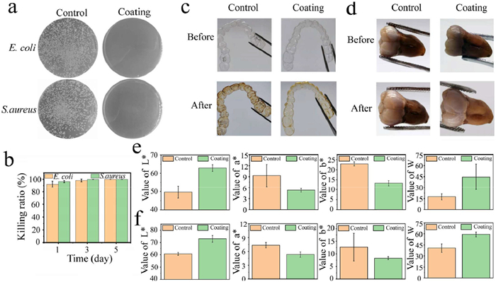
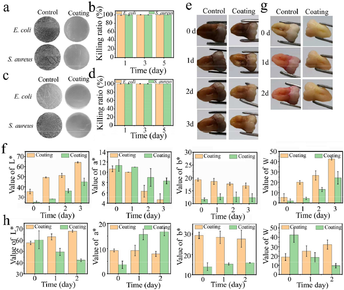
To evaluate antibacterial performance, Lys@GOD@Hb-coated dental orthotics were incubated with E. coli and S. aureus for 1, 3, 5 days, with untreated orthotics as controls. Bacterial viability on/near the orthotics was quantified (Figs. 2a and b, Fig. S4 in Supporting information). Results showed that ROS diffusion from the coating suppressed both Gram-positive and Gram-negative bacteria on and surrounding the orthotics, with no efficacy reduction over time. Notably, the coated group achieved 100% bacterial inhibition by day 5, confirming robust and sustained antimicrobial activity.
Figure 2
Figure 2.
(a) Bacterial colonies of E. coli and S. aureus on agar plates from the surface of orthotics with/without Lys@GOD@Hb nanocoating. (b) Quantitative inhibition rates against surface-adhered bacteria. (c) Prevention of staining: Orthotics with/without coating before and after immersion in glucose-containing soy sauce (simulated oral staining). (d) Teeth-whitening effect: Soy sauce-stained teeth treated in PBS-glucose with/without coated orthotics. (e) Quantitative analysis of stain prevention. (f) Quantitative analysis of teeth whitening. Data: mean ± SD (n = 3 independent experiments).
Based on its antibacterial efficacy, the transparent Lys@GOD@Hb nanofilm further demonstrated dual stain-degradation and whitening functions in simulated oral environments. To validate this, coated/uncoated orthotics were soaked with soy sauce (common stain source) alongside glucose at 37 ℃. Visually, coated orthotics resisted stain deposition (Fig. 2c) and actively whitened adjacent stained teeth (Fig. 2d, the teeth are from Zigong Fourth People's Hospital). The research processes meet the scientific and ethical requirements, which have been approved by the participants and Ethics Committee of Zigong Fourth People's Hospital, No. 2024–032). To quantitatively assess the whitening performance, we employed the standardized CIELAB color space system, which digitally characterizes color perception through three primary parameters: L* (lightness), a* (red-green axis), and b* (yellow-blue axis) [55]. The whiteness index (W), calculated as W = L* − |a*| − |b*|, was specifically developed to quantify the net whitening effect by accounting for changes in all three color dimensions simultaneously [56], which exactly represented the clean degree of orthotics and the surrounding teeth (W = 100 means the same as the unstained group). The quantitative analysis of the color and brightness of these groups were presented in Figs. 2e and f, which present the W value of orthotics with Lys@GOD@Hb coating is 3 times higher than that of orthotics without it, indicating the nanofilm prevented the staining of orthotics during usage. Moreover, the W value of the stained tooth after co-soaking with the Lys@GOD@Hb coated orthotics is far over that of stained tooth co-soaking with pure orthotics, indicating the additional property of Lys@GOD@Hb to whiten the surrounding tooth.
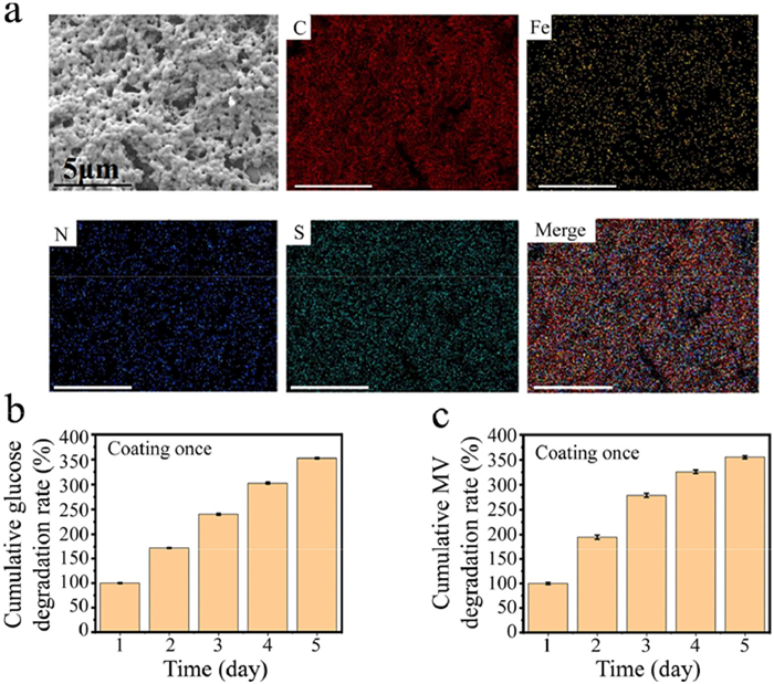
The coating of Lys@GOD@Hb on teeth was performed in the same way as the orthotics. Similar microstructures consisting of nanoparticles with uniform distribution of C, Fe, N, and S were observed by SEM, which confirmed the formation of Lys@GOD@Hb nanofilm on teeth (Fig. 3a). As expected, the Lys@GOD@Hb functionalized teeth also perform glucose consumption and ROS production, which can be sustained for at least 5 days after a single coating (Figs. 3b and c).
Figure 3
Figure 3.
(a) SEM analysis and elemental distribution of C, Fe, N and S on the teeth after nanocoating. Scale bar: 5 µm. (b) Glucose consumption of the teeth after nanocoating was detected by the glucose meter. (c) ROS generation of the teeth after nanocoating, which was detected by the UV–vis spectrum and methyl violet (typical ROS indicator). Data are mean ± SD (n = 3 independent experiments).
To investigate the antimicrobial efficacy of Lys@GOD@Hb-coated teeth, specimens were incubated with Gram-positive (S. aureus) and Gram-negative (E. coli) bacterial suspensions for 1, 3, and 5 days. Subsequent quantification of bacterial colonization was performed for both on-tooth surfaces and periventricular microenvironments, with uncoated teeth serving as the control group. As demonstrated in Figs. 4a–d, the nanocoated specimens exhibited significant and sustained inhibition of bacterial proliferation across both bacterial classifications throughout the 5-day observation period. Notably, this bacteriostatic effect remained consistent regardless of bacterial localization (on-tooth versus adjacent areas).
Figure 4
Figure 4.
(a) Bacterial colonies of E. coli and S. aureus on agar plates cultured from tooth surfaces with/without Lys@GOD@Hb coating. (b) Quantitative antibacterial rates against surface-adhered bacteria. (c) Bacterial colonies cultured from suspensions surrounding coated/uncoated teeth. (d) Quantitative antibacterial rates against surrounding bacteria. (e) Whitening performance of soy sauce-stained teeth with/without coating after 24 h in glucose-containing PBS (simulated oral conditions). (f) Quantitative colorimetric analysis of stain removal. (g) Anti-staining capability of coated teeth incubated with RhB/glucose-PBS for 24 h. (h) Quantitative analysis of anti-pollution efficacy. Data are mean ± SD (n = 3 independent experiments).
The whitening performance of Lys@GOD@Hb-coated teeth was systematically evaluated through chromatic analysis. Visual assessment (Fig. 4e) revealed pronounced bleaching efficacy on soy sauce-stained specimens following glucose-phosphate buffered saline (PBS) immersion. Quantitative colorimetric analysis (Fig. 4f) demonstrated a two-fold enhancement in whitening coefficient (ΔW) for coated versus uncoated teeth within 3 days of treatment, suggesting effective prevention of pigment deposition during routine exposure. To further characterize the anti-staining properties, specimens with differing surface treatments were subjected to Rhodamine B (RhB) challenge. Time-lapse imaging (Fig. 4g) documented progressive RhB adsorption on untreated surfaces versus maintained chromatic stability in coated specimens over 72 h. Corresponding spectrophotometric measurements (Fig. 4h) quantified a three-fold reduction in pigment adsorption (P < 0.01) for nanocoated teeth, confirming the material's capacity to preserve dental surface integrity against chromogenic agents.
In summary, a nanocoating (Lys@GOD@Hb) with whitening and antibacterial functions has been prepared using Lys, Hb and GOD via protein co-assembly, which can be applied on both teeth and oral materials to damage bacteria, whiten teeth and prevent further staining by the Hb and GOD co-mediated catalytic medicine, starvation therapy and hypoxia reversion, without affecting their aesthetics and function. Moreover, the nanocoating would stably anchor on the surface of teeth and oral materials to perform a long-term activity only through a simple process of mouthwash or soaking, which benefits the daily use of the Lys@GOD@Hb.
Declaration of competing interest
We declare that we have no known competing financial interests or personal relationships that could have appeared to influence the work reported in this paper.
CRediT authorship contribution statement
Yi Liu: Writing – original draft, Visualization, Funding acquisition. Xiaolin Yu: Formal analysis, Data curation. Wenyun Mu: Writing – review & editing, Visualization, Formal analysis, Data curation. Minsi Meng: Writing – original draft, Visualization, Formal analysis, Data curation. Baixue Li: Investigation. Jie Liu: Investigation. Haixin Qian: Investigation. Lin Weng: Writing – review & editing, Investigation. Tingting Yu: Investigation. Nan Hu: Investigation. Xin Chen: Supervision, Resources, Funding acquisition, Conceptualization. Yi Hao: Supervision, Resources, Conceptualization.
Acknowledgments
This work was supported by the "Young Talent Support Plan" of Xi'an Jiaotong University (X. Chen) and Foundation of Zigong Academy of Big Data and Artificial Intelligence in Medical Science (No. 2024-YGY-01-01 to Y. Liu)
Supplementary materials
Supplementary material associated with this article can be found, in the online version, at doi:10.1016/j.cclet.2025.111338.
X.Y. Hu, L. Xie, Z.Y. Xu, et al., ACS Appl. Mater. Interfaces 13 (2021) 35315–35327. doi: 10.1021/acsami.1c06774
[56]
H. Zhang, Y.N. Zhu, Y. Li, et al., Adv. Funct. Mater. 31 (2021) 2104799. doi: 10.1002/adfm.202104799
Scheme 1
Schematic illustration of the construction, function and application of the composite nano-coating with persistent antibacterial and whitening activities for both human tooth and dental orthosis. (a) The construction of nano-coating on human teeth and dental orthosis. (b) Mechanism of whitening and stain prevention for teeth and orthosis after coating. (c) Mechanism of antibacterial action and bacteria prevention for teeth and orthosis after coating.
Figure 1
(a) Photographs of plastic dental orthotics before and after nano-coating. (b) SEM analysis and elemental distribution of C, Fe and S on the plastic dental orthotics after nano-coating. Scale bar: 2 µm. (c) Glucose consumption of the plastic dental orthotics after nanocoating was detected by the glucose meter. (d) ROS generation of the plastic dental orthotics after nanocoating, which was detected by the UV–vis spectrum and methyl violet (typical ROS indicator). (e) Time-dependent oxygen release profile showing the hemoglobin-containing nanofilm's sustainable oxygen capture and release capability. Arrows indicate reoxygenation steps that simulate the continuous oxygen exchange occurring in oral environments. Data are mean ± SD (n = 3 independent experiments).
Figure 2
(a) Bacterial colonies of E. coli and S. aureus on agar plates from the surface of orthotics with/without Lys@GOD@Hb nanocoating. (b) Quantitative inhibition rates against surface-adhered bacteria. (c) Prevention of staining: Orthotics with/without coating before and after immersion in glucose-containing soy sauce (simulated oral staining). (d) Teeth-whitening effect: Soy sauce-stained teeth treated in PBS-glucose with/without coated orthotics. (e) Quantitative analysis of stain prevention. (f) Quantitative analysis of teeth whitening. Data: mean ± SD (n = 3 independent experiments).
Figure 3
(a) SEM analysis and elemental distribution of C, Fe, N and S on the teeth after nanocoating. Scale bar: 5 µm. (b) Glucose consumption of the teeth after nanocoating was detected by the glucose meter. (c) ROS generation of the teeth after nanocoating, which was detected by the UV–vis spectrum and methyl violet (typical ROS indicator). Data are mean ± SD (n = 3 independent experiments).
Figure 4
(a) Bacterial colonies of E. coli and S. aureus on agar plates cultured from tooth surfaces with/without Lys@GOD@Hb coating. (b) Quantitative antibacterial rates against surface-adhered bacteria. (c) Bacterial colonies cultured from suspensions surrounding coated/uncoated teeth. (d) Quantitative antibacterial rates against surrounding bacteria. (e) Whitening performance of soy sauce-stained teeth with/without coating after 24 h in glucose-containing PBS (simulated oral conditions). (f) Quantitative colorimetric analysis of stain removal. (g) Anti-staining capability of coated teeth incubated with RhB/glucose-PBS for 24 h. (h) Quantitative analysis of anti-pollution efficacy. Data are mean ± SD (n = 3 independent experiments).

 DownLoad:

 下载:
下载:


