Laboratory of Biomaterials and Translational Medicine, Department of Ultrasound, Center for Nanomedicine, The Third Affiliated Hospital, Sun Yat-sen University, Guangzhou 510630, China
b.
National Engineering Research Center for Tissue Restoration and Reconstruction, South China University of Technology, Guangzhou 510006, China
c.
Department of Urology, The Third Medical Center, Chinese PLA General Hospital, Beijing 100039, China
Received Date:
17 February 2025 Accepted Date:
26 April 2025 Revised Date:
23 April 2025 Available Online:
15 December 2025
Abstract:
Despite the promising potential of organic nanoscintillator-mediated radiodynamic therapy (RDT) in enhancing the effectiveness of immunotherapy, their cutaneous phototoxicity exacerbates the risk for immune-related adverse events (irAEs). Herein, we demonstrate that organic nanoscintillators, when combined with checkpoint blockade immunotherapy and exposed to X-ray-induced RDT, can trigger cutaneous irAEs. To address this challenge, we engineered diselenide-bridged silicon coatings on organic nanoscintillators, fine-tuning the steric hindrance of the protective layer by varying its thickness. This strategy enables radiation-triggered reactive oxygen species (ROS) generation while mitigating off-target phototoxicity through neutralizing ROS. By optimizing the steric hindrance to precisely control energy transfer between the organic nanoscintillators and surrounding oxygen molecules, we effectively reduce phototoxicity and mitigate off-tumor effects through engineered surface protection. Under X-ray irradiation exposure, the steric hindrance is rapidly deactivated through the dissociation of the silicon coating, activating RDT and inducing abundant ROS generation within tumor cells. In an orthotopic 4T1 breast cancer model, intravenous administration of these surface-engineered nanoscintillators, combined with anti-programmed death-1 (anti-PD-1) antibodies, results in robust anti-tumor immune responses, while minimizing cutaneous irAEs. This work offers valuable insights into how surface engineering can modulate the delicate balance between anti-tumor efficacy and off-tumor toxicity in nanoscintillator-mediated RDT.
Cancer immunotherapy has emerged as a cornerstone in the treatment of tumors, demonstrating considerable potential for clinical application [1–4]. However, many patients experience suboptimal responses due to the low immunogenicity and immunosuppressive nature of the tumor microenvironment (TME), limiting treatment efficacy [5–7]. Recently, radiodynamic therapy (RDT) has emerged as a promising strategy to modulate the TME by inducing immunogenic cell death (ICD) [8–10]. Traditional RDT systems, which combine organic photosensitizers (PSs) with scintillators, suffer from complex synthesis and energy loss [11–13]. In contrast, organic phosphorescent nanoscintillators (OPS) can efficiently generate singlet and triplet excitons under X-ray irradiation [11], offering enhanced immune activation potential, while this approach remains underexplored.
The combination of radio-activated therapy (RDT) and immunotherapy is a promising strategy in tumor treatment [14,15]. However, optimizing the safety and durability of immune responses remains a challenge [16], particularly in managing cutaneous immune-related adverse events (irAEs), which are crucial indicators of treatment efficacy [17–20]. Between 30% and 50% of patients undergoing immunotherapy experience cutaneous irAEs, some of which can be life-threatening [21–24]. Incorporating heteroatoms into organic phosphorescent nanoscintillators (OPS) enhances ultraviolet-visible (UV–vis) absorption and intersystem crossing, leading to increased triplet exciton generation and phototoxicity [11]. These excitons induce significant phototoxicity, and when OPS accumulate in the skin, they can cause cutaneous damage and inflammation, thereby amplifying the risk of irAEs in combination with immunotherapy [25–27]. However, there is limited research on balancing immune activation with off-target immunotoxicity in RDT-immunotherapy combinations.
RDT generates cytotoxic reactive oxygen species (ROS) via electron transfer or energy exchange with oxygen molecules [12], leading to phototoxic damage in cutaneous tissues [15,28–30]. Recent advancements highlight the importance of surface-engineering nanoscintillators to control ROS generation by manipulating the energy transfer process [31,32]. Silicon-based coatings, particularly diselenide-bridged silicon, offer stability, biocompatibility [33–36], and susceptibility to dissociation under X-ray exposure [37–41], enabling spatiotemporal control of ROS production. Additionally, diselenide compounds exhibit ROS-scavenging and anti-inflammatory properties, which can help mitigate skin inflammation [42–45]. Consequently, the development of diselenide-bridged silicon-coated RDT systems holds promise for enhancing immune responses while reducing cutaneous immune-related adverse events (irAEs).
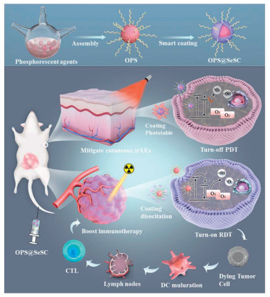
Herein, we designed and synthesized a photoswitchable surface-engineered organic phosphorescent nanoscintillator (OPS@SeSC) to improve cancer immunotherapy and reduce the risk of cutaneous irAEs (Scheme 1). The OPS were encapsulated with X-ray-sensitive diselenide-bridged silicon, forming OPS@SeSC with varying coating thicknesses (2, 5, 15 nm). The diselenide-bridged silicon coating offers photoswitchable steric hindrance, enabling precise control of ROS generation. Among the formulations, OPS@SeSC-5 exhibited superior performance by reducing off-tumor toxicity and controlling ROS production. The X-ray-responsive coating dissociates upon radiation exposure, enhancing tumor-targeted ROS production. Additionally, OPS@SeSC-5 triggers the release of tumor-associated antigens and damage-associated molecular patterns, boosting immunogenicity and promoting anticancer immune responses. Combined with anti-programmed death-1 (anti-PD-1), it demonstrated pronounced abscopal effects. This strategy provides a novel approach for developing a radiation-driven immunotherapeutic enhancer with reduced cutaneous irAEs.
Scheme 1
Scheme 1.
Schematic illustration of a photoswitchable RDT system mediates by surface-engineered organic phosphorescent nano-scintillator (OPS@SeSC) that can controllably generate ROS to potentiate cancer immunotherapy and diminish the risk of cutaneous irAEs.
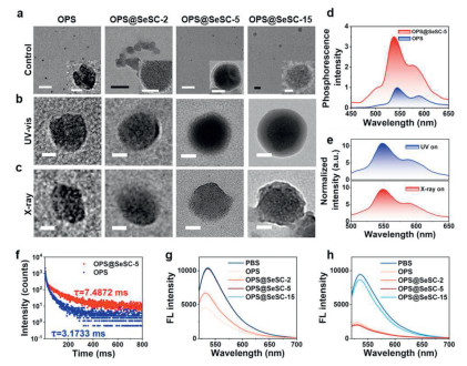
According to previous reports [11], the organic phosphorescent molecule 9,9′-(6-iodophenoxy-1,3,5-triazine-2,4-diyl)bis(9H-carbazole) (ITC) is successfully synthesized and characterized via NMR spectroscopy and elemental analysis (Fig. S1 in Supporting information). ITC was co-assembled with amphiphilic phospholipid molecules to form organic phosphorescent nanoscintillators (OPS), which exhibited a monodisperse spherical morphology with an average size of approximately 40 nm, indicating excellent dispersibility (Fig. 1a). To prepare surface-engineered organic phosphorescent nanoscintillators (OPS@SeSC), a diselenide-bridged silicon coating is applied to the surface of the OPS particles. Transmission electron microscope (TEM) images clearly illustrate homogeneous spherical particles with a distinct core-shell structure, highlighting a 5 nm thick shell layer in contrast to the bare OPS (Fig. 1a and Fig. S2a in Supporting information). The element analysis of OPS@SeSC-5 confirms the presence of C, N, O, I, Se, and Si in the nanoparticles (Fig. S2b in Supporting information). The Fourier transform infrared (FTIR) spectra further support the successful introduction of the diselenide-bridged silicon coating, as evidenced by characteristic Se–Se and C–Se stretching vibrations within the 550–750 cm−1 range (Fig. S2c in Supporting information). To investigate the impact of coating thickness, different OPS@SeSC formulations are prepared by varying the ratio of OPS to diselenide-bridged silicon, yielding coatings with thicknesses of 2 nm (OPS@SeSC-2), 5 nm (OPS@SeSC-5), and 15 nm (OPS@SeSC-15) (Fig. 1a). A decrease in surface zeta potential across the OPS@SeSC samples, compared to bare OPS, indicated successful encapsulation with varying thicknesses of the diselenide-bridged silicon coating (Fig. S2d in Supporting information). Subsequent evaluation of the photoswitchable properties of under UV–vis light and X-ray irradiation demonstrated that all samples maintained their spherical morphology and structural integrity under UV–vis light exposure, indicating the high photostability of the diselenide-bridged silicon coating (Fig. 1b). In contrast, X-ray irradiation leads to rapid degradation of the diselenide-bridged silicon coating in samples with a coating thickness below 5 nm, exposing the OPS core (Fig. 1c). Notably, OPS@SeSC-15, with its thicker coating, fails to effectively degrade and expose the core under X-ray irradiation, leading to ineffective switching. These findings underscore the crucial role of coating thickness in optimizing the photoswitchable performance of the RDT system.
Figure 1
Figure 1.
Characterization of surface-engineered organic phosphorescent nano-scintillator. (a) TEM images of OPS, OPS@SeSC-2, OPS@SeSC-5, OPS@SeSC-15. Scale bar: 500 nm (20 nm in inserted figures). (b, c) TEM images of OPS, OPS@SeSC-2, OPS@SeSC-5, OPS@SeSC-15 with UV–vis light irradiation (10 min) and 2 Gy X-ray irradiation. Scale bar: 20 nm. (d) Phosphorescence spectra of OPS@SeSC-5 under excitation at 340 nm. (e) Radioluminescence spectra of OPS@SeSC-5. (f) Time-resolved decay profile of phosphorescence emission wavelength at 544 nm. (g, h) The fluorescence spectra of SOSG in OPS@SeSC-5 solution (g) with UV–vis light irradiation (10 min), and (h) with 2 Gy X-ray irradiation.
The influence of diselenide-bridged silicon coating thickness on the photophysical properties of OPS@SeSC is systematically evaluated. The UV–vis absorption spectra and fluorescence emission profiles of OPS@SeSC formulations remains consistent with those of the parent OPS, indicating that encapsulation with the diselenide-bridged silicon coating do not alter the inherent chemical characteristics of the organic phosphorescent nanoscintillators (Figs. S3a and b in Supporting information). Upon UV–vis light excitation, OPS@SeSC exhibit dual emission peaks at 544 and 589 nm, indicative of photoluminescence, as confirmed through time-gated photoluminescence spectroscopy (Fig. 1d). The phosphorescence emission observes under X-ray irradiation closely mirrored the emission profile under UV–vis excitation, confirming the scintillation properties of OPS@SeSC (Fig. 1e and Fig. S4 in Supporting information). Notably, the absolute phosphorescent quantum yield (QY) increases significantly with thicker diselenide-bridged silicon coatings (QY: 1.92% for bare OPS, 2.14% for OPS@SeSC-2, 5.32% for OPS@SeSC-5, and 5.35% for OPS@SeSC-15). Moreover, the phosphorescence emission lifetime at 544 nm for OPS@SeSC-5 is significantly extended to 7.4872 ms, compared to 3.1733 ms for bare OPS (Fig. 1f). These results suggest that the diselenide-bridged silicon coating effectively created steric hindrance on the surface of OPS@SeSC, preventing energy transfer between the OPS and surrounding O2 molecules, thereby reducing dynamic fluorescence quenching. Post-X-ray pre-irradiation, phosphorescence emission from OPS@SeSC decreases notably, with minimal effect observed on bare OPS, highlighting that the degradation of the diselenide-bridged silicon coating exposed the OPS core to surrounding O2 molecules, leading to dynamic fluorescence quenching (Fig. S5 in Supporting information). Importantly, the fluorescence quenching efficiency inversely correlates with the thickness of the diselenide-bridged silicon coating, indicating the fine-tuning the steric hindrance of the protective coating by varying its thickness. These findings confirm that the surface-engineered OPS@SeSC introduces steric hindrance that efficiently orchestrated energy transfer between the OPS and surrounding O2 molecules. Furthermore, the photoswitchable steric hindrance can be finely tuned by adjusting the thickness of the diselenide-bridged silicon coating, providing a precise mechanism for controlling photophysical properties.
To evaluate the regulatory effect of photoswitchable steric hindrance on ROS generation, we monitored 1O2 production from surface-engineered OPS@SeSC nanoscintillators under UV–vis and X-ray irradiation using a singlet oxygen sensor green (SOSG) probe. Under UV–vis light, bare OPS produced substantial 1O2 (Fig. 1g, Figs. S6 and S7a in Supporting information), whereas OPS@SeSC-5 and OPS@SeSC-15 exhibited markedly reduced ROS generation (Figs. 1g and h, Figs. S6 and S7a), attributable to the steric hindrance imposed by the diselenide-bridged silicon coating. Notably, the degree of inhibition correlated with coating thickness, with thicker coatings conferring greater suppression. Upon X-ray irradiation, the steric barrier in OPS@SeSC-2 and OPS@SeSC-5 was effectively deactivated, restoring 1O2 production to levels comparable to uncoated OPS, while OPS@SeSC-15 remained inhibited (Fig. 1h and Fig. S7b in Supporting information). These findings demonstrate that the diselenide-bridged silicon coating enables precise, spatiotemporal control of ROS generation, suppressing off-target phototoxicity under ambient light while permitting robust radiodynamic activation.
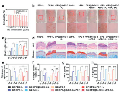
To assess the phototoxicity profile of photoswitchable OPS@SeSC-5 nanoscintillators, we conducted in vitro cytotoxicity assays using MSF and JB6-C30 cell lines. OPS@SeSC-5 and uncoated OPS nanoscintillators exhibited negligible cytotoxicity in the absence of light, with cell viability exceeding 80% at ITC concentrations up to 16 µg/mL (Fig. 2a and Fig. S8 in Supporting information). Upon UV–vis irradiation (50 mW/cm2 for 10 min), cells treated with OPS (4 µg/mL ITC) displayed a marked reduction in viability to 50%, indicative of pronounced phototoxicity. In contrast, OPS@SeSC-5-treated cells maintained high viability post-irradiation, comparable to dark controls, demonstrating that the diselenide-bridged silicon coating effectively suppresses ROS generation under UV–vis light (Figs. S8 and S9 in Supporting information). The in vivo phototoxicity and risk of cutaneous irAEs were further evaluated. All animal experimental protocols implemented in this study received approval from the Ethics Committee for the Use of Experimental Animals at South China University of Technology (the assigned approval/accreditation No 2019031). OPS@SeSC-5 demonstrates exceptional colloidal stability under physiological conditions, attributed to PEGylated phospholipid molecules (Fig. S10 in Supporting information). Biodistribution studies revealed significant skin accumulation of both OPS and OPS@SeSC (Fig. S23d in Supporting information). Mice treated with OPS or Ce6 (positive control) develop severe erythema and edema at irradiated sites, while phosphate buffered saline (PBS)-treated mice remained unaffected (Fig. 2b). The combination of OPS with anti-PD-1 (αPD-1) induces the most severe erythema and edema, suggesting that phototoxicity exacerbates cutaneous irAEs. In contrast, mice treated with OPS@SeSC-5 exhibit minimal erythema due to its turn off phototoxicity. Additionally, treatment with αPD-1 in combination with OPS@SeSC-5 reduce erythema and edema, indicating the decrease in risk of cutaneous irAEs due to the anti-inflammatory effects of the diselenide-bridged silicon coating through ROS-scavenging activity (Fig. S9 in Supporting information) [42,43]. For quantitative assessment, mice are sacrificed, and skin tissue samples (1.5 cm × 1.5 cm) are collected to evaluate lesion severity. Mice treated with OPS or αPD-1 have skin weights 1.75-fold and 1.85-fold higher, respectively, than the PBS group (Fig. 2c). Notably, the combination of OPS and αPD-1 with light irradiation leads to the most significant increase in skin weight (2.13-fold), underscoring the heightened risk of cutaneous irAEs due to phototoxicity. In contrast, a slight elevation in skin weight is observed following treatment with OPS@SeSC-5 combined with αPD-1 under light irradiation, suggesting minimal risk of cutaneous irAEs. Histological examination further confirmed reduced tissue damage and inflammation in OPS@SeSC-5-treated mice (Figs. 2d–f), corroborated by lower tumor necrosis factor-alpha (TNF-α) and interleukin-6 (IL-6) levels (Figs. 2g and h). Collectively, these findings demonstrate that diselenide-bridged silicon coatings on organic nanoscintillators effectively mitigate phototoxicity and off-target effects, offering a promising strategy to minimize cutaneous irAEs in radiodynamic-immunotherapy.
Figure 2
Figure 2.
Evaluation of phototoxicity and cutaneous irAEs. (a) Cell viability of MSF cells treated with OPS or OPS@SeSC-5 with LED (50 mW/cm2) irradiation for 10 min. (b) Skin photosensitivity of mice treated with formulations at 4 days. (c) Weight of the irradiated skin samples 1.5 cm × 1.5 cm in size at 4 days post-light irradiation. (d) H&E-stained and Masson-stained analysis of the irradiated skin samples. Scale bar: 20 µm. (e, f) Corresponding quantification of epidermis thickness and collagen content. (g, h) Levels of pro-inflammatory cytokines, including TNF-α and IL-6 on day 3. All data are the mean ± SD (n = 3). *P < 0.05, **P < 0.01, ***P < 0.001, ****P < 0.0001.
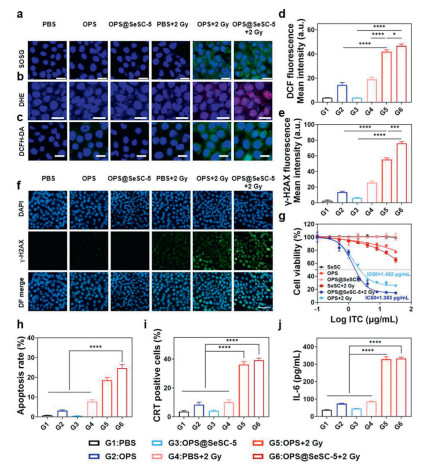
The in vitro assessment of the on-demand RDT process is conducted by subjecting 4T1 cells to X-ray irradiation. Following a 6-h incubation period, efficient internalization of OPS@SeSC-5 by 4T1 cells is observed (Fig. S11 in Supporting information). Subsequently, the efficacy of RT is evaluated using SOSG to monitor the generation of 1O2. Compared to X-ray, OPS, and OPS@SeSC-5 groups, a significant increase in green fluorescence, indicating intracellular 1O2 generation, is detected in cells treated with OPS or OPS@SeSC-5 under X-ray irradiation (Fig. 3a and Fig. S12 in Supporting information), suggesting that the diselenide-bridged silicon coating effectively triggered the on-demand initiation of RDT. Additionally, the levels of O2•− radicals in 4T1 cells treated with OPS@SeSC-5 are elevated compared to those treated with OPS alone, attributed to the radiosensitization effect of selenide (Fig. 3b and Fig. S13 in Supporting information). The increased levels of ROS in 4T1 cells treated with OPS@SeSC-5 following X-ray exposure further confirms that the coating does not compromise the radiotherapeutic potential (Figs. 3c and d, Fig. S14 in Supporting information). Furthermore, the degree of DNA damage is evaluated, revealing that γ-H2AX fluorescence in 4T1 cells treated with OPS@SeSC-5 combined with X-ray irradiation is significantly higher than in cells treated with alternative agents, indicating enhanced DNA double-strand breaks (Figs. 3e and f). Cell viability decreases markedly following treatment with this switchable RDT approach (Fig. 3g). Notably, the combination of OPS@SeSC-5 and X-ray results in more extensive DNA double-strand breaks and higher cytotoxicity compared to OPS with X-ray, which is consistent with the enhanced radiosensitization effect. Treatment with OPS@SeSC-5 and 2 Gy of X-ray irradiation leads to a pronounced increase in apoptosis and necrosis compared to other groups (Fig. 3h and Fig. S15 in Supporting information), demonstrating superior antitumor efficacy. Collectively, these findings highlight the potent anticancer activity of OPS@SeSC-5 through the synergistic combination of RDT and radiotherapy.
Figure 3
Figure 3.In vitro ROS generation and anti-tumor effect of OPS@SeSC-5-mediated RDT. (a) The fluorescence signal of SOSG in the treated 4T1 cells in darkness or with 2 Gy irradiation. SOSG: green; 4′, 6-diamidino-2′-phenylindole (DAPI): blue. (b) The fluorescence signal of dihydroethidium (DHE) in the treated 4T1 cells in darkness or with 2 Gy irradiation. DHE: red; DAPI: blue. (c) The fluorescence signal of 2′, 7′-dichlorodihydrofluorescein diacetate (DCFH-DA) in the treated 4T1 cells in darkness or with 2 Gy irradiation. DCFH-DA: green; DAPI: blue. Scale bar: 20 µm. (d) Quantitative data on the average fluorescence intensity of DCFH-DA under different treatments. (e) Quantitative data on the average fluorescence intensity of γ-H2AX under different treatments. (f) Representative immunofluorescence images of γ-H2AX in the treated 4T1 cells after 2 Gy of radiation. γ-H2AX: green; DAPI: blue. Scale bar: 50 µm. (g) Cell viabilities of 4T1 cells. (h) Late apoptosis rate of 4T1 cells under different treatments. (i) CRT expression in the treated 4T1 cells. (j) The secretion levels of IL-6 in BMDCs. Data represent mean ± SD. *P < 0.05, ***P < 0.001, ****P < 0.0001.
Recent studies have demonstrated that RDT can effectively induce tumor ICD, characterized by the release of danger-associated molecular patterns (DAMPs) such as calreticulin (CRT), high-mobility group box 1 (HMGB1), and adenosine triphosphate (ATP) [9,46]. This process primes antigen-presenting cells (APCs), triggering specific antitumor immune responses [47–49]. Treatment of 4T1 cells with OPS@SeSC-5 under X-ray irradiation resulted in a significant increase in CRT exposure compared to other treatment groups (Fig. 3i and Fig. S16 in Supporting information), along with elevated HMGB1 release and ATP secretion (Figs. S17 and S18 in Supporting information), suggesting a potent ICD response. Subsequent co-culture of bone marrow-derived macrophages (BMDMs) with treated cancer cells revealed that OPS@SeSC-5 combined with X-ray produced the highest proportion of mature dendritic cells (DCs) relative to other treatments (Fig. S19 in Supporting information). In parallel, an upregulation of pro-inflammatory cytokines is observed in mature DCs treated with OPS@SeSC-5 and X-ray (Fig. 3j and Fig. S20 in Supporting information). These results indicate that RT utilizing switchable OPS@SeSC-5 not only enhances DC maturation but also facilitates tumor antigen presentation, thereby potentially augmenting the antitumor immune response.
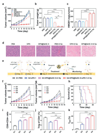
OPS@SeSC-5 exhibits minimal hemolysis, confirming its hemocompatibility (Fig. S22 in Supporting information). Tumor accumulation and biodistribution are evaluated by quantifying iodine(I) content using ICP-MS. OPS@SeSC-5 demonstrates prolonged systemic circulation with a half-life of approximately 8 h, attributed to the PEGylated phospholipid coating (Fig. S23a in Supporting information). Notably, time-dependent tumor accumulation of OPS@SeSC-5 increases post-injection, peaking at 10 h, indicating this as the optimal window for X-ray irradiation (Figs. S21 and S23b in Supporting information). Furthermore, substantial accumulation in liver tissues is observed, suggesting the involvement of the reticuloendothelial system in its clearance, consistent with previous reports on nanoparticle biodistribution (Fig. S23c in Supporting information) [50,51]. The in vivo anticancer efficacy of OPS@SeSC-5 is assessed. X-ray irradiation alone demonstrates limited tumor growth inhibition relative to the PBS control, highlighting the suboptimal therapeutic efficacy of standalone radiotherapy (Figs. 4a and b, Fig. S24 in Supporting information). However, OPS@SeSC-5 combined with X-ray irradiation significantly suppresses tumor growth, whereas OPS@SeSC-5 without X-ray shows negligible effects, underscoring the enhanced antitumor efficacy of OPS@SeSC-5-mediated RDT. Moreover, OPS@SeSC-5 exhibits superior efficacy over OPS in combination with X-ray, attributable to the radiosensitizing effect of diselenide. Hematoxylin and eosin (H&E) staining further reveals that OPS@SeSC-5 with X-ray leads to the highest levels of apoptosis and necrosis in tumor tissues (Fig. 4d). Furthermore, mice treated with OPS@SeSC-5 also promote a significant release of HMGB-1 in comparison to other treatments, indicating the ICD effects (Fig. 4c). Given the increased tumor immunogenicity, the potential to stimulate an antitumor immune response is explored. Treatment with OPS@SeSC-5 and X-ray induced greater DC maturation in the draining lymph nodes compared to OPS with X-ray or X-ray alone, while PBS and OPS@SeSC-5 without X-ray had minimal impact on DC activation (Figs. S25 and S26 in Supporting information). These findings suggest that radiotherapy-induced ICD may potentiate a robust antitumor immune response. Consistent with DC maturation, the combined treatment induces the highest levels of pro-inflammatory cytokines, including IL-6 and TNF-α, further supporting its immunostimulatory effects (Fig. S27 in Supporting information). Importantly, no significant systemic toxicity is observed throughout the treatment period, demonstrating the safety of OPS@SeSC-5-mediated RDT (Figs. S28 and S29 in Supporting information). These results collectively demonstrate that photoswitchable surface-engineered organic phosphorescent nanoscintillator to enhance cancer immunotherapy and mitigate the risk of cutaneous irAEs.
Figure 4
Figure 4.
Therapeutic efficacy of OPS@SeSC-5-mediated RDT and synergistic RDT-immunotherapy in vivo. (a) Tumor growth curves and (b) tumor weights in each group. (c) HMGB-1 expressions in tumor. (d) Representative images of H&E-stained tumor slices after various treatments. Scale bar: 100 µm. (e) Scheme of RDT-immunotherapy treatment options in vivo. (f, g) Distant tumor and proximal tumor growth curves after different treatments (n = 3). (h) Survival rates (n = 5). (i, j) Ratio of CD8+/CD4+ cell (i) and population of Treg cell (j) in distant tumor after 10-day treatment. (k) Effective memory T cell (TEM) levels in the spleen after 20-day treatment. Data are represented as mean ± SD. **P < 0.01, ***P < 0.001, ****P < 0.0001.
Tumor immunogenicity is a crucial strategy for enhancing the efficacy of cancer immunotherapy [52–54]. In this study, a bilateral 4T1 tumor model is utilized to explore the potential of boosting systemic antitumor immune responses and suppressing the growth of distant tumors through RDT-induced immunogenicity (Fig. 4e). Monotherapy with αPD-1 checkpoint blockade results in rapid progression of both primary and distant tumors, reflecting a limited therapeutic response to immune checkpoint inhibition (Figs. 4f and g, Fig. S30 in Supporting information). Conversely, while OPS@SeSC-5 combined with X-ray irradiation effectively suppresses primary tumor growth, it exhibits reduced efficacy against distant tumors. Remarkably, the combination of OPS@SeSC-5, X-ray, and αPD-1 significantly inhibits the growth of both primary and distant tumors, suggesting the induction of systemic antitumor immunity and an abscopal effect. This synergistic combination therapy leads to sustained antitumor responses and a marked improvement in overall survival (Fig. 4h). Mechanistically, treatment with OPS@SeSC-5, X-ray, and αPD-1 increases T-cell infiltration in distant tumors (Fig. 4i and Fig. S31 in Supporting information) [55,56]. This combined approach yields a 23.43-fold increase in CD8+ T-cell populations within distant tumors, signifying the generation of robust, tumor-specific T-cell responses [57]. Moreover, higher levels of CD4+ T cells are observed, which are linked to promoting antitumor immunity, alongside a decrease in regulatory T cells (Foxp3+CD4+ T cells), highlighting the reversal of the immunosuppressive TME (Fig. 4j and Fig. S32 in Supporting information). Further analysis demonstrates sustained antitumor immunity, with an increase in effector memory CD8+ T cells in the spleen (Fig. 4k and Fig. S33 in Supporting information). Additionally, OPS@SeSC-5 combined with X-ray and αPD-1 treatment triggers elevated secretion of pro-inflammatory cytokines, such as IL-6 and TNF-α, indicating a robust activation of the pro-inflammatory immune response (Fig. S34 in Supporting information). Importantly, histological assessment of major organs demonstrates no significant toxicity or tissue damage, confirming the biocompatibility of the combination treatment (Fig. S35 in Supporting information). In summary, these findings illustrate that OPS@SeSC-5-mediated RDT can synergistically enhance the efficacy of immunotherapy by promoting systemic antitumor immunity and reshaping the immunosuppressive TME, offering a promising strategy for improving cancer treatment outcomes.
Conventional PSs cannot be directly activated by X-rays due to the energy mismatch between X-rays and PSs [8]. PSs conjugated with noble metal scintillators generate ROS through X-ray-induced UV–vis luminescence [10,11]. However, inefficient energy transfer often reduces therapeutic efficacy [12,13]. To enhance RDT performance, we incorporated heavy halogens like bromine and iodine into phosphorescent PSs, improving X-ray absorption and promoting long-lived triplet exciton formation [11]. Organic phosphorescent nanoscintillators also trigger Auger reactions, producing over 75% triplet excitons under X-ray exposure, significantly improving RDT efficiency [12], while the accumulation in the skin poses a risk of cutaneous irAEs [58]. To address this limitation, we engineered diselenide-bridged silicon coatings on organic nanoscintillators, offering photoswitchable steric hindrance that allows precise modulation of energy transfer between the nanoscintillators and surrounding oxygen molecules. By adjusting the coating thickness, energy transfer can be finely tuned to optimize radiation-triggered ROS generation, thereby stimulating immune responses while minimizing off-target phototoxicity. This strategy enhances the safety and efficacy of cancer immunotherapy. Overall, our work provides new insights into developing photoswitchable, surface-engineered RDT systems that effectively balance anti-tumor efficacy with reduced off-target toxicity.
Declaration of competing interest
The authors declare that they have no known competing financial interests or personal relationships that could have appeared to influence the work reported in this paper.
CRediT authorship contribution statement
Kai Li: Writing – original draft, Methodology, Investigation, Data curation, Conceptualization. Hui Fang: Methodology, Investigation, Data curation. Feixia Ruan: Formal analysis, Data curation. Xiaochun Xie: Investigation, Data curation. Huicong Zhou: Validation, Data curation. Zhenjun Luo: Validation, Data curation. Dan Shao: Supervision, Resources. Mingqiang Li: Supervision, Resources. Qing Yuan: Writing – review & editing, Supervision, Project administration, Funding acquisition. Fangman Chen: Writing – review & editing, Supervision, Project administration, Funding acquisition. Yu Tao: Writing – review & editing, Supervision, Project administration, Funding acquisition.
Acknowledgment
This research was supported by the Science and Technology Program of Guangzhou (No. 2023A03J0218).
Supplementary materials
Supplementary material associated with this article can be found, in the online version, at doi:10.1016/j.cclet.2025.111261.
Scheme 1
Schematic illustration of a photoswitchable RDT system mediates by surface-engineered organic phosphorescent nano-scintillator (OPS@SeSC) that can controllably generate ROS to potentiate cancer immunotherapy and diminish the risk of cutaneous irAEs.
Figure 1
Characterization of surface-engineered organic phosphorescent nano-scintillator. (a) TEM images of OPS, OPS@SeSC-2, OPS@SeSC-5, OPS@SeSC-15. Scale bar: 500 nm (20 nm in inserted figures). (b, c) TEM images of OPS, OPS@SeSC-2, OPS@SeSC-5, OPS@SeSC-15 with UV–vis light irradiation (10 min) and 2 Gy X-ray irradiation. Scale bar: 20 nm. (d) Phosphorescence spectra of OPS@SeSC-5 under excitation at 340 nm. (e) Radioluminescence spectra of OPS@SeSC-5. (f) Time-resolved decay profile of phosphorescence emission wavelength at 544 nm. (g, h) The fluorescence spectra of SOSG in OPS@SeSC-5 solution (g) with UV–vis light irradiation (10 min), and (h) with 2 Gy X-ray irradiation.
Figure 2
Evaluation of phototoxicity and cutaneous irAEs. (a) Cell viability of MSF cells treated with OPS or OPS@SeSC-5 with LED (50 mW/cm2) irradiation for 10 min. (b) Skin photosensitivity of mice treated with formulations at 4 days. (c) Weight of the irradiated skin samples 1.5 cm × 1.5 cm in size at 4 days post-light irradiation. (d) H&E-stained and Masson-stained analysis of the irradiated skin samples. Scale bar: 20 µm. (e, f) Corresponding quantification of epidermis thickness and collagen content. (g, h) Levels of pro-inflammatory cytokines, including TNF-α and IL-6 on day 3. All data are the mean ± SD (n = 3). *P < 0.05, **P < 0.01, ***P < 0.001, ****P < 0.0001.
Figure 3In vitro ROS generation and anti-tumor effect of OPS@SeSC-5-mediated RDT. (a) The fluorescence signal of SOSG in the treated 4T1 cells in darkness or with 2 Gy irradiation. SOSG: green; 4′, 6-diamidino-2′-phenylindole (DAPI): blue. (b) The fluorescence signal of dihydroethidium (DHE) in the treated 4T1 cells in darkness or with 2 Gy irradiation. DHE: red; DAPI: blue. (c) The fluorescence signal of 2′, 7′-dichlorodihydrofluorescein diacetate (DCFH-DA) in the treated 4T1 cells in darkness or with 2 Gy irradiation. DCFH-DA: green; DAPI: blue. Scale bar: 20 µm. (d) Quantitative data on the average fluorescence intensity of DCFH-DA under different treatments. (e) Quantitative data on the average fluorescence intensity of γ-H2AX under different treatments. (f) Representative immunofluorescence images of γ-H2AX in the treated 4T1 cells after 2 Gy of radiation. γ-H2AX: green; DAPI: blue. Scale bar: 50 µm. (g) Cell viabilities of 4T1 cells. (h) Late apoptosis rate of 4T1 cells under different treatments. (i) CRT expression in the treated 4T1 cells. (j) The secretion levels of IL-6 in BMDCs. Data represent mean ± SD. *P < 0.05, ***P < 0.001, ****P < 0.0001.
Figure 4
Therapeutic efficacy of OPS@SeSC-5-mediated RDT and synergistic RDT-immunotherapy in vivo. (a) Tumor growth curves and (b) tumor weights in each group. (c) HMGB-1 expressions in tumor. (d) Representative images of H&E-stained tumor slices after various treatments. Scale bar: 100 µm. (e) Scheme of RDT-immunotherapy treatment options in vivo. (f, g) Distant tumor and proximal tumor growth curves after different treatments (n = 3). (h) Survival rates (n = 5). (i, j) Ratio of CD8+/CD4+ cell (i) and population of Treg cell (j) in distant tumor after 10-day treatment. (k) Effective memory T cell (TEM) levels in the spleen after 20-day treatment. Data are represented as mean ± SD. **P < 0.01, ***P < 0.001, ****P < 0.0001.

 DownLoad:

 下载:
下载:


