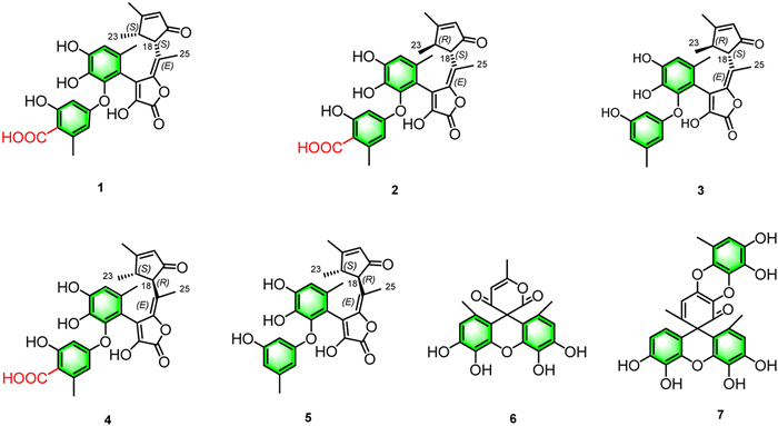
Figure 1.
The structures of compounds 1–7.

Diphenyl ethers (DPEs) are well-documented in microorganisms and plants, exhibiting a wide range of bioactive properties, including antibacterial, antioxidant, antitumor, antihemolytic, neuroprotective, and anti-Alzheimer effects [1–3]. Among these, gerfelin, a notable diphenyl ether, has been identified as a novel inhibitor of geranylgeranyl diphosphate synthase, isolated from Beauveria felina QN22047 [4]. Similarly, γ-butyrolactone, a five-membered heterocycle with ester functionality, has garnered significant attention in drug discovery due to its presence in numerous biologically active molecules [5]. Several Food and Drug Administration (FDA)-approved drugs containing γ-butyrolactone serve diverse purposes, including as diuretics, anticancer agents, contraceptives, and treatments for heart disease and glaucoma [6]. The conjugated cyclopentenone (CP) moiety, another important pharmacophore, plays a pivotal role in anticancer agents such as cyclopentenone prostaglandins (CyPGs) and clavulones [7,8]. Despite the growing discoveries of both natural and synthetic DPEs [3,9], γ-butyrolactones [10], and CP derivatives [11], hybrids of DPEs fused with conjugated γ-butyrolactones and CPs, as well as spiro-DPEs, remain undescribed.
Aspergillus is a genus of ubiquitous fungi with both significant pathological and therapeutic importance [12,13]. Recent studies have identified 361 new secondary metabolites from Aspergillus species between 1915 and 2020 [14]. The search for novel bioactive structures in endophytic fungi has become a major research focus, with Aspergillus serving as a valuable reservoir of bioactive secondary metabolites [15,16]. These species produce a wide range of metabolites, including polyketides, alkaloids, terpenoids, and sterols, with over half exhibiting notable bioactivities [17,18]. Although DPEs, γ-butyrolactones, and CPs have been identified in Aspergillus species [3,9–11], significant breakthroughs in the discovery of their hybrids or spiro-diphenyl ethers have yet to be achieved.
As part of our ongoing study on the endophytic fungi associated with Bufo gargarizans [19,20], Aspergillus sp. F1–8A was isolated from the parotoid gland secretions of Bufo gargarizans. Screening of various culture media (Fig. S1 in Supporting information) led to the isolation of five previously undescribed diphenyl ethers hybrids (1–5) and two spiro-diphenyl ethers (6, 7) (Fig. 1). To the best of our knowledge, asplactones A–E (1–5) represent the first naturally occurring hybrids composed of diphenyl ethers, conjugated γ-butyrolactones, and CPs. Aspviolaceol A (6) and B (7) are the first known natural spiro-diphenyl ethers. Compounds 1, 2, 4, 6, and 7 exhibited strong antioxidant activity both in vitro and in vivo. This report details the isolation, structural characterization, proposed biosynthetic pathways, and antioxidant activities of these novel diphenyl ether derivatives.
Asplactone A (1) was isolated as a light yellow powder with a molecular formula of C28H26O10, deduced from high-resolution electrospray ionization mass spectrometry (HR-ESI-MS) analysis. The molecular ion peak at m/z 523.1594 [M + H]+ (calcd. for C28H27O10, 523.1599) indicated 16 degrees of unsaturation, consistent with the carbon-13 nuclear magnetic resonance (13C NMR) data. The infrared (IR) spectrum displayed absorption bands corresponding to hydroxyl (3731 cm−1) and γ-lactone (1744 cm-1) functional groups (Fig. S2 in Supporting information). The 1H NMR (Table S2 in Supporting information) and 13C NMR (Table S3 in Supporting information) spectra displayed characteristic signals for a C-8 substituted gerfelin moiety, including δH 6.16 (1H, d, J = 2.5 Hz, H-1), 6.06 (1H, d, J = 2.5 Hz, H-5), 6.68 (1H, s, H-10), 2.04 (3H, s, Me-26), 2.34 (3H, s, Me-27), and δC 141.7 (C-2), 108.6 (C-3), 160.9 (C-4), 162.9 (C-6), 142.8 (C-7), 114.6 (C-8), 127.6 (C-9), 147.7 (C-11), 136.2 (C-12), and 172.6 (C-28). Key 1H–1H COSY correlations (Fig. S8 in Supporting information) revealed two independent spin systems [Me-27/H-1 and Me-26/H-10] (Fig. 2). Heteronuclear multiple bond correlation (HMBC) analysis (Fig. S10 in Supporting information) revealed correlations from Me-27 to C-1 and C-2, and from Me-26 to C-10 and C-8, which aided in establishing the planar structure of the diphenyl ether core (rings A and B) (Figs. 1 and 2) [21].
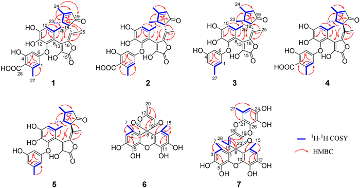
Additional NMR signals included two methyl singlets [δH 1.97 (s, 3H, Me-24) and 1.66 (s, 3H, Me-25)], one methyl doublet [δH 0.96 (3H, d, J = 7.0 Hz), Me-23], two methine protons [δH 2.21 (1H, dq, J = 15.0, 7.0 Hz, H-22) and 3.33 (1H, d, J = 7.0 Hz, H-18)], and an olefinic proton (δH 5.88, s, H-20) (Table S2). The 13C NMR data revealed the presence of a ketone carbonyl (δC 206.0, C-19), an ester carbonyl (δC 163.6, C-15)], six olefinic carbons [δC 119.4 (C-13), 139.5 (C-14), 144.7 (C-16), 116.5 (C-17), 129.0 (C-20), and 182.7 (C-21)], and two methine carbons [δC 51.0 (C-18) and 43.6 (C-22)] (Table S3). 1H–1H correlation spectroscopy (COSY) correlations indicated an independent spin system connecting H-20, Me-24, H-22, Me-23, and H-18 (Fig. 2). Moreover, HMBC correlations from Me-25 to C-16, C-17, C-18, and C-22; from Me-24 to C-19, C-20, and C-22; from Me-23 to C-18 and C-21; and from H-22 to C-19 and C-20 (Fig. 2), along with the remaining 7 degrees of unsaturation, supported the structural assignment of a conjugated γ-butyrolactone (ring C) and a CP (ring D) moiety. The connectivity of the diphenyl ether core with the conjugated γ-butyrolactone and CP units was further established by HMBC correlations from Me-26 to C-13 and from Me-25 to C-16 and C-18 (Fig. 2).
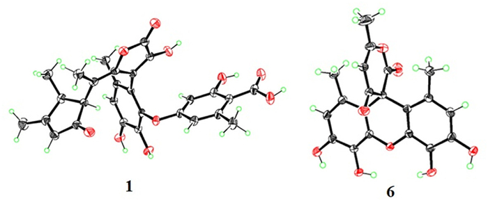
The cis-configuration of H-18 and H-22 was evidenced by nuclear Overhauser effect spectroscopy (NOESY) correlations between H-22 (δH 2.21) and H-18 (δH 3.33), as well as between Me-25 (δH 1.66) and Me-23 (δH 0.96) (Figs. S11, S12, and S64 in Supporting information). Finally, the 18S, 22S configurations of 1, along with the E-geomtry of the Δ16,17 double bond, were unequivocally determined through single-crystal X-ray diffraction (Fig. 3).
Asplactone B (2) was isolated as a light yellow powder. Its molecular formula was determined to be C28H26O10 based on HR-ESI-MS (m/z 545.1411 [M + Na]+, calcd. for C28H26O10Na, 545.1418). The IR spectrum indicated the presence of hydroxyl (3731 cm−1) and γ-lactone (1746 cm−1) functional groups (Fig. S15 in Supporting information). High performance liquid chromatography (HPLC) analysis baseline-separated of compounds 1 and 2 were on an RP-18 column (Fig. S2A in Supporting information), with similar ultraviolet (UV) spectra (Figs. S2B and C in Supporting information). Analysis of the 1D (Tables S2 and S3) and 2D NMR (Fig. 2 and Figs. S20–S24 in Supporting information) spectra revealed that the NMR spectra for 2 were similar to those of 1. However, the upfield shift of H-18 (δH 3.03), the downfield shift of H-22 (δH 2.66), and H3–23 (δH 1.12) in the 1H NMR (Table S2), as well as upfield chemical shifts of C-23 (compound 1: δC 14.1; compound 2: δC 13.3) in the 13C NMR (Table S3), compared to compound 1 [H-18 (δH 3.33), H-22 (δH 2.21), and H3–23 (δH 0.96)], along with the opposite specific optical rotation values (1: −73.30, 2: +51.67), suggest that compound 2 is a C-22 epimer of 1 and adopts a 22R-configuration. The trans-configuration of H-18 and H-22 was further supported by the NOESY correlations (Fig. S23 in Supporting information) between H3–23 (δH 1.12) and H-18 (δH 3.03), and between H3–25 (δH 1.60) and H-22 (δH 2.66) (Figs. S24 and S64 in Supporting information). The stereochemistry of 2 was ultimately determined to be 18S, 22R by comparing electronic circular dichroism (ECD) calculations with experimental circular dichroism (CD) data (Fig. 4A and Table S4 in Supporting information).
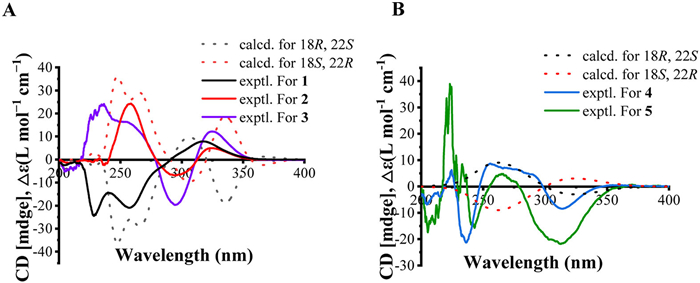
Asplactone C (3) was obtained as a light yellow powder with a molecular formula of C27H26O8, corresponding to 15 degrees of unsaturation and a molecular weight 44 Da less than that of compound 2 (vide supra). This was corroborated by its HR-ESI-MS ion at m/z 477.1559 [M–H]- (calcd. for C27H25O8, 477.1555). Analysis of the 1D and 2D NMR data revealed that the structure of 3 closely resembled that of 2, except for the absence of a carboxyl group (δC 172.5), which was further supported by the appearance of an additional proton at H-3 (δH 6.00) and the upfield shift of H3–27 (δH 2.09) (Tables S2 and S3). In the HPLC analysis, the retention time of compound 3 was adjacent to that of compounds 1 and 2 (Fig. S2A), with a slightly different UV profile (Fig. S2B). Additionally, the ECD profile of 3 closely matched that of compound 2, and its stereochemistry was confirmed as 18S, 22R by comparing ECD calculations with the experimental CD data (Fig. 4A and Table S5 in Supporting information).
By comparing the configurations of compounds 1–3 with their corresponding CD spectra, it can be concluded that the positive Cotton effect observed in the 220–280 nm range, coupled with the negative Cotton effect in the 280–310 nm range, suggests the 22R-configuration. In contrast, the 22S-configuration is implied in the opposite scenario. Additionally, the positive Cotton effect observed in the 310–350 nm range is indicative of the 18S-configuration (Fig. 4A).
Asplactone D (4) was isolated as a light yellow powder, and its molecular formula was determined to be the same as that of compounds 1 and 2, based on the HR-ESI-MS ions at m/z 523.1593 [M + H]+ (calcd. for C28H27O10, 523.1599). The IR spectral data (Fig. S39 in Supporting information) and UV profile of 4 (Figs. S2B and C) closely resembled those of compounds 1 and 2. HPLC analysis over an Eclipse XDB-C18 column indicated that compounds 1, 2, and 4 could be baseline-separated (Fig. S2A). However, the slight upfield shift of H-18 (δH 3.26) and the significant upfield shift of H3–25 (δH 1.08) in the 1H NMR (Table S2), compared to compound 1 [H-18 (δH 3.33) and H3–25 (δH 1.66)], suggest that compound 4 is a C-18 epimer of 1 and likely adopts an 18R-configuration. This hypothesis is further supported by the notable downfield shift of C-18 (compound 1: δC 51.0; compound 4: δC 56.7) and the upfield shift of C-25 (compound 1: δC 16.2; compound 4: δC 10.7) in the 13C NMR (Table S3). Additionally, the opposite configuration of 4 compared to 3 was confirmed by the reversed specific optical rotation values (3: +20.00, 4: −28.00). The negative Cotton effect observed around 220–250 nm and 310–350 nm, couple with the positive Cotton effect in the 250–300 nm range, indicates the 22S-configuration for compound 4 (Fig. 4B). Ultimately, the 18R, 22S configuration of 4 was established through ECD calculations for the 18R, 22S and 18S, 22R stereoisomers (Fig. 4B). However, compared to compounds 1–3, the geometry of the Δ16,17 double bond in 4 remained uncertain and required further confirmation (Table S3). To address this, the 13C NMR chemical shifts for both the 4Z and 4E diastereomers of 4 were calculated at the B3LYP/6–31+G (d, p) level in polarizable continuum model (PCM) dimethylsulfoxide. The 4E configuration was determined by the 100% DP4+ probability and the excellent correlation coefficient (R2) between the experimental 13C NMR for 4 and the calculated data for 4E diastereomer (Fig. S51 and Tables S6 and S7 in Supporting information).
Asplactone D (5) was isolated as a light yellow powder with a molecular formula of C27H26O8 (15 degrees of unsaturation), which is 44 Da less than that of compound 4 (vide supra). This was confirmed by its HR-ESI-MS ion at m/z 477.1559 [M − H]− (calcd. for C27H25O8, 477.1555). Comparisons of the 1D and 2D NMR spectra (Tables S2 and S3) suggest that compound 5 is a 3-deacetyl derivative of 4. This was further supported by the disappearance of the δC 172.7 signal (Table S3) and the appearance of an extra proton at H-3 (δH 5.94) in 5 (Table S2), as well as a significant upfield shift of Me-27 signal from δH 2.37 (s) to 2.08 (s) in comparison to 4. The absolute configuration of 5 was established as 18R, 22S by comparing its experimental ECD profiles with those of 4 and with the ECD spectra calculated for 18R, 22S and 18S, 22R stereoisomers (Fig. 4B)
Aspviolaceol A (6) was isolated as a yellow powder, with a molecular formula of C20H16O8, determined by HR-ESI-MS at m/z 385.0920 [M + H]+ (calcd. for C20H17O8, 385.0918), indicating 13 degrees of unsaturation. The 13C NMR spectrum (Fig. S68 in Supporting information) displayed 13 carbon signals, suggesting partial symmetry in the structure of 6. Compared to violaceol I [22], the 1H NMR (Fig. S67 in Supporting information) of 6 exhibits an additional methyl signal at δH 2.29 (3H, s, H3–20) and an olefinic proton (δH 6.13, s, H-18) (Table S2). Furthermore, the 13C NMR spectrum of 6 showed six extra carbon signals compared to violaceol I, including one ester carbonyl (δC 172.5, C-16), one ketone carbonyl (δC 194.4, C-19), two olefinic carbons [C-17 (δC 170.1) and C-18 (δC 109.9)], one sp3 quaternary carbon (δC 58.2, C-8), and one methyl group (δC 20.4, C-20) attached to the Δ17,18 double bond (Table S3). A 6-methyl-pyran-2, 4-dione structure was proposed based on the HMBC correlations (Fig. S72 in Supporting information) from Me-20 to C-17/C-18 and H-18 to C-8/C-17/C-19/C-20. Moreover, the pyranone moiety was fused with violaceol I through C-8 spirocyclization, as established by the key HMBC correlations of H-18 (δH 6.13, s) and H-3 (δH 6.40, s) to C-8 (δC 58.2) (Fig. 2). The proposed structure for compound 6 was eventually confirmed by single-crystal X-ray diffraction (Fig. 3), and the P-1 space group indicates that 6 is archiral.
Aspviolaceol A (7) was obtained as a reddish-brown powder with a molecular formula of C28H22O10, indicating 18 degrees of unsaturation, as determined by HR-ESI-MS at m/z 517.1156 [M − H]− (calcd. for C28H21O10, 517.1140). Further analysis of the 1H (Fig. S76 in Supporting information) and 13C NMR (Fig. S77 in Supporting information) data revealed that rings A to C in compound 7 closely resembled those of violaceol I [22], while the remaining moiety of 7 closely resembled gibellulin A [23]. However, the C-1 and C-9 positions of the violaceol I moiety in 7 were fused with gibellulin A via a C-8 spirocyclization [23], as evidenced by the absence of two aromatic protons at δH 6.15 (m) [22], the significant upfield shift of C-8 from δC 111.5 to δC 59.1, and the absence of a singlet aromatic proton at δH 6.33 compared to violaceol I and gibellulin A, respectively [23]. Furthermore, the hydroxy group in gibellulin A was oxidized into a ketone carbonyl, as indicated by the notable downfield shift of C-20 from δC 145.0 to δC 195.2 in 7. Finally, the unprecedented heterodimer 7, constructed via a spirocyclization reaction between violaceol I and the oxidized gibellulin A, was confirmed by the four spin-coupling fragments in its 1H-1H COSY spectrum (Fig. 2 and Fig. S79 in Supporting information), along with the HMBC correlations from Me-28 to C-8/C-16/C-17, and from H-17 to C-8/C-19 (Fig. 2).
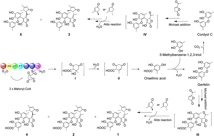
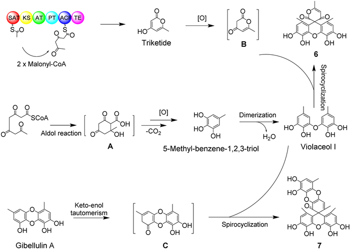
Interestingly, the metabolites of Aspergillus sp. F1–8A, associated with toad venom, are completely different from the chemical components of toad venom itself, suggesting that the presence of the endophytic fungus introduces additional compounds and potential bioactivities to the medicinal material. Biogenetically, the precursor of compounds 1–5 originate from malonyl-CoA, which, under the catalysis of polyketide sythase (PKS), is converted to orsellinic acid [24]. This intermediate undergoes dehydration with 5-methyl-benzene-1,2,3-triol to form gerfelin, which is subsequently decarboxylated to yield cordyol C [25]. Both gerfelin and cordyol C can undergo a Michael addition reaction with 5-acetylfuran-2(3H)-one, resulting in the formation of intermediates iii and iv, respectively. These intermediates then undergo aldol reactions with (S or R)-3,4-dimethylcyclopent-2-en-1-one to produce compounds 1, 2, 4 and compounds 3, 5, respectively (Scheme 1). For the biosynthesis of spiro-diphenyl ethers 6 and 7, two 5-methyl-benzene-1,2,3-triol units [26] act as the upstream precursor, undergoing dehydration to form violaceol I [22]. Violaceol I then undergoes spirocyclization with triketone B [24] to yield aspviolaceol A (6). Additionally, gibellulin A undergoes keto-enol tautomerism to form intermediate C, which then undergoes spirocyclization with violaceol I to produce aspviolaceol B (7) (Scheme 2).
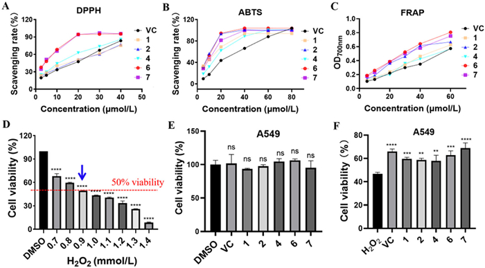
Compounds 1–7 were evaluated for their in vitro antioxidant ability and cytoprotective effects, with the exception of 3 and 5, which were not tested due to insufficient quantities. Antioxidant ability was assessed using the 1,1-diphenyl-2-picrylhydrazyl (DPPH) and 2,2′-azino-bis(3-ethylbenzothiazoline-6-sulfonate) (ABTS) radical scavenging assays, and the ferric reducing antioxidant power (FRAP) method [27]. Compounds 1, 2, 4, 6, and 7 exhibited significant DPPH• (Fig. 5A and Table S8 in Supporting information) and ABTS•+ scavenging abilities (Fig. 5B and Table S8), as well as strong ferric reducing power (Fig. 5C), comparable to or exceeding the positive control (ascorbic acid or vitamin C (VC)) (Fig. 5 and Table S8). A549 cells treated with 0.9 mmol/L H2O2 showed a viability of 50% ± 0.44% (Fig. 5D), establishing this concentration as suitable for subsequent experiments. Further investigation revealed that pretreatment with compounds 1, 2, 4, 6, and 7 (all at 20 µmol/L) had no significant effect on A549 cell viability (Fig. 5E), but significantly improved the viability of A549 cells damaged by 0.9 mmol/L H2O2, matching or surpassing the effects of vitamin C (Fig. 5F) [28]. These findings suggest that compounds 1, 2, 4, 6, and 7 exhibit excellent in vitro antioxidant ability and cytoprotective effects against H2O2-induced cellular damage.
In summary, this study marks the first discovery of asplactones A–E (1–5), which represent the first examples of diphenyl ethers fused with unusual conjugated γ-butyrolactone and CP moieties, implying that 1–5 are representatives of a new subclass within the diphenyl ether family. Furthermore, these are the first documented natural products with a hybrid structure that integrates both CP and γ-butyrolactone moieties. Additionally, aspviolaceols A (6) and B (7) represent the only two examples of spiro-diphenyl ethers found in nature to date. This work marks a significant milestone in decades of research on Aspergillus, showcasing the remarkable chemical ingenuity of this genus. The discovery of these unique diphenyl ether hybrids and spiro-diphenyl ethers has expanded the structural diversity of the natural product library and their excellent antioxidant properties and cytoprotective effects provide promising insights for further pharmacological investigation.
The authors declare that they have no known competing financial interests or personal relationships that could have appeared to influence the work reported in this paper.
Hai-Ying Yu: Writing – original draft, Methodology, Investigation, Data curation. Yu-Wei Huang: Methodology, Investigation, Formal analysis, Data curation. Li-Ping Lin: Writing – review & editing, Visualization, Validation, Supervision, Resources, Project administration, Methodology, Investigation, Funding acquisition, Formal analysis, Data curation, Conceptualization. Ren-Xiang Tan: Supervision, Project administration.
This work was financially supported by the National Natural Science Foundation of China (No. 82073721) and Major Basic Research Project of the Natural Science Foundation of the Jiangsu Higher Education Institutions of China (No. 23KJA310003).
Supplementary material associated with this article can be found, in the online version, at doi:
B. Liu, Y.X. Wang, N. Chen, et al., Mini-Rev. Org. Chem. 21 (2024) 590–598. doi: 10.2174/1570193x20666230707140919
X. Tang, M. Xie, Y.X. Sun, et al., Chin. Chem. Lett. 20 (2009) 435–438. doi: 10.1016/j.cclet.2008.12.027
Y.B. Ji, W.J. Chen, T.Z. Shan, et al., Chem. Biodivers. 17 (2020) e1900640. doi: 10.1002/cbdv.201900640
S. Zenitani, S. Tashiro, K. Shindo, et al., J. Antibiot. 56 (2003) 617–621. doi: 10.7164/antibiotics.56.617
Q. Zhang, J.X. Pang, T.Z. Wang, et al., Chin. Chem. Lett. 34 (2023) 108121. doi: 10.1016/j.cclet.2022.108121
J. Hur, J. Jang, J. Sim, Int. J. Mol. Sci. 22 (2021) 2769. doi: 10.3390/ijms22052769
S.H. Burstein, Prostaglandins Other Lipid Mediat. 148 (2020) 106408. doi: 10.1016/j.prostaglandins.2020.106408
M. Conti, Anticancer Drugs 17 (2006) 1017–1022. doi: 10.1097/01.cad.0000231471.54288.00
C. Zhou, Y. Ge, D. Lan, et al., Fitoterapia 176 (2024) 106039. doi: 10.1016/j.fitote.2024.106039
K.M. Kitzinger, J.S. Johnson, Org. Lett. 25 (2023) 7446–7450. doi: 10.1021/acs.orglett.3c03100
W.S. Song, H. Qi, S.Y. Zhang, et al., J. Asian Nat. Prod. Res. 24 (2022) 1058– 1063. doi: 10.1080/10286020.2021.2023505
A.M. Fu, C.M. Chen, Q. Li, et al., Chin. Chem. Lett. 35 (2024) 109100. doi: 10.1016/j.cclet.2023.109100
H. Zhang, C.P. Li, L.L. Wang, et al., Chin. Chem. Lett. 35 (2024) 109351. doi: 10.1016/j.cclet.2023.109351
C. Sun, Y. Ha, X. Liu, et al., Molecules 29 (2024) 459. doi: 10.3390/molecules29020459
H. Jangid, S. Garg, P. Kashyap, et al., Front. Microbiol. 15 (2024) 1379602. doi: 10.3389/fmicb.2024.1379602
K. Xu, C. Li X.L. Yuan, et al., Mar. Drugs 18 (2020) 54. doi: 10.3390/md18010054
X. Bai, Y. Sheng, Z. Tang, et al., J. Fungi 9 (2023) 261. doi: 10.3390/jof9020261
R. Orfali, M.A. Aboseada, N.M. Abdel-Wahab, et al., RSC Adv. 11 (2021) 17116–17150. doi: 10.1039/d1ra01359a
J. Guan, P.P. Zhang, X.H. Wang, et al., J. Nat. Prod. 87 (2024) 238–251. doi: 10.1021/acs.jnatprod.3c00907
P. Li, Z.J. Zhang, Y.T. Guo, et al., Phytochemistry 222 (2024) 114073. doi: 10.1016/j.phytochem.2024.114073
J.F. Sanchez, Y.M. Chiang, E. Szewczyk, et al., Mol. Biosyst. 6 (2010) 587–593.
L.J. Fremlin, A.M. Piggott, E. Lacey, et al., J. Nat. Prod. 72 (2009) 666–670. doi: 10.1021/np800777f
F.Y. Lv, X.M. Li, L.P. Chi, et al., J. Oceanol. Limnol. 38 (2020) 1225–1232. doi: 10.1007/s00343-020-0052-3
C. Zaehle, M. Gressler, E. Shelest, et al., Chem. Biol. 21 (2014) 719–731. doi: 10.1016/j.chembiol.2014.03.010
T. Bunyapaiboonsri, S. Yoiprommarat, K. Intereya, et al., Chem. Pharm. Bull. 55 (2007) 304–307. doi: 10.1248/cpb.55.304
M.L. Nielsen, J.B. Nielsen, C. Rank, et al., FEMS Microbiol. Lett. 321 (2011) 157–166. doi: 10.1111/j.1574-6968.2011.02327.x
I. Gulcin, Arch. Toxicol. 94 (2020) 651–715. doi: 10.1007/s00204-020-02689-3
B. Ghodsi-Moghadaml, A. Asoodeh, Int. J. Pept. Res. Ther. 25 (2019) 1065–1074. doi: 10.1007/s10989-018-9754-1

Figure 4 Stereochemical assignments of compounds 1–5 through comparative analysis of experimental CD spectra and computed spectra of potential stereoisomers.

Figure 5 Antioxidant ability and cytoprotective effects of the tested compounds. DPPH (A) and ABTS (B) free radical scavenging ability, expressed as scavenging rate (%), with VC used as the control. (C) Ferric reducing ability, expressed as optical density (OD) at 700 nm, with VC as the control. (D) Effects of H2O2 at various concentrations (0.7, 0.8, 0.9, 1.0, 1.1, 1.2, 1.3, and 1.4 mmol/L) on the viability of A549 cells. (E) Cytotoxicity of the tested compounds and VC (all at 20 µmol/L) on A549 cells, with DMSO as the negative control (ns, no significance). (F) Protective effects of the tested compounds (20 µmol/L) against H2O2-induced oxidative damage in A549 cells, with 0.9 mmol/L H2O2 treatment as the model group. Results are expressed as mean ± SD (n = 3). **P < 0.01, ***P < 0.001, ****P < 0.0001 vs. the DMSO control or the model group.

 扫一扫看文章

扫一扫关注我们
 DownLoad:

 下载:
下载:


