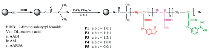
Figure 1.
Preparation of five polymer brushes-modified resins.
Two-site synergistic binding strategy for improving adsorption and separation performance of antibodies
Xia Liu , Wenzhuo Dong , Mengqian Jia , Dexiu Zhang , Jingyi Niu , Jiwei Shen , Chaozhan Wang , Yinmao Wei

Antibodies are of immune proteins secreted by effector B cells, and have been widely utilized as drugs for treating cancer, cardiovascular diseases, and autoimmune diseases, etc. At present, the sale of antibody drugs in the biopharmaceutical market continues to rise annually, with expectation of exceeding $300 billion by 2025 [1–3]. In industry, the production of monoclonal antibodies (mAbs) involves upstream processing and downstream purification processes. The upstream processing includes cell culture and antibody expression, while the downstream process commonly comprises antibody capturing from the culture medium and subsequent purification. Of the total production cost, the downstream process generally accounts for approximately 50%−80% [4], to which antibody capturing step attributes the majority. The capturing methods used to include protein A affinity chromatography [5,6] and biomimetic chromatography using peptide and peptidomimetics as ligands of adsorbents [7,8]. Compared with the two methods, HCIC and BAC have the advantages of adsorbents easy to prepare, low cost, and the ability to overcome the harsh elution conditions in protein A and biomimetic affinity chromatography, so they attract much attentions recently.
In HCIC and BAC of antibodies, the separation mechanism and performance largely depend on the structure of adsorbents. At present, two classes of adsorbents have been developed in view of the presence of N-glycosyl sites on the Fc fragment and hydrophobic binding site at the CH2-CH3 junction of antibody. (1) Boronate affinity (BA) adsorbents. The boronic groups on the surface of adsorbent can form stable cyclic ester through covalent bond with the glycosyl of antibody molecular in alkaline solutions. In acidic solutions, the cyclic ester can reversibly dissociate to release the antibody [9]. This pH-dependent reaction endows the adsorbent with unique adsorption selectivity towards antibodies [10–12]. However, the co-existing glycoproteins can also be captured based on the same mechanism [13,14], lowering the specific in BAC. (2) Hydrophobic charge induced (HCI) adsorbents. In principle, the antibodies can easily bind with the ligands on the surface of adsorbent through hydrophobic interactions under neutral pH, whereas they are released through electrostatic repulsion under acidic conditions [15]. Currently, the popular HCI ligands mainly include 4-mercaptoethylpyridine [16], 5-aminobenzimidazole [17], 2-mercapto-1-methylimidazole [18], 2-mercaptoimidazole [19], 5-aminoindole [20], and 2-mercaptobenzimidazole [21]. Despite much progress, the specific of HCI ligands is still limited because the ligands can also adsorb some proteins according to HCIC mechanism [22,23]. With respect to the preparation of adsorbents, the usual method is modifying either BA or HCI ligands on the surface of the matrix via multi-step chemical reactions. However, the method often leads to a low adsorption capacity due to the low binding density of ligands. Specifically, the created adsorbents normally allow for a single HCIC or BAC mechanism, providing a relatively low specific to antibodies. Consequently, the current HCIC and BAC suffer from a limited purity and recovery of antibodies.
To address these issues, this work proposes a two-site synergistic binding strategy integrating HCIC and BAC mechanism based on the structural characteristics of both hydrophobic binding region and sugar chain on the Fc region of antibody. The strategy is accomplished on a novel polymer brushes-grafted resins (Fig. 1), which were easily constructed by using surface-initiated ARGET ATRP to polymerize different ratios of 3-acrylamide phenylboronic acid (AAPBA), 5-acrylamidobenzimidazole (AABI) and acrylamide (AM) on the surface of agarose gel (Seplife 6FF, cross-linking degree of 6%, particle size of 45–165 µm, average particle size of 90 µm). Herein, 5-aminobenzimidazole acting as the HCI ligand has an excellent recognition ability to antibodies [17,24]. The surface-initiated ARGET ATRP not only overcomes the drawbacks of the traditional free radical polymerization method [25], but also avoids the toxic residue of Cu in the adsorbent prepared by normal ATRP [26]. ATPR time exceeded 24 h, enabling the grafted chain length to reach the maximum for five resins [27–29]. Moreover, the chain units in the copolymer brushes were tried to be illustrated by detecting the dependence of the concentration of reactants on reaction time, taking P1 resin as an example only (Fig. S1 and Table S1 in Supporting information). It is found that AAPBA and AABI have a similar reactivity ratio due to the similar structure. Therefore, the copolymerization reaction belongs to ideal azeotropic co-polymerization [30]. According to the theory of the segment sequence number distribution function [30], the chain unit, C1, accouters for the majority among all the possible units for P1 (Fig. S2 in Supporting information), which are co-polymerized by 1:1 of AAPBA to AABI. Similarly, C2 and C3 are respectively inferred to be the major chain units among all the possible units in the P2 and P3 due to the similar structure of AAPBA, AABI and AM (Fig. S3 in Supporting information). It should be noted that two side groups in the chain units, phenylboronic acid (PBA) and benzimidazole (BI) can bind with N-glycosyl and hydrophobic binding sites of antibody, respectively, while AM has no interaction with antibody, only playing a role to regulate the distance between PBA and BI groups in the polymer chain. P4 and P5 resins, on which the polymer chains are homopolymers from AAPBA and AABI monomers, correspond to single HCIC and BAC mechanisms, respectively, and serves as controls for P1-P3. Overall, five resins with different chain structures were designed to explore the synergistic binding of PBA and BI groups with antibodies.
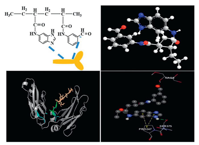
Firstly, molecular docking was performed to investigate how and on which resin BI and PBA groups can simultaneously bind with antibodies in a two-site manner. Herein, IgG serves as the receptor and the predominant chain units as the ligands (C1-C3). Due to the lack of an algorithm for B atoms in the currently available software, the real interactions between the ligands and IgG cannot be calculated. Considering that benzaldehyde can react with the o-dihydroxy in glycosyl to form an acetal, and the acetal structure is similar to the five-membered cyclic ester formed by PBA and glycosyl, we replaced PBA on the chain unit with benzaldehyde to obtain a simulated ligand to solve this problem. The docking of C1 unit corresponding to P1 is shown in Fig. 2, while the docking of P2 and P3 are shown in Fig. S4 (Supporting information). It can be observed that: (1) For the P1 resin with 1:1 of AAPBA to AABI, the predominant unit (C1) can perfectly fit spatially into the hydrophobic region and the groove formed by the N-acetylglucosamine, suggesting a two-site synergistic binding with an IgG molecular. Where, BI allows for the hydrophobic and electrostatic interaction with Pro247 and Ser375 [31], and benzaldehyde for the hydrogen bond interaction with N-acetylglucosamine in IgG. Such a synergistic bonding can enhance the affinity of resin to IgG according to the chemical coordination theory. (2) For P2 and P3 resins (the ratios of AABI, AM to AAPBA is 1:1:1 for P2, and 1:2:1 for P3), the predominant units are C2 and C3, respectively, where an IgG molecular is bonded via either glycosyl with PBA or hydrophobic region with BI. In other words, an IgG molecular is bonded in a single-site manner. This is attributed to the inserting of AM between AABI and AAPBA, leading to a large space interval between BI and PBA groups so that two groups cannot match spatially with the two sites in an IgG molecular. Significantly, ARGET ATRP is a random copolymerization reaction, so there are possibly other possible units in P2 and P3 resins, i.e., C6 composed of closely adjacent two PBI groups and one PBA group or C7 of two PBA groups and one PBI. As does C1, C6 and C7 also form the synergy interaction with IgG, but most IgG molecules are essentially bonded in a single-site manner on account of small proportion of C6 and C7 units in P2 and P3. In a word, although the simulated ligands are different from the actual ligands, the docking results can still illustrate that BI and PBA groups in the chain unit of P1 resin can well match the hydrophobic region and glycosyl in terms of spatial distance to be able to form an effective synergistic binding with IgG.
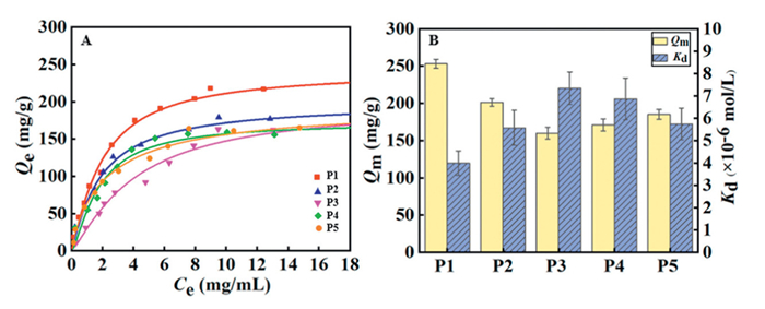
Secondly, the adsorption isotherms of IgG on five resins were determined through static adsorption experiments, and were fitted by linear Langmuir equation to obtain the dissociation constant (Kd) and adsorption capacity (Qm) of adsorbent (Fig. 3). Of five resins, P1 presents a much stronger affinity (Kd 3.9 × 10⁻⁶ mol/L) than P2 and P3 as well as P4 and P5 resins. This result conforms to the expectation with the molecular docking. The chain units in P1, in which the closely adjacent BI and PBA groups occupy the majority, allow for the synergistic binding with IgG and, thus, raise the affinity of P1 to IgG. In contrast, BI and PBA in the polymer chain of P2 and P3 are spatially separated by AM and thereby cannot form the synergistic binding with IgG, consequently the adsorption of antibody is more likely dominated by the single-site manner arising from HCIC or BAC. In terms of adsorption capacity, P1 (Qm = 253 mg/g) is much larger than P2-P5 resins and the reported protein A, G and L, biomimetic peptide and small molecule ligands-bonded adsorbents [31–41]. This result is in line with the observations that the polymer modification can improve the adsorption capacities of adsorbents [42–45]. Moreover, the Kd of IgG on P1 resin is in the range of 10−6 mol/L, which is within the reported range for the reported boron affinity chromatography and hydrophobic charge-induced interaction chromatography in the literature (Table S2 in Supporting information).
In order to further verify the two-site synergistic binding interaction, the competitive adsorption experiments were carried out using 20 mmol/L glucose, 5% polyethylene glycol (PEG), and a mixture of 20 mmol/L glucose and 5% PEG in adsorption solution. As shown in Fig. 4, there are three main observations. (1) PEG in the solution can weaken the binding interaction between IgG and BI group because hydrophobic PEG can competitively interact with hydrophobic region at the CH2-CH3 junction of IgG according to the viewpoint proposed by Debnatha et al. [46], leading to the diverse changes in the adsorption capacity of five resins. P4 resin, which only provides pure HCI mechanism, shows a significant decrease in Qe of IgG with a reduction rate of 61.9%. For P2 and P3 resins, IgG molecules are equally adsorbed by individual BI and PBA groups in polymer chains. As a result, their adsorption capacities also decline because of the inhibition of HCI interaction by PEG, but the reduction degrees (17.6% for P2 and 37.3% for P3) are rather smaller compared with P4 resin since PBA groups can still adsorb IgG unaffected in P2 and P3. (2) Glucose in the solution can compete with IgG for PBA sites, weakening the adsorption of IgG [47]. As a result, P5 resin with pure BAC mechanism yields a considerable decrease rate of 67.9% in Qe. However, P2 and P3 resins show a slight decrease of 13.1% and 29.1% in Qe since HCI interaction remains unchanged on the adsorption of IgG. Compared with P2-P5 resins, PEG or glucose in solution has almost no effect on Qe of P1 resin (around 253 mg/g). The reason can be attributed to the fact that the synergistic effect is less affected by the individual inducing factors of BA or HCI. (3) Under existence of both glucose and PEG, both HCI and BA interactions are weakened. Consequently, the Qe of P2 and P3 resins decrease much greater (35.2% for P2 and 51.2% for P3) than those under the presence of PEG or glucose alone. In contrast, the decrease in Qe of P1 resin is relatively small with a decrease rate of 22.7%. In conclusion, P1 resin indeed imposes the synergistic interactions on IgG by BI and PBA groups, which improves the specific of the adsorbent to IgG.

Additionally, the adsorption isotherms of IgG on five resins were measured to obtain Qm and Kd in the pH range of 4.0–8.0 (Figs. S5 and S6 in Supporting information). In terms of Qm and Kd, P4 and P5 resins with pure HCI and BA mechanism appear a similar affinity dependency on pH. Where, the affinity and adsorption capacity for IgG are weak at initial pH 4.0, gradually goes up with pH, and eventually reaches the maximum at pH 8.0 [48,49]. This result indicates that the interaction between HCI and BA with antibodies will not offset each other at any pH. As a result, P1-P3 resin exhibits the similar adsorption behaviors as P4 and P5, following the adsorption under alkaline solution and the desorption under acidic solution. Furthermore, P1 resin invariably has the strongest affinity among five resins in the tested pH range, further confirming the contribution of the two-site synergy.
According to the pH effect, a solid-phase extraction method was optimized in terms of purity and recovery of IgG. Herein, a mixture of IgG, ovalbumin (OVA) and bovine serum albumin (BSA) (1:1:4) was employed as a model and the elution was analyzed by sodium dodecyl sulfate polyacrylamide gel electrophoresis (SDS-PAGE) (Fig. S7 in Supporting information). As the adsorption pH was set at 8.0 and the elution pH ranged from 3.5 to 5.0, P1 resin exhibits an excellent enrichment selectivity with a maximum purity of approximately 98.0% and a recovery of about 97.0%. Two reasons are responsible for the excellent adsorption performance. Firstly, the two-site synergistic adsorption that enhances the binding specific of IgG allows IgG to be preferentially adsorbed while the interfering proteins to be effectively excluded through the "crowing-out" effect. As a result, the anti-interference of resin towards competitive substrates is improved. Secondly, ARGET ATRP increases the density of functional groups on the resin surface, providing more specific binding sites for IgG. Whereas for P2-P5 resins, the single-site binding allows IgG but also minor hydrophobic BSA and glycoprotein OVA for the adsorption based on BAC and HCI mechanisms, leading to a lower recovery and purity of IgG. In this sense, P1 is the best among five resins for the adsorption and separation of IgG, and the adsorption and elution solution pH are chosen as pH 8.0 and pH 4.5, respectively. At this pH, IgG can achieve high purity and recovery without losing activity.
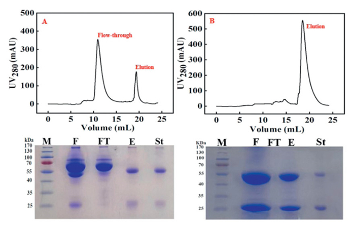
Moreover, liquid chromatography method was performed to purify antibodies from serum and cell culture supernatant using ÄKTA protein purification system. Herein, human serum from the blood donors was provided by the Chang'an Hospital of Northwest University (Xi'an, China). All experiments were approved by the Academic Research Ethics Committee of Northwest University (Xi'an, China) and obtained the informed consent from the blood donors. Serum is rather complex and usually serves as a model to evaluate the separation property of adsorbent. Human serum mainly consists of human serum albumin (HSA), IgG, transferrin (TRF), alpha-fetoprotein (AFP), and haptoglobin (HP) [50]. Of these proteins, IgG, TRF and HP are glycoproteins and HSA is a hydrophobic protein. The serum dilutions and cell culture were injected into five columns, and the chromatograms are shown in Fig. 5 for P1, and in Figs. S8 and S9 (Supporting information) for P2-P5. All SDS-PAGEs indicate that the impurity proteins are mainly in the flow-through fractions while IgG is in the elution fractions. P1 resin gives a purity of 95.8% and a recovery of 95.9% for IgG, being the highest among five resins (Fig. S10 in Supporting information). Cell culture is currently the mainstream source for mAb. As expected, P1 resin also yields the highest purity and recovery up to 98.3% and 97.5% for mAb, respectively (Fig. S10). In comparison with the reported methods (Table S3 in Supporting information), the purities and recoveries of IgG and mAb are comparable to or higher than those with protein A, G and L that are considered to have the largest specific so far, and those with HCIC ligands. In terms of adsorption capacity, the adsorbent in this work almost is the largest among the two categories. Most importantly, our method has a simpler and milder elution (pH 4.5, no additive in elution buffer), which is more favorable to simply the post-treatment and maintain the activity of antibody. The excellent separation performance indicates a promising potential of the proposed strategy and P1 resin in the production of antibody drugs.
Finally, the stability of resin was investigated at extreme pH 2.0 and pH 14.0. After immersed in the solutions with pH 2.0 and pH 14.0 for 3 days, respectively, the P1 resin was collected, washed and subjected to the measurement of adsorption capacity for IgG. The measured value drops slightly for the treated resin over the initial one. Upon treated for 6 days, the resin approximately remains the adsorption capacity as for 3 days (Fig. S11 in Supporting information). The result indicates that the resin maintains largely stable under short-term strong acidic and alkaline conditions.
In summary, this work presents a two-site synergistic binding strategy towards IgG on a polymer brushes-grafted adsorbent. The closely adjacent BI and PBA as side groups of the polymer units can spatially match the N-glycosylation sites and hydrophobic binding site in antibody, generating the synergistic binding with IgG. This strategy follows the same adsorption and elution patterns but can enhance the affinity and specific of adsorbent to antibody compared with the single-site binding mechanism in HCIC and BAC. Furthermore, the polymer modification endows the adsorbent with higher adsorption capacity. Benefiting from the properties, IgG from serum and mAb from cell culture achieve higher recoveries and purities, demonstrating the significant potential of the strategy in the purification of antibody drugs. In addition, the strategy provides a new concept to design the selective resins for antibody as well as for the interesting membrane receptors that have glycosyl and drug binding site in biological research.
The authors declare that they have no known competing financial interests or personal relationships that could have appeared to influence the work reported in this paper.
Xia Liu: Writing – original draft, Methodology, Investigation, Formal analysis, Data curation. Wenzhuo Dong: Validation, Investigation, Data curation. Mengqian Jia: Methodology, Investigation, Formal analysis. Dexiu Zhang: Methodology, Formal analysis. Jingyi Niu: Validation, Formal analysis. Jiwei Shen: Supervision, Project administration. Chaozhan Wang: Writing – review & editing, Resources, Funding acquisition. Yinmao Wei: Writing – review & editing, Supervision, Resources, Project administration, Conceptualization.
This work was supported by the National Natural Science Foundation of China (Nos. 22274129, 21974106 and 22074117).
Supplementary material associated with this article can be found, in the online version, at doi:
J. Wang, L.G. Zhuo, P. Zhao, et al., Chin. Chem. Lett. 33 (2022) 3502–3506.
M.F. Mercogliano, S. Bruni, F.L. Mauro, R. Schillaci, Cancers 15 (2023) 1987. doi: 10.3390/cancers15071987
H. Lin, H.F. Hong, L.P. Feng, et al., Chin. Chem. Lett. 32 (2021) 4041–4044.
L. Zhang, S. Parasnavis, Z.J. Li, J. Chen, S. Cramer, J. Chromatogr. A 1602 (2019) 317–325.
G. Rigi, S. Ghaedmohammadi, G. Ahmadian, Biotechnol. Appl. Bioc. 66 (2019) 454–464. doi: 10.1002/bab.1742
N.A. Owens, P.H. Anborgh, I. Kolotilin Sci. Rep. 14 (2024) 8714.
T. Sugita, M. Katayama, M. Okochi, et al., Biochem. Eng. J. 79 (2013) 33–40.
M.J.B. Matos, J. Gonçalves, U. Rothbauer, et al., Sep. Purif. Technol. 265 (2021) 118476.
M.B. Espina-Benitez, J. Randon, C. Demesmay, V. Dugas, Sep. Purif. Rev. 47 (2018) 214–228. doi: 10.1080/15422119.2017.1365085
S.A.S.L. Rosa, R.D. Santos, M.R. Aires-Barros, A.M. Azevedo, Sep. Purif. Technol. 160 (2016) 43–50.
P.F. Guo, X.M. Wang, M.M. Wang, et al., Nanoscale 11 (2019) 9362–9368. doi: 10.1039/c9nr01111k
R.D. Santos, S.A.S.L Rosa, M.R. Aires-Barros, A. Tover, A.M. Azevedo, J. Chromatogr. A 1335 (2014) 115–124.
X.J. Zhou, Chun-E Mo, M. Chen, Y.P. Huang, Z.S. Liu, J. Chromatogr. A 1581 (2018) 8–15.
H.J. Zhu, H. Yao, K.X. Xia, et al., Chem. Eng. J. 346 (2018) 317–328.
M.T. Li, Q.L. Zhang, D.Q. Lin, S.J. Yao, J. Chromatogr. B 1134-1135 (2019) 121850.
J. Chen, J. Tetrault, Y.Y. Zhang, et al., J. Chromatogr. A 1217 (2010) 216– 224.
W. Shi, D.Q. Lin, H.F. Tong, J.X. Yun, S.J. Yao, J. Chromatogr. A 1425 (2015) 97–105.
H.L. Lu, D.Q. Lin, D. Gao, S.J. Yao, J. Chromatogr. A 1278 (2013) 61–68.
W. Phottraithip, D.Q. Lin, F. Shi, S.J. Yao, Biotechnolo. Bioprocess Eng. 18 (2013) 1169–1175. doi: 10.1007/s12257-013-0223-6
G.F. Zhao, G.Y. Peng, F.Q. Li, Q.H. Shi, Y. Sun, J. Chromatogr. A 1211 (2008) 90–98.
H.F. Xia, D.Q. Lin, L.P. Wang, Z.J. Chen, S.J. Yao, Ind. Eng. Chem. Res. 47 (2008) 9566–9572. doi: 10.1021/ie800662r
Y.D. Luo, Q.L. Zhang, S.J. Yao, D.Q. Lin, J. Chromatogr. A 1533 (2018) 77–86. doi: 10.1159/000492024
Q.H. Shi, F.F. Shen, S. Sun, Biotechnol. Progr. 26 (2010) 134–141. doi: 10.1002/btpr.295
J.L. Gu, H.F. Tong, D.Q. Lin, J. Chromatogr. A 1460 (2016) 61–67.
W. Jakubowski, K. Matyjaszewski, Macromolecules 38 (2005) 4139–4146. doi: 10.1021/ma047389l
L.F. Zhang, Z.P. Cheng, Z.B. Zhang, D.Y. Xu, X.L. Zhu, Polym. Bull. 64 (2010) 233–244.
Y.M. Wei, J.J. Ma, C.Z. Wang, J. Membr. Sci. 427 (2013) 197–206.
Y.N. Chen, M.F. He, C.Z. Wang, J. Mater. Chem. A 2 (2014) 10444–10453. doi: 10.1039/c4ta01512f
W. Wang, M.F. He, C.Z. Wang, Anal. Chim. Acta 886 (2015) 66–74.
Z.R. Pan, Polymer Chemistry, 5th edition, Chemical Industry Press, Beijing, 2011, pp. 120–129.
J. Yan, Q.L. Zhang, H.F. Tong, D.Q. Lin, S.J. Yao, J. Sep. Sci. 38 (2015) 2387–2393. doi: 10.1002/jssc.201500178
S. Hober, K. Nord, M. Linhult, J. Chromatogr. A 848 (2007) 40–47.
Z.H. Wang, Q. Liang, K. Wen, S.X. Zhang, J.Z. Shen, J. Chromatogr. B 971 (2014) 10–13. doi: 10.1186/1478-811X-12-10
B. Akerstrom, L. Bjorck, J. Biol. Chem. 264 (1989) 19740–19746.
A.C.A. Roque, M.A. Taipa, C.R. Low, J. Chromatogr. A 1064 (2005) 157–167.
J.M. Haigh, A. Hussain, M.L. Mimmack, C.R. Lowe, J. Chromatogr. B 877 (2009) 1440–1452.
L. Lund, P. Gustavsson, R. Michael, et al., J. Chromatogr. A 1225 (2012) 158–167.
X. Zou, Q. Zhang, H. Lu, D.Q. Lin, S.J. Yao, Chem. Eng. J. 368 (2019) 678–686.
H.F. Tong, D.Q. Lin, W.N. Chu, et al., J. Chromatogr. A 1429 (2016) 258–264.
M. Rahman, J.Z. Wang, H.J. Xia, J.W. Liu, Q. Bai, Chem. Eng. J. 391 (2020) 123561.
H.F. Tong, D.Q. Lin, Y. Pan, S.J. Yao, Biochem. Eng. J. 56 (2011) 205–211.
M.Q. Chen, Z.Y. Lin, H. Qian, Chin. Chem. Lett. 19 (2008) 1495–1498.
B. Kaur, P. Rana, P. Singh, et al., Coord. Chem. Rev. 518 (2024) 216057.
Y.L. Fu, Y. Kong, Y. Wang, et al., Sep. Purif. Technol. 318 (2023) 123817.
X. Li, X. Ding, J.K. Zhou, et al., Chin. Chem. Lett. 35 (2024) 109158.
B. Debnatha, B. Das, J. Mol. Liq. 310 (2020) 113088.
Q. Huang, H.J. Yu, L. Wang, et al., Eur. Poly. 157 (2021) 110651.
W. Schwartz, D. Judd, M. Wysocki, et al., J. Chromatogr. A 908 (2001) 251–263.
11 R.S. Santos, S.A.S.L. Rosa, M.R. Aires-Barros, A.M. Azevedo, Sep. Purif. Technol. 160 (2016) 43–50. doi: 10.18468/estcien.2016v6n2.p43-52
E. Gianazza, I. Miller, L. Palazzolo, C. Parravicini, I. Eberini, J. Proteom. 140 (2016) 62–80.

Figure 2 Simulated structure diagram of polymer chain unit, docking spatial structure and molecular docking model of IgG with simulated ligands (The blue and yellow regions represent the BI binding region and the sugar chain, respectively).

Figure 4 Qe of IgG on five resins under competitive conditions. Initial concentration of IgG, 15 mg/mL; Adsorbent dose: 10 mg.

Figure 5 Chromatographic separation of IgG from human serum (A) and mAb from mouse-derived cell culture supernatant (B) on P1 resin. Protein marker (M), Feedstock (F), Flow-through fractions (FT), Elution fraction (E), IgG standard (St). Gradient mode: the column (5 mL) was pre-equilibrated with 10 mL of 20 mmol/L phosphate buffer (pH 8.0); 1.0 mL of sample was injected; 10 mL of the same buffer was run, followed by 10 mL of 20 mmol/L acetate buffer (pH 4.5). Finally, 20 mL of 0.1 mol/L NaOH were employed to regenerate the column. Flow rate: 0.7 mL/min; UV detection wavelength: 280 nm.

 扫一扫看文章

扫一扫关注我们
 DownLoad:

 下载:
下载:
