Department of Occupational and Environmental Health, School of Public Health, Wuhan University, Department of Radiation and Medical Oncology, Zhongnan Hospital of Wuhan University, Wuhan 430071, China
b.
Research Center of Public Health, Renmin Hospital of Wuhan University, Wuhan 430060, China
c.
Department of Chemistry, The Hong Kong University of Science and Technology, Hong Kong, China
d.
School of Nursing, Wuhan University, Wuhan 430071, China
e.
Hubei Provincial Center for Disease Control and Prevention & NHC Specialty Laboratory of Food Safety Risk Assessment and Standard Development, Wuhan 430079, China
f.
Hubei Key Laboratory of Biomass Resource Chemistry and Environmental Biotechnology, Wuhan University, Wuhan 430071, China
bfyuan@whu.edu.cn (B.-F. Yuan). 1 These authors contributed equally to this work.
Received Date:
11 November 2024 Accepted Date:
13 February 2025 Revised Date:
23 January 2025 Available Online:
15 December 2025
Abstract:
Perfluorooctanoic acid (PFOA) is a highly bioaccumulative environmental endocrine disruptor and a persistent organic pollutant. Epigenetic modifications in DNA and RNA are crucial for regulating gene expression and are involved in numerous physiological processes. However, research on the effects of PFOA on epigenetic modifications is still limited. In this study, we systematically investigated the alterations in epigenetic modifications in both DNA and RNA from the heart, liver, spleen, lung, kidney, and brain of C57BL/6N mice following exposure to PFOA at doses of 0, 0.5, and 5 mg kg−1 d−1, utilizing liquid chromatography-tandem mass spectrometry (LC-MS/MS). The results indicated that exposure to PFOA inhibited weight gain in mice, and significant changes were observed in the organ coefficients of the liver, spleen, lungs, and heart in the high PFOA exposure group. Modifications in DNA and RNA exhibited tissue specificity. Orthogonal partial least squares discriminant analysis revealed that the control group and the high PFOA exposure group clustered well, suggesting that PFOA exposure significantly impacts epigenetic modifications in DNA and RNA. Specifically, PFOA exposure significantly affected the levels of 5-hydroxymethylcytosine (5hmC) in genomic DNA in the heart, lung, kidney, and liver tissues. For RNA modifications, significant changes were observed, with the levels of 12, 13, 10, 6, 12, and 14 modifications in the heart, liver, spleen, lung, kidney, and brain, respectively, altered in response to PFOA exposure. Our study highlights the significance of PFOA exposure in altering DNA and RNA modifications, providing a new perspective on understanding the toxicology of PFOA from an epigenetic standpoint.
Perfluorooctanoic acid (PFOA) is a type of persistent organic pollutant known for its widespread use in industrial and consumer products as a surfactant and lubricant, primarily due to its remarkable resistance to water, oil, and contamination [1–4]. However, as its applications have expanded, awareness has grown regarding the potential adverse effects of PFOA on both the environment and human health [5–7]. Consequently, many countries and regions have implemented measures to restrict or ban PFOA and its derivatives. Notably, on December 29, 2022, China included PFOA in the List of Key Controlled New Pollutants (2023 Edition). Furthermore, China's recently established Standards for drinking water quality (GB 5749-2022) has incorporated an indicator for PFOA into its water quality reference index, establishing a permissible limit of 80 ng/L. In November 2023, PFOA was classified as “carcinogenic to humans" (Group 1) by the International Agency for Research on Cancer [8].
All hydrogen atoms in the carbon chain of PFOA have been replaced by fluorine atoms, resulting in the formation of highly stable carbon-fluorine bonds [9]. This structural characteristic makes PFOA resistant to degradation, enabling its persistence and accumulation in the environment over extended periods [10]. PFOA is mainly absorbed through the digestive and respiratory tracts, with a minor absorption occurring through the skin [11,12]. Once ingested, PFOA exhibits toxicity to multiple organs throughout the body and has a long half-life due to its persistence and accumulation [1]. The liver and blood are the primary tissues where PFOA is distributed [13,14]. Previous studies have established an association between PFOA and a range of adverse health effects, including thyroid dysfunction [15,16], metabolic disorders [17], reproductive toxicity [18,19], neurodevelopmental issues [20], renal disorders [21,22], immunotoxicity [23], and various forms of cancer [24,25]. Additionally, PFOA is associated with its ability to mimic fatty acids and disrupt endocrine functions [26].
Epigenetic modifications in both DNA and RNA play a vital role in the regulation of gene expression and are implicated in various physiological processes [27–31]. 5-Methylcytosine (5mC) is the most common DNA modification in mammals, often referred to as the fifth base due to its critical roles in gene expression, embryogenesis, and tumorigenesis [32,33]. 5-Hydroxymethylcytosine (5hmC) is also gaining attention as the sixth base of mammalian genomes [34]. Apart from being an intermediate in the oxidation of 5mC, 5hmC is a stable epigenetic modification that directly regulates gene expression in both physiological and pathological contexts [34,35]. RNA, like DNA, undergoes numerous post-transcriptional modifications that impact key pathways such as RNA processing and metabolism [36]. N6-methyladenine (m6A) is one of the most prevalent RNA modifications, influencing stem cell differentiation, cancer progression, and immune responses [37–39]. Pseudouridine Ψ enhances RNA stability and translation efficiency [40], while inosine (I) aids in RNA editing and codon-anticodon pairing [41–44]. N7-methylguanosine (m7G) is vital for mRNA stability, nuclear export, and translation initiation [45,46]. So far, over 160 distinct RNA modifications have been identified in tRNA, rRNA, and mRNA [47–50], and aberrant RNA modifications are linked to various human diseases [27,51–53].
There is growing evidence that PFOA has a potential impact on epigenetic modifications [54,55]. Previous studies have indicated that exposure to PFOA alters DNA methylation, particularly affecting genes related to lipid metabolism, which in turn mediates changes in lipid levels [56]. A cohort study showed that prenatal exposure to PFOA could influence the DNA methylation status of infants at birth [57]. Additionally, animal studies demonstrated that mice exposed to PFOA exhibited increased liver weight and reduced overall DNA methylation in the liver [58]. Despite several studies investigating the epigenetic effects of PFOA, many questions and controversies remain in this area [59]. Notably, research on the epigenetic effects of PFOA regarding RNA modifications is still lacking, highlighting the need for further experimental studies to characterize these effects fully.
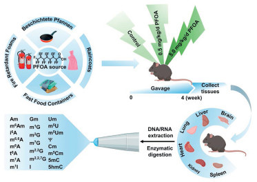
In the current study, we systematically examined the changes in nucleic acid modification content in DNA and RNA from the heart, liver, spleen, lung, kidney, and brain of C57BL/6N mice following exposure to PFOA, utilizing liquid chromatography-tandem mass spectrometry (LC-MS/MS) (Fig. 1). We established a mouse model and randomly assigned the mice into three groups, one control group and two experimental groups, with each group consisting of 10 mice. The control group received corn oil containing 0.1% DMSO as a placebo, while the experimental groups were treated with PFOA dissolved in corn oil with 0.1% DMSO at concentrations of 0.5 mg kg−1 d−1 (low) and 5.0 mg kg−1 d−1 (high), respectively. After daily gavage treatment for four weeks, tissue samples were collected for DNA and small RNA (< 200 nt) extraction. This experiment received approval from the Experimental Animal Control and Use Committee (IACUC) of Wuhan University Animal Experiment Center.
Figure 1
Figure 1.
Schematic illustration of PFOA exposure in mice and mass spectrometry profiling of DNA and RNA modifications. The procedure entails the collection of tissue samples from both control and PFOA-exposed groups, followed by the isolation of DNA and small RNA (< 200 nt). Subsequently, enzymatic digestion is performed on the isolated DNA and small RNA, and LC-MS/MS analysis is conducted to detect and quantify the modifications present in the DNA and small RNA.
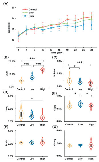
We weighed each mouse daily throughout the rearing period and plotted their growth curves following PFOA exposure by calculating the average weight of each group. The results indicate that the average body weight of mice in the low PFOA exposure group did not significantly differ from that of the control group (Fig. 2A). In contrast, mice in the high PFOA exposure group exhibited progressively retarded body weight growth in the later stages of exposure (Fig. 2A), which aligns with findings from previous studies [60]. This growth inhibition may be attributed to the interference of PFOA with biological pathways related to amino acid, lipid, fatty acid, bile acid, and androgen metabolism, potentially contributing to slower growth [61]. Additionally, we calculated the organ coefficient, which represents the ratio of tissue weight to the body weight of the mice at the time of sacrifice (Table S1 in Supporting information). A significant increase in the organ coefficient of liver was observed (Fig. 2B), consistent with previous findings [62]. We also quantified PFOA levels in mouse liver tissues, which demonstrated that exposure led to increased PFOA levels (Table S2 in Supporting information). In contrast, spleen atrophy was noted (Fig. 2C), likely resulting from macrophage overactivation due to PFOA exposure [63,64]. Additionally, decreased organ coefficients for the lung and heart were recorded at high PFOA exposure levels (Figs. 2D and E), while no significant changes were observed in the brain and kidney (Figs. 2F and G).
Figure 2
Figure 2.
Effect of PFOA exposure on the growth of mice. (A) Growth curves of mice under different treatment. (B-G) Organ coefficients of different tissues of mice at sacrifice (N = 10). *P < 0.05, **P < 0.01, ***P < 0.001.
We subsequently employed LC-MS/MS to measure the changes in the levels of nucleic acid modifications in DNA and small RNA (< 200 nt) from the heart, liver, spleen, lung, kidney, and brain of C57BL/6N mice following PFOA exposure. Small RNAs (< 200 nt) are primarily composed of tRNA (~90%) [65]. They exhibit a rich variety of modifications and are dynamic, responding to environmental exposure. Therefore, we focused on small RNAs in this study. To quantify these modifications, we first constructed calibration curves for DNA and RNA modifications. The details of the nucleoside standards used in the experiment are provided in Table S3 (Supporting information). Specifically, we assessed the levels of 5mC and 5hmC in DNA, as well as 21 types of modifications in small RNA, including N1-methyladenosine (m1A), m6A, N6-isopentenyladenosine (i6A), N6-threonylcarbamoyl-adenosine (t6A), N6, N6-dimethyladenosine (m6, 6A), N6, 2′-O-dimethyladenosine (m6Am), 2′-O-methyladenosine (Am), 1-methylguanosine (m1G), N2-methylguanosine (m2G), m7G, N2, N2-dimethyl-guanosine (m2, 2G), N2, N2, 7-trimethylguanosine (m2, 2, 7G), 2′-O-methylguanosine (Gm), 5, 2′-O-dimethylcytidine (m5Cm), 2′-O-methylcytidine (Cm), 5-methyluridine (m5U), 5, 2′-O-dimethyluridine (m5Um), 2′-O-methyluridine (Um), I, 1-methylinosine (m1I), and Y (Figs. S1-S9 in Supporting information). Mass spectrometry detection was performed in positive ion mode and multiple reaction monitoring (MRM), with the optimized MRM parameters for nucleosides provided in Table S4 (Supporting information). The results indicated that the coefficients of determination (R2) for the calibration curves of the nucleosides were all higher than 0.99, demonstrating good linearity. Additionally, the limits of detection (LODs) and limits of quantification (LOQs) ranged from 0.01 fmol to 418.99 fmol and from 0.04 fmol to 1396.65 fmol, respectively (Tables S5 and S6 in Supporting information). Furthermore, the relative errors (REs) and relative standard deviations (RSDs) were less than 18.2% and 15.5%, respectively, indicating good accuracy and precision (Tables S7 and S8 in Supporting information).
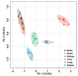
We initially analyzed nucleic acid modifications in various tissues of mice. Principal component analysis (PCA) of nucleic acid modifications across six tissues from mice of control group revealed that, with the exception of the brain and heart, different tissues could be effectively clustered based on their levels of nucleic acid modifications (Fig. 3). These results suggest that the levels of nucleic acid modifications exhibit tissue specificity. The observed tissue-specific levels of RNA modifications may indicate distinct expression patterns of RNA modification regulators in different tissues, which contribute to the unique biological functions associated with each tissue.
Figure 3
Figure 3.
PCA of nucleic acid modifications across six mouse tissues (N = 10).
We next examined the impact of PFOA exposure on nucleic acid modifications from different tissues of mice with orthogonal partial least squares discriminant analysis (OPLS-DA). The results indicated that the high PFOA exposure group could be well differentiated from the control group across six tissues of mice, based on nucleic acid modification levels (Fig. 4). The results suggests that high PFOA exposure leads to significant alterations in nucleic acid modifications from different tissues. In the case of the low PFOA exposure group, differentiation from the control group was also observable in spleen, liver and kidney (Fig. S10 in Supporting information). To assess potential overfitting, we modeled the data using 1, 000 resampling iterations and conducted cross-validation with CV-ANOVA. The results yielded P-values below 0.05, indicating the absence of overfitting in the OPLS-DA model. Additionally, the Q2 values exceeded 0.597, and the R2Y values were greater than 0.772, demonstrating the reliability of the model.
Figure 4
Figure 4.
OPLS-DA model and score plots of PFOA high-dose exposure group and control group across six tissues of mice (N = 10).
We then investigated the relative changes in nucleic acid modifications across various mouse tissues following PFOA exposure. The results showed that PFOA did not significantly affect the levels of 5mC in mouse DNA, but notable changes in 5hmC levels were observed in the heart, lung, kidney, and liver tissues (Fig. 5). Specifically, the levels of 5hmC in the heart and kidney increased with rising PFOA concentrations, whereas a significant decrease in 5hmC was observed in the liver at high PFOA exposure concentrations. These results indicated that the impact of PFOA on 5hmC level varied across different mouse tissues (Fig. S11 and Table S9 in Supporting information).
Figure 5
Figure 5.
Relative alterations in the levels of DNA and RNA modifications among control, low, and high groups across different tissues (N = 10). One-way ANOVA was used to compare differences among the three groups. The P-value indicates the results of the analysis, showing whether significant differences exist among the groups. A gray dot indicates no significant difference, while the colored dot indicates a significant difference.
We subsequently assessed the changes in modification levels of small RNA (< 200 nt). Significant changes were observed in 12, 13, 10, 6, 12, and 14 modifications in the heart, liver, spleen, lung, kidney, and brain, respectively. Specifically, the levels of i6A, t6A, m2, 2G, m1G, m2G, m7G, m1A, m6A, m6, 6A, I, m1I, m5U and Um in the brain, t6A, m2, 2, 7G, m2, 2G, m1G, m2G, m7G, Gm and m5Um in the heart, m2, 2, 7G, m2, 2G, m1G, m2G, m7G, Gm, m5Um, m5U, Um, and Cm in the spleen, m1G, m7G, m2, 2G m6, 6A, Um, and Cm in the lung, m2, 2, 7G, m2, 2G, m1G, m2G, m7G, Gm, m5Cm, Cm, Y and Um in the liver, and m2, 2, 7G, Gm, Am and Um in the kidney were significantly increased in the PFOA exposure group compared to the control group. Additionally, we observed significant decreases in certain modifications in PFOA exposure group relative to the control group, including Y in the brain, m6, 6A, m6Am, m1I and Am in the heart, m6Am, m1I and m1A in the liver, and t6A, m6Am, I, m6, 6A, Y, m6A, m1A and m1I in the kidney (Fig. 5, Figs. S12–S17 and Tables S10–S15 in Supporting information). RNA modifications have been implicated in a variety of human diseases [27,66,67]. However, there are limited studies examining the role of RNA modifications in diseases development associated with PFOA exposure. The dysregulation of nucleic acid modifications across various tissues in response to PFOA exposure observed in the current study may offer new insights into the mechanisms of carcinogenesis linked to PFOA.
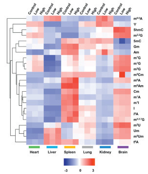
We also conducted cluster analysis based on the average levels of each modification in DNA and small RNA from each tissue across different groups. The overall results revealed that each nucleic acid modification exhibited distinct expression patterns among the tissues of mice in the control, low, and high PFOA exposure groups (Fig. 6). Notably, the signature profiles in the heart were similar to those in the kidney (Fig. 6). Despite the observed changes in modification levels following PFOA exposure, significant differences were evident among the tissues regarding these changes. These results indicate that modifications in different tissues respond differently to PFOA exposure. Additionally, changes in epigenetic modifications may correlate with organ weight. Although the exact mechanisms by which nucleic acid modifications directly influence organ weight are not fully understood, existing research suggests that epigenetic modifications play critical roles in organ development [27,37,68], potentially affecting organ size and quality, especially under certain pathological conditions or experimental treatments.
Figure 6
Figure 6.
The mean levels of each modification in DNA and small RNA across different groups (control, low, and high PFOA exposure) for each tissue. Data were row-normalized with a range from -3 to 3, where blue represents lower levels and red represents higher levels of modification. Rows were clustered to highlight patterns of modification across groups.
In summary, we conducted a comprehensive analysis using LC-MS/MS to investigate the impact of PFOA on DNA and RNA modifications in mouse tissues. Our findings revealed that PFOA exposure altered the levels of DNA and RNA modifications across various mouse tissues. To the best of our knowledge, this is the first study to demonstrate a correlation between PFOA exposure and modifications in both DNA and RNA. These results underscore the significant influence of PFOA on epigenetic modifications in DNA and RNA. Furthermore, with the well-established mapping methods for various nucleic acid modifications [69–77], future research could further explore the mechanisms by which PFOA alters the distribution of these modifications. This would establish a foundation for understanding the adverse effects of PFOA on human health from the perspective of nucleic acid modification. Additionally, since nucleic acid modifications can serve as sensitive biomarkers for various diseases [78–80], they may also indicate exposure to environmental pollutants, including PFOA.
Declaration of completing interests
The authors declare that they have no known competing financial interests or personal relationships that could have appeared to influence the work reported in this paper.
CRediT authorship contribution statement
Shu-Yi Gu: Writing – review & editing, Writing – original draft, Validation, Methodology, Formal analysis, Data curation. Tian Feng: Validation, Data curation. Fang-Yin Gang: Data curation. Si-Yu Yu: Data curation. Wan Chan: Formal analysis. Zhao-Cheng Ma: Methodology, Investigation, Formal analysis, Data curation. Yao-Hua Gu: Data curation. Bi-Feng Yuan: Writing – review & editing, Writing – original draft, Supervision, Project administration, Investigation, Funding acquisition, Formal analysis, Conceptualization.
Acknowledgments
The work is supported by the National Key R & D Program of China (Nos. 2022YFA0806600, 2022YFC3400700), the Fundamental Research Funds for the Central Universities (No. 2042024kf1045), the National Natural Science Foundation of China (No. 22277093), the Key Research and Development Project of Hubei Province (No. 2023BCB094), the Interdisciplinary Innovative Talents Foundation from Renmin Hospital of Wuhan University (No. JCRCGW-2022-008).
Supplementary materials
Supplementary material associated with this article can be found, in the online version, at doi:10.1016/j.cclet.2025.110957.
[1]
I. Rosato, T. Bonato, T. Fletcher, E. Batzella, C. Canova, Environ. Res. 242 (2024) 117743. doi: 10.1016/j.envres.2023.117743
Figure 1
Schematic illustration of PFOA exposure in mice and mass spectrometry profiling of DNA and RNA modifications. The procedure entails the collection of tissue samples from both control and PFOA-exposed groups, followed by the isolation of DNA and small RNA (< 200 nt). Subsequently, enzymatic digestion is performed on the isolated DNA and small RNA, and LC-MS/MS analysis is conducted to detect and quantify the modifications present in the DNA and small RNA.
Figure 2
Effect of PFOA exposure on the growth of mice. (A) Growth curves of mice under different treatment. (B-G) Organ coefficients of different tissues of mice at sacrifice (N = 10). *P < 0.05, **P < 0.01, ***P < 0.001.
Figure 5
Relative alterations in the levels of DNA and RNA modifications among control, low, and high groups across different tissues (N = 10). One-way ANOVA was used to compare differences among the three groups. The P-value indicates the results of the analysis, showing whether significant differences exist among the groups. A gray dot indicates no significant difference, while the colored dot indicates a significant difference.
Figure 6
The mean levels of each modification in DNA and small RNA across different groups (control, low, and high PFOA exposure) for each tissue. Data were row-normalized with a range from -3 to 3, where blue represents lower levels and red represents higher levels of modification. Rows were clustered to highlight patterns of modification across groups.

 DownLoad:


 下载:
下载:


