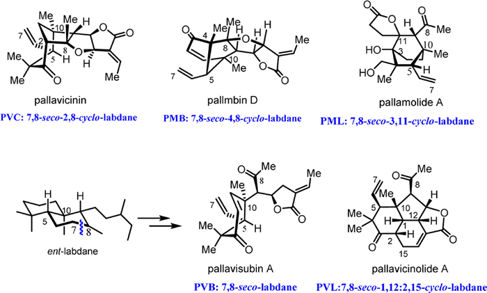
Figure 1.
Representative PDs of subtypes PVC, PMB, PML, PVL, and PVB; revised structures of pallavicinin, pallmbin D, and pallavicinolide A; reported pallamolide A structure; and previously undescribed pallavisubin A structure.
Revision of the absolute configurations of Pallavicinia diterpenoids and further discovery of their Diels−Alder cycloadducts
Jiao-Zhen Zhang , Cheng-Min Zhang , Yong-Jie Wang , Pei-Lin Wu , Rui-Feng Liu , Ye Li , Ming-Zhu Zhu , Shuang-Zhi Yuan , Ze-Jun Xu , Hong-Xiang Lou

Pallavicinia diterpenoids (PDs) are structurally complex 7,8-seco-labdanes that are exclusively present in the liverworts of Pallavicinia species as bioactive constituents and taxonomic markers of the genus Pallavicinia [1] and have inspired several remarkable total syntheses [2–11]. Originated from a labdane-type diterpene followed by the oxidative cleavage of C7–C8 and facilitated by the increase in the oxidation state and modification [12], subtypes named as pallavicinins (PVCs), pallmbins (PMBs), pallamolides (PMLs), and pallavicinolides (PVLs) were reconstructed through the cyclization of C2–C8, C4–C8, and C11–C3 and intramolecular [4 + 2] cycloaddition, respectively (Fig. 1). However, the scaffold diversity and emerging chiral centers due to rearrangements challenge the structural identification accuracy.
Since the first report published in 1994 [13], dozens of PDs have been identified via nuclear magnetic resonance (NMR) measurements and single-crystal X-ray diffraction (SCXRD) with Mo Kα radiation or determined by a simple comparison with the previously reported spectroscopic data [14–16], which are not sufficiently conclusive to determine their absolute configurations.
Our prior research efforts resulted in the unambiguous identification of a series of Pallavicinia dimers from P. subciliata [17] and P. ambigua [18,19] using SCXRD with Cu Kα radiation. These dimers are intermolecular Diels–Alder (DA) adducts of PDs from the same species. Based on the absolute configurations of the previously reported PDs, four PMBs, pallambins A–D, were reinvestigated, and their corresponding enantiomers were identified via electronic circular dichroism (ECD) calculations, X-ray diffraction, and chemical conversion [18]. These findings prompted us to reinvestigate other PDs reported in the literatures.
Herein, we revised the absolute configurations of PVCs and PVLs using a combination of SCXRD with Cu Kα radiation and ECD and NMR calculations. Additionally, five unprecedented PVL and PVC based DA cycloadducts and three previously undescribed 7,8-seco-labdanes, pallavisubin A–C from P. subciliata, which can be considered key intermediates in a biogenesis pathway, were identified.
Pallavicinin (1) was the first reported PVC structure. It was isolated for the first time from P. subciliata of Taiwan province in China in 1994, in which the stereochemistry of 1 was confirmed by SCXRD with Mo Kα radiation, and the negative Cotton effect was observed in an ECD spectrum [13]. In 1998, it was reported together with 18-hydroxpallavicinin (2), and their absolute configurations were determined by SCXRD with Cu Kα radiation; however, the Flack parameters of the produced crystals were not published [14]. Further investigating P. subciliata in this study, we obtained compounds 1 and 2 again, and their NMR spectra, optical rotations, and ECD curves were in good agreement with those reported in the literature (Fig. 2, Figs. S1–1–S1–10, Tables S1 and S3 in Supporting information), from which we concluded that the isolated compounds were identical to the literature structures. However, the absolute configurations of 1 and 2 were accurately determined as 2R, (4S), 5S, 8S, 9R, 10R, 11R, 12R by performing SCXRD with Cu Kα radiation (CCDC 2337893, Flack parameter: 0.14 (11) for 1, Flack parameter: 0.85 (11) for the enantiomer of 1; and CCDC 2337894, Flack parameter: = −0.06 (7) for 2, Flack parameter: = 1.06 (7) for the enantiomer of 2), confirming that the absolute configuration reported in the literature should be revised to the opposite one (Fig. 2) and that the studied compound is an ent–labdane diterpenoid.
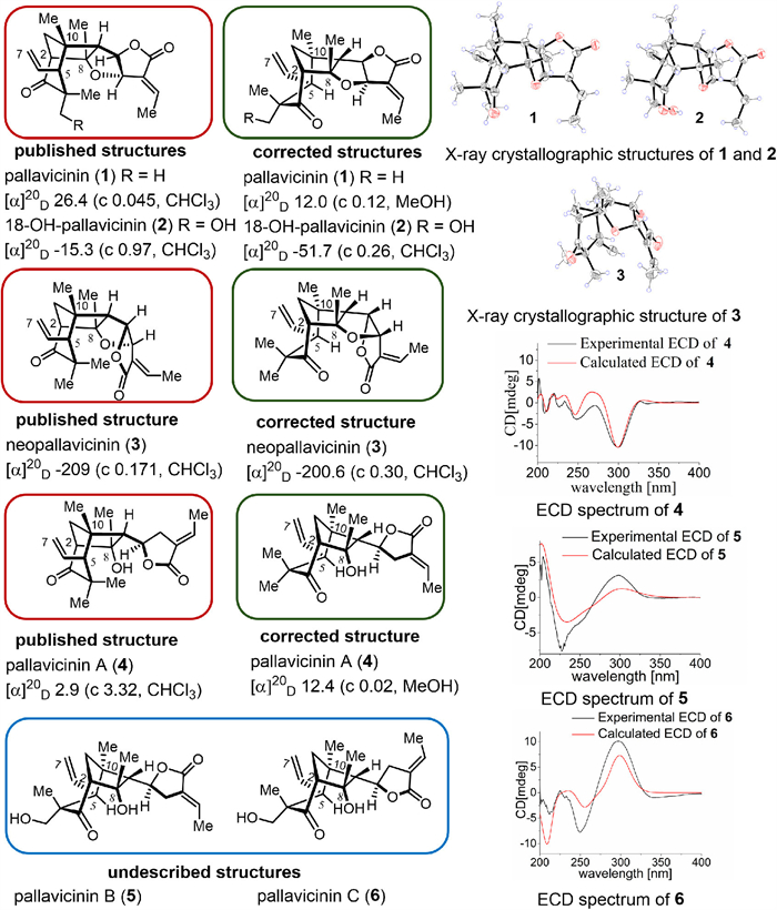
Neopallavicinin (3), a diastereomer of pallavicinin, was first isolated from P. subciliata in 1999 [20]. Its configuration was determined via a spectroscopic comparison with the previously reported pallavicinin structure (Tables S2 and S3 in Supporting information) [13]. In 2005, it was isolated from P. ambigua along with 1 and 2, and its configuration was determined through SCXRD with Mo Kα radiation and a CD spectral comparison. In this study, we suspected that the compound was also misconfigured and validated our idea using SCXRD with Cu Kα radiation (CCDC 2354333, Flack parameter: 0.08(13) for 3, Flack parameter: 0.91(13) for the enantiomer of 3). Considering the same biosynthetic pathway, the absolute structure of 3 was unequivocally determined as 2R, 5S, 8S, 9R, 10R, 11S, 12S (Fig. 2).
According to previous works, the total syntheses of racemic pallavicinin and neopallavicinin have been initially conducted by Wong’s group in 2006 [2]. In 2015, Jia et al. realized the enantioselective syntheses of (–)-pallavicinin and (+)-neopallavicinin [7]. However, the reported optical rotation was doubtful as the configurations determined by SCXRD were consistent with those of the natural products discussed above. The same group has reported the syntheses of revised pallavicinin and neopallavicinin enantiomers in 2024 [11], based on our corrections of pallambins A–D [18].
Pallavicinin A (4) is a PVC with a seco-furan ring, and its NMR spectrum and optical rotation are identical to those reported by Asakawa in 1998 (Tables S2 and S3, Figs. S4–1–S4–10 in Supporting information). Unlike the known configuration, the absolute configuration of this compound was identified as 2R, 5S, 8S, 9R, 10R, 11S by perfectly matching the experimental and calculated ECD curves (Fig. 2). Thus, the absolute configurations of four reported PVCs are certainly corrected.
Pallavicinins B and C (5 and 6) are previously undescribed PVC configurations possessing the same planar structure as that of 4. Their only difference from 4 is the presence of an oxygenated methylene group at C-4. Their relative and absolute configurations were determined by the nuclear Overhauser enhancement spectroscopy (NOESY) correlations as well as ECD calculations of possible configurations (Figs. S2 and S8 in Supporting information). Hence, the stereoisomers of 5 and 6 were defined as 2R, 4S, 5S, 8S, 9R, 10R, 11S and 2R, 4S, 5S, 8S, 9R, 10R, 11R, respectively (Fig. 2), supporting their same biosynthetic origin.
The first reported PMBs was pallambin D, which was initially isolated from liverwort P. subciliata by Asakawa et al. [14] who reported X-ray crystallographic data without Frack parameters or physicochemical properties. Afterwards, pallambins A–D were fractionated from Chinese liverwort P. ambigua by our group, and their absolute configurations were determined via CD based on neopallavicinin [15]. Recently, our group redetermined the absolute configurations of these four compounds obtained from the same plant, and SCXRD data revealed that the initial structures were revised to their enantiomers (Fig. 3) [18].
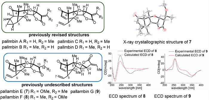
The unprecedented molecular skeletons of pallambins A–D have attracted considerable attention in the synthesis community. Wong and Carreira sequentially reported the total syntheses of (±) pallambins C and D and (±) pallambins A and B in 2012 [4] and 2015 [6]. In the following year, Baran conducted the syntheses of pallambins C and D without studying optical rotations [8]. In 2019, Jia et al. completed the enantioselective total syntheses of pallambins A–D [9]. However, the optical rotations provided in this work are unreasonable negative and agree with the reference [16], and the synthesized pallambins A–D tend to be enantiomers of the present revised natural products, as the optical rotations of key intermediates in the synthetic process are positive. Furthermore, the syntheses of (+) pallambins A–D have been recently realized by Jia et al. [11].
Pallambins E and F (7 and 8) are diastereoisomers that represent the hydrogenated and methoxylated derivatives of pallambins C and D, respectively. SCXRD with Cu Kα radiation (CCDC 2337892, Flack parameter: 0.06 (16)) was employed to confirm the configuration of 7 as 4S, 5S, 8S, 9R, 10R, 11S, 12S, 13R, 14R. 8 was determined as 4S, 5S, 8S, 9R, 10R, 11S, 12S, 13R, 14S based on the differences between the chemical shift values, NOESY results, and calculated ECD curves (Fig. 3).
Pallambin G (9) shares the same planar structure with pallambin D. Meanwhile, the H-11 and H-12 of 9 are β-oriented. Finally, its absolute configuration is 4S, 5S, 8S, 9R, 10R, 11R, 12R, as determined from a good match between the calculated and experimental ECD curves (Fig. 3).
PVLs pallavicinolides A and B (10 and 11) were initially isolated from P. subciliata in 1998, as their NMR data and optical rotations (Fig. 4, Tables S4 and S5 in Supporting information) were identical to those published previously [14]. However, the absolute configurations of these two compounds have not been reported in literature [14]. Wong et al. implemented the total synthesis of (±)-pallavicinolide A in 2009, and its stereochemistry was determined via derivatization and SCXRD [3]. Jia et al. synthesized (+)-pallavicinolides B and C in 2024 [11]. The fitting results of the ECD experimental and computational curves ultimately determined the absolute conformations of 10 and 11 as 1R, 2S, 5S, 9R, 10R, 11R, 12S and 1R, 2S, 4S, 5R, 9R, 10S, 11R, 12S, respectively (Fig. 4). Pallavicinolide C is a C-4 isomer of pallavicinolide B [14], it has not been obtained in this study, from a biosynthetic perspective, it should also be revised as the enantiomer of reported configuration.
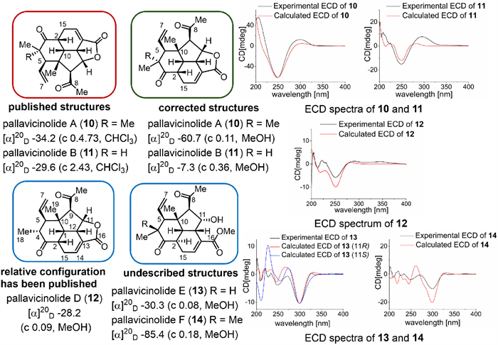
Pallavicinolide D (12) was identified as the C-4 epimer of 11. Comprehensive analysis of the NMR data (Tables S11 and S12, Supporting information) indicated that the relative configuration of 12 was consistent with that of intermediate 41 reported in previous synthetic study [11]. The absolute configuration of 12 was determined to be 1R, 2S, 4R, 5R, 9R, 10S, 11R, 12S based on the agreement between the ECD spectra (Fig. 4).
Pallavicinolide E (13) and pallavicinolide F (14) are PVLs with open 16,11-γ-lactone rings (Fig. S1 in Supporting information). Compound 14 is structurally similar to compound 13 with only one additional methyl group at C-4. The relative configurations of compounds 13 and 14 were determined from the NOESY spectra (where the cross-peaks of H3–18/H-5/H-2 demonstrated that their configurations of H-2 were α-oriented (Fig. S2)) and ECD calculations, which showed that H-11 was β-oriented (Fig. 4). The absolute configurations of 13 and 14 were established as 1R, 2R, 4R, 5R, 9R, 10S, 11R, 12S and 1R, 2R, 5S, 9R, 10R, 11R, 12S, respectively, by comparing the experimental and calculated ECD data (Fig. 4).
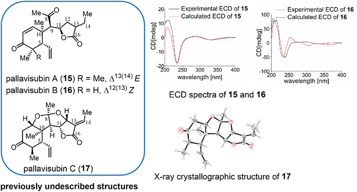
Pallavisubins A and B (15 and 16), two undescribed 7,8-seco-labdanes without rearrangements, were obtained, and their configurations were determined as 5S, 9R, 10R, 11R, and 4R, 5R, 9R, 10R, 11R, respectively, via ECD and 13C NMR calculations (Fig. 5; Figs. S9, S10 and Tables S18–S23 in Supporting information). Structurally, they are key intermediates in the formation of PDs, contributing to a better understanding of their biosynthetic pathways. Based on the structure of 16, the C=O group at C-8 undergoes condensation with C-12 to form pallavisubin C (17), an unprecedented 7,8-seco-labdane ketal. The characteristic 13C NMR signal (δC-8 119.0) confirmed the existence of the ketone acetal. The absolute configuration of 17 determined by SCXRD is 1S, 4R, 5R, 8S, 9R, 10R, 11R, 12R (CCDC 2337889) (Fig. 5).
Interestingly, dimeric labdane derivatives from the same plant support this conclusion. PDdimer A (18) was identified as a 7,8-seco-labdane dimer formed from PVC and PVL through the intermolecular [4 + 2] cycloaddition, and the heterodimer with PVL was first obtained from P. subciliata. Carefully resolving its NMR signals (Figs. S18–1–S18–6 in Supporting information), the structural unit of PVC was identical to that of 5, and the other unit was highly similar to 11 [14] except for the different configuration at the C-4′ position supported by the NOESY correlation of H3–19′/H-4′ (Fig. S4 in Supporting information). Unlike the previously PD-derived dimers, the intracyclic double bond of PVL reacted with PVC as a proto-dienophile to generate a new six-membered ring, in which C12-C13′ and C15-C14′ were the newly formed carbon bonds (Fig. S3 in Supporting information). Moreover, the NOESY signal of H3–17/H-12/H-12′ confirmed that they were close in space, and the endo-DA reaction occurred to form compound 18 (Fig. S4). The absolute configuration of 18 was 2R, 4S, 5S, 8S, 9R, 10R, 11S, 12R, 1′R, 2′S, 4′R, 5′R, 9′R, 10′S, 11′R, 12′S, 13′R, 14′S, as determined via SCXRD with Cu Kα radiation (CCDC 2292684) (Fig. 6).
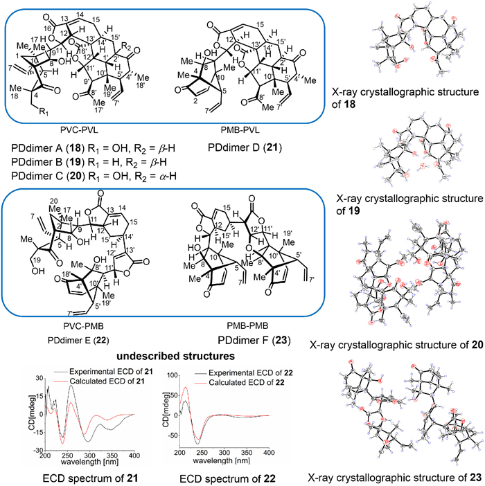
PDdimer B (19) possesses a structure similar to that of 18, with the only difference being that the group linked to C-4 is a gem–dimethyl group (Fig. S3). By conducting SCXRD with Cu Kα radiation (CCDC 2292679), the absolute configuration of 19 was delineated as 2R, 5S, 8S, 9R, 10R, 11S, 12R, 1′R, 2′S, 4′R, 5′R, 9′R, 10′S, 11′R, 12′S, 13′R, 14′S (Fig. 6).
PDdimer C (20) and 18 are a pair of diastereomers that share the same PVC configuration. The NOE correlations of H-2′/H-9′/H-5′/H3–18′ in 20 exhibit the opposite H-2′ configuration as compared with that of 18 in the PVL unit, which is consistent with the change of chemical shifts for this unit (Fig. S3). SCXRD with Cu Kα radiation (CCDC 2292682) was performed to determine the absolute configuration of 20: 2R, 4S, 5S, 8S, 9R, 10R, 11S, 12R, 1′R, 2′R, 4′R, 5′R, 9′R, 10′S, 11′R, 12′S, 13′R, 14′S (Fig. 6).
PDdimer D (21) was found to be a dimer that shared an identical PVL unit with dimer 20, and another unit was PMB whose gross structure and relative configuration were similar to those of pallaviambin C (Figs. S3 and S4) [18]. A comparison of the experimental and calculated ECD data revealed that the absolute configuration of compound 21 was 4S, 5S, 8S, 9R, 10R, 11R, 12S, 1′R, 2′R, 4′R, 5′R, 9′R, 10′S, 11′R, 12′S, 13′R, 14′S (Fig. 6).
PDdimer E (22) is a PVC–PMB heterodimer that has not been previously identified. These two units formed a six-membered ring connected by the C15–C14′ and C12–C15′ bonds (Fig. S3). In this case, the pro-dienophile site was Δ14′(15′), which was different from the Δ13′(14′) position in dimers 18–21. The relative configuration of 22 was deduced from the NOESY signals of H2–19a/H-11, H2–15′b/H3–17/H-12/H-9/H3–20, and H3–18′/H-5′/H3–19′/H-11′/H-5′ (Fig. S4). By ECD calculation, the absolute configuration of compound 22 was determined as 2R, 4S, 5S, 8S, 9R, 10R, 11S, 12S, 4′S, 5′S, 8′S, 9′R, 10′R, 11′S, 14′R (Fig. 6).
PDdimer F (23) is also a PD-derived dimer, in which both units are PMBs similar to those in pallaviambin [18]. The NOESY signals of H-11′/H3–19′/H-5′ suggest that H-11′ and H-12′ may exhibit different configurations (Fig. S4) [18]. Finally, SCXRD with Cu Kα radiation (CCDC 2337891) confirmed that the absolute configuration of 23 was 4S, 5S, 8S, 9R, 10R, 11R, 12R, 4′S, 5′S, 8′S, 9′R, 10′R, 11′R, 12′R, 13′S, 14′R (Fig. 6).
Biosynthetically, the formation of PDs has been disputed, and labdane diterpenoids were considered common precursors until, even though some ent–labdanes were obtained together with PDs from P. ambigua [19]. In this study, by conducting an absolute configuration revision of PDs, a plausible biogenetic pathway was proposed (Fig. 7), in which an ent–labdane precursor undergoes an oxidative cleavage of the C7–C8 bond to produce the key intermediate (15) and reconstruction of the C2–C8, C4–C8, and C3–C11 bonds to produce PVCs, PMBs, and PMLs, respectively. PVLs and PDdimers are generated via intra-molecular and inter-molecular DA reactions. The reconstruction and formation of the rings lead to skeletal and stereo diversification and complication.

The anti-inflammatory effects of isolated PDs are preliminarily investigated by evaluating the inhibition of nitric oxide (NO) production induced by lipopolysaccharide (LPS) in RAW264.7 cells. The maximum inhibition rate (MIR) and median effective concentration (EC50) against NO production are tested. As shown in Table S36 and Fig. S24 (Supporting information), 1 (MIR = 85.31% (12.5 µmol/L), EC50 = 16.38 µmol/L) and 22 (MIR = 82.08% (25 µmol/L), EC50 = 5.88 µmol/L) exhibited significant inhibition on NO production. Compounds 5, 10–14, 18, 20, and 21 had moderate inhibitory ability, with MIR values of 56.12%–79.04% [21].
In conclusion, we have revised the absolute configurations of four PVCs and three PVLs reported previously. The pioneering identification of the DA cycloadducts supports our conclusions, considering that they have the same biosynthetic origins. These results underscore the decisive role of some “inconspicuous” parameters, such as optical rotations and Flack parameters. The absences of Flack parameters in literature are the primary causes of the incorrect absolute configurations. Thus, the SCXRD with Cu Kα radiation and comprehensive crystal data are important for the report of natural products. Moreover, instead of blindly following the literature, the recognition and evaluation of known structures based on biosynthetic points are important in the fields of natural products and total synthesis.
The authors declare that they have no known competing financial interests or personal relationships that could have appeared to influence the work reported in this paper.
Jiao-Zhen Zhang: Writing – review & editing, Writing – original draft, Funding acquisition. Cheng-Min Zhang: Writing – original draft, Investigation. Yong-Jie Wang: Investigation. Pei-Lin Wu: Investigation. Rui-Feng Liu: Investigation. Ye Li: Investigation. Ming-Zhu Zhu: Investigation. Shuang-Zhi Yuan: Investigation. Ze-Jun Xu: Methodology. Hong-Xiang Lou: Writing – review & editing, Supervision, Funding acquisition.
This work was supported by the National Natural Science Foundation of China (Nos. 82293682, 82293684, and 82173703).
Supplementary material associated with this article can be found, in the online version, at doi:
P.S. Grant, M.A. Brimble, Chem. Eur. J. 27 (2021) 6367–6389. doi: 10.1002/chem.202004574
X.S. Peng, H.N.C. Wong, Chem. Asian. J. 1–2 (2006) 111–120. doi: 10.1002/asia.200600061
J.Q. Dong, H.N.C. Wong, Angew. Chem. Int. Ed. 48 (2009) 2351–2354. doi: 10.1002/anie.200806335
X.S. Xu, Z.W. Li, Y.J. Zhang, et al., Chem. Commun. 48 (2012), 8517–8519. doi: 10.1039/c2cc34310j
M. Markovič, M. Ďuranová, P. Koóš, et al., Tetrahedron 69 (2013) 4185–4169. doi: 10.1016/j.tet.2013.03.100
C. Ebner, E.M. Carreira, Angew. Chem. Int. Ed. 54 (2015) 11227–11230. doi: 10.1002/anie.201505126
B. Huang, L. Guo, Y. Jia, Angew. Chem. Int. Ed. 54 (2015) 13599–13603. doi: 10.1002/anie.201506575
L.P. Martinez, S. Umemiya, S.E. Wengryniuk, et al., J. Am. Chem. Soc. 138 (2016) 7536–7539. doi: 10.1021/jacs.6b04816
X. Zhang, X. Cai, B. Huang, et al., Angew. Chem. Int. Ed. 58 (2019) 13380–13384. doi: 10.1002/anie.201907523
Y. Zhang, L. Chen, Y. Jia, Angew. Chem. Int. Ed. 63 (2024) e202319127. doi: 10.1002/anie.202319127
L. Chen, P. Chen, X. Zhang, et al., Chem 10 (2024) 2473–2483. doi: 10.1016/j.chempr.2024.04.003
A. Almeida, L. Dong, G. Appendino, et al., Nat. Prod. Rep. 37 (2020) 1207–1228. doi: 10.1039/c9np00030e
C.L. Wu, H.J. Liu, H.L. Uang, Phytochemistry 35 (1994) 822–824. doi: 10.1016/S0031-9422(00)90616-2
M. Toyota, T. Saito, Y. Asakawa, Chem. Pharm. Bull. 46 (1998) 178–180. doi: 10.1248/cpb.46.178
Z.J. Li, H.X. Lou, W.T. Yu, et al., Helv. Chim. Acta 88 (2005) 2637–2640. doi: 10.1002/hlca.200590204
L.N. Wang, J.Z. Zhang, X. Li, et al., Org. Lett. 14 (2012) 1102–1105. doi: 10.1021/ol3000124
S.G. Liu, C.Y. Zhang, J.C. Zhou, et al., Org. Chem. Front. 9 (2022) 1790–1796. doi: 10.1039/d1qo01891d
C. Zhang, Y. Li, Z. Chu, et al., Chin. Chem. Lett. 35 (2024) 108206. doi: 10.1016/j.cclet.2023.108206
Y. Li, X.B. Li, J.C. Zhou, et al., Phytochemistry 212 (2023) 113702. doi: 10.1016/j.phytochem.2023.113702
H.J. Liu, C.L. Wu, J. Asian Nat. Prod. Res. 1 (1999) 177–182. doi: 10.1080/10286029908039862
M.X. Zhou, X. Wei, A.L. Li, et al., BMC Complem. Altern. Med. 16 (2016) 360. doi: 10.1186/s12906-016-1347-y

Figure 1 Representative PDs of subtypes PVC, PMB, PML, PVL, and PVB; revised structures of pallavicinin, pallmbin D, and pallavicinolide A; reported pallamolide A structure; and previously undescribed pallavisubin A structure.

 扫一扫看文章

扫一扫关注我们
 DownLoad:
 下载:
下载:


