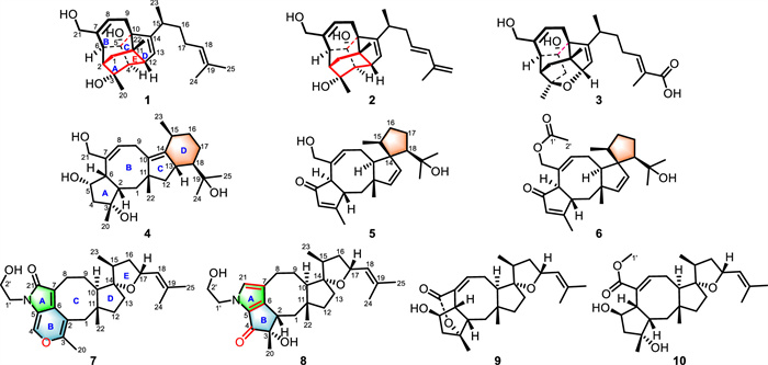
Figure 1.
Structures of compounds 1–10.
Ophiobolin-type sesterterpenoids with unprecedented chemical architectures from Bipolaris oryzae and their inflammatory activity
Meijia Zheng , Yingjie Liu , Chunmei Chen , Qin Li , Xinran Zhang , Xiaotian Zhang , Weiguang Sun , Yonghui Zhang , Hucheng Zhu

Natural products are a crucial source for drug discovery and development [1–5], which have been used for the prevention and treatment of a variety of diseases [6–9]. Sesterterpenoids are a relatively small group within the terpenoid family, but their sources are widespread, including fungi, plants, insects, and sponges [10–13]. Many sesterterpenoids have diverse structures and significant bioactivities [14–16], such as niduene D, possessing unprecedented 5/5/5/5/6 pentacyclic ring skeleton demonstrated potent resensitization of SW620/AD300 cells to paclitaxel (PTX) by lowering the half maximal inhibitory concentration (IC50) values of PTX up to 5.26-fold from 340 nmol/L to 1.79 µmol/L [17]. At the same time, sesterterpenoids have also attracted great attention of biology communities [18,19], such as the bioinspired total syntheses of unprecedented sesterterpenoids, orientanoids A–C, were effectively achieved in 7–8 steps in overall yields of 2.3%–6.4% from the commercially available santonin without using any protecting groups [20].
Ophiobolins are a type of sesterterpenoid possessing a 5/8/5-fused tricyclic skeleton. Since ophiobolin A was firstly isolated and identified from Ophiobolus miyabeanus in 1958 [21], >120 ophiobolins have been reported [22]. Ophiobolin-type sesterterpenoids have intriguing molecular architectures and wide array of biological effects [23–26]. For example, bipolarolide A, with an intriguing and complex 5/6/6/6/5 caged pentacyclic skeleton, had the most potent 3–hydroxy-3-methyl glutaryl coenzyme A reductase (HMGR) inhibitory activity (IC50 = 2.5 ± 0.1 µmol/L) [27]. In 2024, Jia and co-workers achieved the first total synthesis of bipolarolides A and B [28].
Our group has a longstanding interest in the ophiobolin-derived sesterterpenoids from the genus Bipolaris [27,29–32]. In the course of our continuing search for structurally interesting and bioactive ophiobolin-derived sesterterpenoids, the phytopathogenic fungus, Bipolaris oryzae, was investigated. At present, there is not much research on the secondary metabolites of Bipolaris oryzae, mainly producing ophiobolins [33–35]. In this paper, ten new ophiobolin-derived sesterterpenoids, bipolarpenoids A–J (1–10), were isolated from the ethyl acetate extract, which possess four new classes of skeletons. Among them, 1 and 2 represent the first examples of sesterterpenoids with a new 5/6/6/5/6 carbon-skeleton featuring a caged pentacyclo[8.4.0.01,5.04,9.07,11]tetradecane motif, and biosynthetically the core structure is constructed by the fusion of ring A on the western hemisphere and ring D on the eastern. In this article, we report the isolation, structural elucidation, proposed biosynthetic pathways, and bioactivity evaluation of these new compounds (Fig. 1).
Bipolarpenoid A (1) was obtained as a white powder. Its molecular formula, C25H36O3, was assigned according to high resolution electrospray ionization mass spectrometry (HRESIMS) data in conjunction with nuclear magnetic resonance (NMR) analysis, requiring eight indices of hydrogen deficiency. The 13C NMR data (Table S2 in Supporting information) and distortionless enhancement by polarization transfer (DEPT) spectrum of 1 displayed a total of 25 signals, ascribable to five methyls, five methylenes (including one oxygenated), eight methines (including three olefinic), seven nonprotonated carbons (including two oxygenated and three olefinic). A comparison of the NMR data of 1 with those of bipolarolide A [27], implied that 1 also possessed the same 5/6/6/5-fused (A/B/C/D rings) carbon skeleton. The obvious differences were the absence of one ketone carbonyl, and the presence of one methine in 1. The above analyses, along with the indices of hydrogen, revealed that the key structural feature of 1 is the presence of an additional ring. Considering the two methine groups at C-4 (δC 61.0) and C-12 (δC 52.5), the linkage of C-4 and C-12 via a single carbon–carbon bond was proposed, resulting in the formation of an unexpected ring (E ring). This speculation was supported by the key heteronuclear multiple-bond correlation (HMBC) correlations (Fig. 2A) from H-4 to C-11, C-12, and C-13, and from H-12 to C-5, along with the weak 1H–1H correlated spectroscopy (COSY) correlation of H-4/H-12 (the dihedral angle around 90° between H-4 and H-12). Finally, the 1H–1H COSY correlations of H-15/H2-16/H2-17/H-18 indicated that C-17 (δC 26.1) should be a methylene group in 1 instead of the ketone carbonyl in bipolarolide A. Thus, the planar structure of 1 was determined to possess an unprecedented and highly caged pentacyclo[8.4.0.01,5.04,9.07,11]tetradecane scaffold (Fig. 2B).
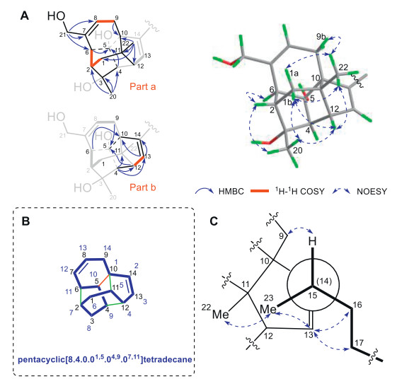
Compound 1 is a novel caged, rigid, and sterically congested sesterterpenoid, and its relative configuration was deduced by analyses of its nuclear Overhauser effect spectroscopy (NOESY) correlations (Fig. 2A). First, H3-22 was assumed to be β-oriented and the key NOESY correlation of H3-22/H-9b suggested the S* configuration of C-10, and indicated that the α-orientation of OH-5, facing the outside of the caged structure. Moreover, NOESY correlations of H-12/H-1b/H3-20, H-12/H3-22, H3-22/H2-1, and H3-20/H-2 indicated that H-2, H-12, and H3-20 were assigned to be β-oriented, while H-4 and H-6 could face the outside of the caged structure, assigned to be α-oriented. In addition, the relative configuration of C-15 was determined by analyzing the Newman projection of C-14–C-15 (Fig. 2C). The observed NOESY correlations of H-13/H3-23, H-13/H-16, H-13/H-17, H-15/H-9, and H3-23/H3-22, suggested the S* configuration of C-15. Meantime, according to the biosynthetic origin of all ophiobolins, the configuration of C-15 should be 15S* [36]. Comparison of the experimental and computed electronic circular dichroism (ECD) curves (Fig. S1 in Supporting information) determined the absolute configuration of 1 as 2S,3R,4S,5S,6S,10S,11S,12R,15S.
Bipolarpenoid B (2) was isolated as a colorless oil, and its molecular formula was confirmed to be C25H34O3 based on the HRESIMS and 13C NMR data. The 1D and 2D NMR data of 2 were very similar to those of 1, especially in the core scaffold, with the difference occurring at the side chain. The 1H–1H COSY correlations of H-15/H2-16/H-17/H-18 and HMBC correlations from H2-25 to C-18, C-19, and C-24 confirmed that the trisubstituted double bond at Δ18,19 in 1 was replaced by the conjugated double bonds at Δ17(18),19(25) in 2. The relative configuration of 2 was determined to be the same as that of 1 by analyzing their NOESY correlations, and the E-configuration of the double bond Δ17,18 was deduced from the large coupling constant (3J17,18 = 15.8 Hz). Furthermore, the similar experimental ECD spectra (Fig. S1) and specific rotation of 1 and 2 indicated that they share the same absolute configuration.
Bipolarpenoid C (3) has a molecular formula of C25H34O5, based on its HRESIMS data at m/z 437.2298 [M + Na]+, requiring nine indices of hydrogen deficiency. Its NMR data suggested that 3 was a analogue of bipolarolide B [27], except that a hydroxymethyl group in bipolarolide B was replaced by a carboxyl group (δC 172.0, C-25) in 3. This deduction was confirmed by the HMBC correlations (Fig. S2 in Supporting information) from H-18 and H3-24 to C-25. The NOESY spectrum of 3 disclosed that its relative configuration was consistent with bipolarolide B. Finally, the similar experimental ECD spectra (Fig. S4 in Supporting information) of 3 and bipolarolide B assigned the absolute configuration of 3 to be 2S,3R,5R,6S,10R,11R,12S,15S.
Bipolarpenoid D (4) was also isolated as a colorless oil. Its molecular formula was determined to be C25H40O4 on the basis of the HRESIMS and 13C NMR data, implying six indices of hydrogen deficiency. The 13C NMR data (Table S3 in Supporting information) along with the DEPT spectrum provided the resonances for five methyls, seven methylenes (including one oxygenated), seven methines (including one oxygenated and one olefinic), six nonprotonated carbons (including two oxygenated and three olefinic). These typical signals suggested that 4 was most likely an ophiobolin-type sesterterpenoid with a tetracyclic system. Further analyses of the 1H–1H COSY and HMBC correlations of 4 elucidated the typical 5/8/5-fused (A/B/C rings) carbon skeleton of 4. In addition, the 1H–1H COSY correlations (Fig. S2) of H2-12/H-13/H-18/H2-17/H2-16/H-15/H3-23, along with HMBC correlations from H3-23 to C-14, C-15, and C-16 and from H-15 to C-13, established the presence of an additional six-membered cyclohexane (D ring). Then, the HMBC correlations (Fig. S2) from H3-25 to C-18, C-19, and C-24 revealed that C-19 was linked to C-18. Thus, the planar structure of 4 was established, featuring an unprecedented 5/8/5/6-fused carbon skeleton. The NOESY correlations (Fig. S3 in Supporting information) of H3-22/H-6/H-2 and H-2/H-5/H-6 revealed that they are cofacial and were assigned as β-oriented. NOESY correlation of H-2/H3-20 suggested H3-20 was β-oriented. In addition, NOESY correlations of H3-22/H-13 and H-13/H3-23 suggested H-13 and H3-23 were β-oriented, while correlations of H3-22/H-12a, H-18/H-12b, and H3-24/H-13 revealed the α-orientation of H-18. Finally, the absolute configuration of 4 was determined by ECD calculations. As shown in Fig. S5 (Supporting information), the experimental ECD of 4 was consistent with the calculated ECD of 4, establishing the absolute configuration of 4 as 2S,3R,5S,6S,11R,13S,15S,18R.
Bipolarpenoid E (5) was assigned as the molecular formula of C25H36O3, based upon the 13C NMR data as well as HRESIMS analysis, requiring eight indices of hydrogen deficiency. Detailed analyses of its 2D NMR data confirmed that this compound was also an ophiobolin-type sesterterpenoid possessing an 5/8/5-fused carbon skeleton. The 1H−1H COSY correlations (Fig. S2) of H-18/H2-17/H2-16/H-15/H3-23, along with the HMBC correlations from H3-23 to C-14, C-15, and C-16 and from H2-16, H2-17, and H-18 to C-14, suggested the presence of a new five-membered cyclopentane (D ring), as well as the fusion of rings C and D to construct the unusual spiro[4.4]nonane core. The HMBC correlations from H3-25 to C-18, C-19, and C-24 revealed that the hydroxyisopropyl group was located at C-18. The NOESY correlations (Fig. S3) of H-2/H3-22 and H3-22/H3-23 revealed they were β-oriented and suggested the R* configuration of C-14. Then, NOESY correlations of H-9a/H-10/H-18 suggested H-10 and H-18 were α-oriented. In addition, NOESY correlation of H-10/H-6 revealed H-6 was α-oriented. The absolute configuration of 5 was determined to be 2S,6R,10R,11S,14R,15S,18R, through the comparison of experimental and calculated ECD curves (Fig. S5).
Bipolarpenoid F (6), isolated as a white powder, was deduced to have a molecular formula of C27H38O4 based on its 13C NMR data and HRESIMS analysis, requiring nine indices of hydrogen deficiency. The 1D (Table S3) and 2D NMR data of 6 showed close structural resemblances to those of 5, except for the presence of an additional acetoxyl group in 6. An acetoxyl group was located at C-21 based on the HMBC correlation (Fig. S2) from H2-21 to C-1ʹ (δC 172.3). The similarities of their NOESY data (Fig. S3) suggested that 6 and 5 possessed identical relative configurations. Finally, the absolute configuration of 6 was determined by the similar ECD spectra with those of 5 (Fig. S5).
Bipolarpenoid G (7) was obtained as a colorless oil, and its molecular formula, C27H37NO4, was deduced from the HRESIMS and 13C NMR data. Careful analyses of its 1D (Table S4 in Supporting information) and 2D NMR data demonstrated that 7 was a nitrogenous sesterterpenoid derivative, bearing great similarity to the known compound bipolarolide F [27], except for B ring. The HMBC correlations (Fig. S2) from H3-20 to C-2 and C-3 and from H-4 to C-3, C-5, and C-6, in combination of the molecular formula and the chemical shifts of C-3 (δC 154.0) and C-4 (δC 130.2) inferred the presence of the 4H-pyran ring (B ring) in 7. Furthermore, the density functional theory (DFT) was used to calculate the 13C NMR chemical shifts of 7 at the B3LYP-D3(BJ)/6-31G* level in MeOH. Accordingly, the correlation coefficient (R2) for 7 was 0.9988 (Fig. S6 in Supporting information), and the corresponding mean absolute error (MAE) was 1.4 ppm (Table S6 in Supporting information), which further supported our proposed planar structure. The key NOESY correlations (Fig. S3) of H3-22/H-9a, H-10/H-12b, H3-22/H-15, H3-23/H-13a, H3-23/H-17, and H-17/H-13b determined the relative configuration of 7. To determine the absolute configuration, the ECD curves (Fig. S5) of 7 and ent–7 were calculated, and the former showed excellent agreement with the experimental one. Therefore, the absolute configuration of 7 was deduced to be 10R,11R,14S,15S,17R.
Bipolarpenoid H (8) was isolated as a colorless oil with a molecular formula of C27H39NO4, as assigned by its HRESIMS and 13C NMR data. Analyses of its NMR spectroscopic data demonstrated that 8 was also a nitrogenous sesterterpenoid derivative, similar to bipolarolide G [27], with the distinct difference lying in the structure of rings A and B. The HMBC correlations (Fig. S2) from H3-20 to C-2, C-3, and C-4 indicated that the methylene group (C-4) in bipolarolide G was replaced by a carbonyl group (δC 193.5) in 8. Meanwhile, the HMBC correlations from H-21 and H-2 to C-5 and C-6 suggested that the α,β-unsaturated lactam ring in bipolarolide G was replaced with a pyrrole ring (A ring) in 8. NOESY correlation (Fig. S3) of H3-22/H-2 revealed that they are cofacial and were assigned as β-oriented. Due to the formation of a rigid plane between rings A and B in 8, combined with NOESY correlation of H-2/H3-20 suggested H3-20 should be β-oriented. NOESY correlations of H-2/H-1a and H-10/H-1b revealed the α-orientation of H-10. Furthermore, NOESY correlations H3-22/H-9, H-9/H-15, H3-23/H-17, and H-17/H-13b, indicated the S* configuration of C-14 and the β-orientation of H-17 and H3-23. Finally, the absolute configuration of 8 was determined as 2S,3S,10R,11R,14S,15S,17R based on ECD calculations (Fig. S5).
Bipolarpenoid I (9) was determined to have a molecular formula of C25H36O4, based on the analysis of HRESIMS and 13C NMR data, corresponding to eight indices of hydrogen deficiency. Its 1H and 13C NMR data (Table S4) closely resembled those of ophiobolin A lactone [37], with the major differences being that the C-3 resonance in 9 (δC 93.7) was shifted downfield compared to that (δC 80.2, C-3) in ophiobolin A lactone, while the C-5 resonance in 9 (δC 72.4) was shifted upfield compared to C-5 (δC 81.4) in ophiobolin A lactone. Thus, the lactone ring was formed between C-21 and C-3 in 9. Finally, 9 was successfully subjected to single-crystal X-ray diffraction using Cu Kα radiation (Fig. S7 in Supporting information) with a Flack parameter of −0.01(2), which confirmed the skeleton and absolute configuration of 9 as 2S,3R,5R,6S,10R,11R,14S,15S,17R.
Bipolarpenoid J (10) was deduced to have a molecular formula of C26H40O5, based on its 13C NMR data and HRESIMS analysis. A detailed comparison of the NMR data manifested that the structure of 10 was similar to 9, with the primary difference being the presence of an additional methoxy group (δC 52.7), which were further supported by the HMBC correlation (Fig. S2) from H3-1ʹ (δH 3.72) to C-21 (δC 172.6). The relative configuration of 10 was consistent with 9 by its NOESY correlations (Fig. S3) and H − H coupling constants. Finally, the absolute configuration of 10 was determined to be 2S,3R,5R,6S,10R,11R,14S,15S,17R by comparison of its experimental ECD curve with that of 9 (Fig. S8 in Supporting information).
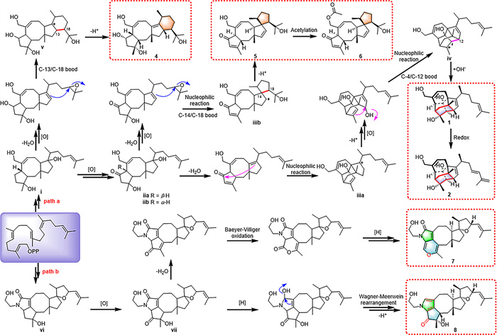
The unusual sesterterpenoids (1, 2, and 4–8) give rise to questions about their biosynthetic origin. The putative biosynthetic pathways were proposed, as shown in Scheme 1. Starting from GFPP, a series of via a series of enzymatic reactions and then undergoes two unusual pathways (paths a and b). In path a, ⅰ undergoes multi-step oxidizations to afford intermediates ⅱa and ⅱb (6–epi isomers) [36]. Subsequently, ⅱa undergoes dehydration, and nucleophilic reactions to afford intermediate ⅲa bearing a 5/6/6/5 fused carbon skeleton [27]. Further deprotonation, oxidization at C-12, and nucleophilic reactions of ⅲa produce ⅳ, which possesses a new single carbon−carbon bond between C-4 and C-12. Finally, a hydroxylation of iv generates 1, which further undergoes a redox reaction to afford 2. In addition, ⅱb undergoes dehydration, oxidization, and nucleophilic reactions to form intermediate ⅲb with a new 5/8/5/5 fused carbon skeleton. Then, an additional deprotonation reaction led to the formation of 5, which further undergoes an acetylation reaction to afford 6. On the other hand, ⅰ directly undergoes dehydration, oxidization, and nucleophilic reactions to form intermediate ⅴ with a new 5/8/5/6 fused carbon skeleton. Afterward, the same deprotonation reaction led to the formation of 4. In path b, an oxidization reaction of ⅵ generates ⅶ; on one hand, ⅶ undergoes dehydration, Baeyer-Villiger oxidation, and reduction reactions to produce 7; on the other hand, ⅶ might undergo a series of reduction, Wagner-Meerwein rearrangement, and deprotonation reactions to form 8.
Lipopolysaccharide (LPS) induced RAW264.7 polarization of mouse macrophages is a classic inflammatory model [38]. So, we used LPS to induce nitric oxide (NO) release in RAW264.7 cells to mimic the inflammatory response, and further determined the anti-inflammatory activity. As shown in Fig. S9 (Supporting information), the results showed that compound 9 have the best anti-inflammatory activity of ten compounds with an IC50 value of 3.4 ± 0.5 µmol/L (Fig. S9A). Additionally, the cell counting kit-8 (CCK-8) kit assay showed that 9 with no cytotoxic effects on RAW264.7 cells in the range of 10–40 µmol/L (Fig. S9B). Quantitative real-time polymerase chain reaction (qRT-PCR) further validated the anti-inflammatory activity of 9, and the results showed that 9 could reduce the mRNA levels of pro-inflammatory cytokines, including interleukin-6 (IL-6), tumor necrosis factor-α (TNF-α), and IL-1β (Fig. S9C). The nuclear factor-κB (NF-κB) pathway is an important signaling pathway that regulates various physiological processes such as immunity, inflammatory response, and cell apoptosis [39]. NF-κB p65 is a key transcription factor in macrophage activation and can be transferred to the nucleus when stimulated by LPS. To evaluate whether 9 can inhibit the NF-κB pathway, we performed immunofluorescence staining on the nuclei of RAW264.7 cells, as shown in Fig. S9D, 9 can inhibit the nuclear translocation of NF-κB p65. Meanwhile, through examined the signaling proteins in RAW264.7 cell lines after being stimulated by LPS, we found that 10 µmol/L of 9 reduced the phosphorylated-inhibitor of κB-α (p-IκB-α) signal intensities (Fig. S9E). These results suggest that 9 may inhibit the inflammatory response by downregulating the NF-κB pathway.
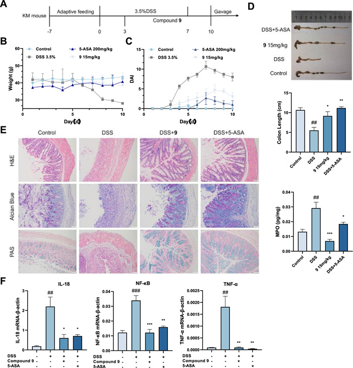
Among the chemically induced models of colitis, dextran sodium sulfate (DSS) is one of the most commonly used modeling agents that can induce severe colitis when taken orally by drinking water, with characteristics similar to some of the symptoms of a human ulcerative colitis (UC) attack [40]. Therefore, we used an acute DSS colitis model to evaluate the in vivo anti-inflammatory activity of 9. All animal experiments were performed in strict accordance with the requirements of the Animal Research Committee and approved by the Institutional Animal Care and Use Committee (IACUC) of Huazhong University of Science and Technology. Except for the control group, each mouse was given 3.5% DSS solution and treated with 9 (15 mg/kg) or 5-amino salicylic acid (5-ASA) (200 mg/kg) for 7 days by oral administration (Fig. 3A). The body weight of mice was recorded daily, and colon samples were collected at the end of the experiment for subsequent assays. As shown in Figs. 3B–D, compared with the model group, 9 treatment slowed down weight loss, decreased disease activity index (DAI) score, increased colon length, and decreased the number of white blood cells in the DSS-induced mice. The results of histopathological analysis are shown in Fig. 3E. Compared with the control group, hematoxylin and eosin (H&E) staining showed that the structure of colonic crypts was disrupted, goblet cells decreased, inflammatory cells infiltrated, and submucosal edema occurred in DSS treated mice. Alcian blue (AB) staining and periodic acid Schiff (PAS) staining showed that neutral and acidic mucin in goblet cells in DSS treated mice were significantly reduced, while the colon structure in the 9 treatment group was almost intact with fewer inflammatory cells. The histopathological section results showed that 9 significantly improved DSS induced acute colitis. Extract RNA from the colon for qRT-PCR detection, the mRNA levels of IL-18, NF-κB, and TNF-α were significantly increased in the DSS-induced colitis model, but treatment with 9 significantly inhibited this trend (Fig. 3F). This result is consistent with the detection results at the cellular level, and compound 9 may inhibit inflammatory response and treat colitis through the NF-κB pathway.
Additionally, the immunosuppressive activities of compounds 1–10 were evaluated using the concanavalin A (Con A)-induced murine lymphocyte proliferation assay (Table S5 in Supporting information). As a result, compounds 5 and 10 exhibited inhibitory effects against Con A-induced T cells with IC50 values of 13.8 ± 2.1 and 7.4 ± 1.3 µmol/L, respectively. Cyclosporin A (CsA) was used as the positive control.
In summary, successive chromatographic separation of an extract of the phytopathogenic fungus Bipolaris oryzae growing on fermented rice medium afforded ten new ophiobolin-derived sesterterpenoids (1–10). Compounds 1 and 2 represent the first examples of sesterterpenoids featuring a caged pentacyclo[8.4.0.01,5.04,9.07,11]tetradecane core. In bioactivity assays, compound 9 showed potential anti-inflammatory effect in RAW264.7 macrophages and ulcerative colitis mice. This study not only found new ophiobolin congeners, but also provided a promising source for the discovery of new anti-inflammatory lead compounds.
The authors declare that they have no known competing financial interests or personal relationships that could have appeared to influence the work reported in this paper.
Meijia Zheng: Writing – original draft. Yingjie Liu: Writing – original draft. Chunmei Chen: Data curation. Qin Li: Investigation, Funding acquisition. Xinran Zhang: Investigation. Xiaotian Zhang: Data curation. Weiguang Sun: Data curation. Yonghui Zhang: Funding acquisition. Hucheng Zhu: Supervision, Funding acquisition.
This work was financially supported by the National Key Research and Development Program of China (No. 2021YFA0910500), the National Natural Science Foundation of China (Nos. U22A20380, 82173706, and 82104028), the Fundamental Research Funds for the Central Universities (No. 2024BRA018), and the Science and Technology Major Project of Hubei Province (No. 2021ACA012). We thank the Analytical and Testing Center and Medical Subcenter at Huazhong University of Science and Technology for assistance in the acquisition of the NMR, ECD, UV, and IR spectra. The computation is completed in the HPC Platform of Huazhong University of Science and Technology.
Supplementary material associated with this article can be found, in the online version, at doi:
B. Shen, Cell 163 (2015) 1297–1300.
D.J. Newman, G.M. Cragg, J. Nat. Prod. 79 (2016) 629–661. doi: 10.1021/acs.jnatprod.5b01055
B. Chopra, A.K. Dhingra, Phytother. Res. 35 (2021) 4660–4702. doi: 10.1002/ptr.7099
Y.J. Wang, P. Tang, W.C. Tu, et al., Chin. Chem. Lett. 36 (2025) 109955.
H.B. Wang, X.X. Bai, Y.H. Huang, et al., Chin. Chem. Lett. 34 (2023) 107671.
Y. Fang, C. Yang, Z.Q. Yu, et al., Acta Pharm. Sin. B 11 (2021) 621–631.
A.J. Siddiqui, S. Jahan, R. Singh, et al., Biomed. Res. Int. 2022 (2022) 5425485.
Z.F. Zhong, C.T. Vong, F.Y. Chen, et al., Med. Res. Rev. 42 (2022) 1246–1279. doi: 10.1002/med.21876
S. Ali, M. Alam, F. Khatoon, et al., Biomed. Pharmacother. 147 (2022) 112658.
J.J. Yu, Y.X. Jin, S.S. Huang, et al., J. Fungi 8 (2022) 9.
D.S. Li, M.J. Yang, R.F. Mu, et al., Chin. Chem. Lett. 34 (2023) 107469.
H.B. Yu, H.Y. Chen, S. Duan, et al., Chem. Biodivers. 19 (2022) e202200049.
K. Guo, T.T. Zhou, S.H. Luo, et al., J. Med. Chem. 67 (2024) 513–528. doi: 10.1021/acs.jmedchem.3c01759
K. Guo, Y. Liu, S.H. Li, Nat. Prod. Rep. 38 (2021) 2293–2314. doi: 10.1039/d1np00021g
X.T. Zhang, M.J. Zheng, A.M. Fu, et al., Chin. J. Chem. 41 (2023) 3115–3132. doi: 10.1002/cjoc.202300275
K.K. Li, K.R. Gustafson, Nat. Prod. Rep. 38 (2021) 1251–1281. doi: 10.1039/d0np00070a
A.M. Fu, C.M. Chen, Q. Li, et al., Chin. Chem. Lett. 35 (2024) 109100.
P. Chen, L.J. Liang, Y.F. Zhu, et al., Chin. Chem. Lett. 35 (2024) 109229.
P. Zhang, J.Z. Qi, Y.C. Duan, et al., J. Fungi 8 (2022) 1080.
C.Y. Zheng, J.X. Zhao, C.H. Yuan, et al., Chem. Sci. 14 (2023) 13410–13418. doi: 10.1039/d3sc04238c
M. Nakamura, K. Ishibashi, Nippon Nogei Kagaku Kaishi 32 (1958) 739–744. doi: 10.1271/nogeikagaku1924.32.10_739
Y.Y. Zheng, Q. Li, M.L. Gu, et al., J. Nat. Prod. 87 (2024) 1965–1974. doi: 10.1021/acs.jnatprod.4c00385
Q.X. Wang, L. Bao, X.L. Yang, et al., Fitoterapia 90 (2013) 220–227.
M. Arai, H. Niikawa, M. Kobayashi, J. Nat. Med. 67 (2013) 271–275. doi: 10.1007/s11418-012-0676-5
W.J. Ding, C. Uvarani, F.F. Wang, et al., Mar. Drugs 18 (2020) 575. doi: 10.3390/md18110575
Y.D. Wang, J. Yang, L. Hu, et al., J. Agric. Food Chem. 71 (2023) 11982–11992. doi: 10.1021/acs.jafc.3c03352
M.T. Liu, W.G. Sun, L. Shen, et al., Angew. Chem. Int. Ed. 58 (2019) 12091–12095. doi: 10.1002/anie.201905966
B. Li, C.Z. Tan, T.H. Ma, et al., Angew. Chem. Int. Ed. 63 (2024) e202319306.
M.T. Liu, Y. He, L. Shen, et al., Chin. J. Nat. Med. 17 (2019) 935–944.
M.T. Liu, W.G. Sun, L. Shen, et al., J. Nat. Prod. 82 (2019) 2897–2906. doi: 10.1021/acs.jnatprod.9b00744
X.Y. Duan, X.S. Tan, L.H. Gu, et al., Bioorg. Chem. 99 (2020) 103816.
L. Shen, M.T. Liu, Y. He, et al., Front. Microbiol. 11 (2020) 856.
P. Phuwapraisirisan, K. Sawang, P. Siripong, et al., Tetrahedron Lett. 48 (2007) 5193–5195.
Q.X. Wang, J.L. Yang, Q.Y. Qi, et al., Bioorg. Med. Chem. Lett. 23 (2013) 3547–3550.
D.F. Xue, Q.X. Wang, Z.H. Chen, et al., Bioorg. Med. Chem. Lett. 25 (2015) 1464–1470.
K. Narita, R. Chiba, A. Minami, et al., Org. Lett. 18 (2016) 1980–1983. doi: 10.1021/acs.orglett.6b00552
E. Li, A.M. Clark, D.P. Rotella, et al., J. Nat. Prod. 58 (1995) 74–81. doi: 10.1021/np50115a009
M. Shayan, J. Padmanabhan, A.H. Morris, et al., Acta Biomater. 75 (2018) 427–438.
T. Lawrence, Cold Spring Harbor Perspect. Biol. 1 (2009) a001651. doi: 10.1101/cshperspect.a001651
K. Sugihara, N. Kamada, Nutrients 13 (2021) 1533. doi: 10.3390/nu13051533

Figure 2 (A) Key 2D correlations of 1 and 2. (B) The nomenclature of the unusual bridged system (numbering blue) of 1 and 2. (C) Newman projection with a view along the C-14–C-15 bond of 1 and 2.

Figure 3 Effect of compound 9 on DSS-induced ulcerative colitis. (A) Experimental design. (B) Changes in body weight of mice. (C) Assessment of the activity index of colitis disease. (D) Colonic length of mice. (E) Colonic H&E, Alcian blue, and PAS staining and histopathological score. (F) Result of IL-18, NF-κB, and TNF-α mRNA expression in colonic tissues. Data were presented as the mean ± standard deviation (SD) (n = 3). ##P < 0.01, ###P < 0.001 vs. the control; *P < 0.05, **P < 0.01, ***P < 0.001 vs. the model. MPO, myeloperoxidase.

 扫一扫看文章

扫一扫关注我们
 DownLoad:

 下载:
下载:
