
Scheme 1.
(A) Structure of BChE-specific chemiluminescent probe CL-BChE. (B, C) Schematic representation of the application of CL-BChE in CL-BChE tumor imaging and pesticide residue detection in real vegetable samples.
Debut of a responsive chemiluminescent probe for butyrylcholinesterase: Application in biological imaging and pesticide residue detection
Shuaige Bai , Shuai Huang , Ting Luo , Bin Feng , Yanpeng Fang , Feiyi Chu , Jie Dong , Wenbin Zeng

As one of the two main types of cholinesterase in the human body, butyrylcholinesterase (BChE) is generated in the liver and mainly distributed in plasma, muscle tissue, and brain tissue. There are strong evidences that BChE can serve as a more specific biomarkers for many diseases [1]. Considering liver diseases, for instance, a marked decrease in serum BChE activity was found with hepatocyte dysfunction, and restoring with hepatocyte recovery [2,3]. Another typical example is biochemical analyses of tissue homogenates from brain regions of Alzheimer’s disease (AD) patients, in which an increase in butyrylcholinesterase activity can be observed. This also implies that BChE may be able to become a reliable option for the early diagnosis of AD [4,5]. In addition, the normal function of BChE can be disrupted by the irreversible binding of some pesticides (e.g., organophosphate and carbamate), thereby providing extended opportunities to access the amount of pesticide residues by detecting the activity of BChE [6]. Therefore, detection and imaging of BChE in biological matrixes is highly desired for the diagnosis of related diseases and the evaluation of pesticide residues.
To date, the widely used method for BChE is Ellman’s colorimetric method [7] involving artificial substrate thiocholine ester. However, such an indirect method suffers from several interferences such as free thiols in biological materials, hemoglobin absorbance at about 420 nm, and poor stability of detection reagents. Recently, fluorescence techniques and their accompanying probes have been a candidate tool for the detection and imaging of biological analysts owing to its advantages of ease-to-use, non-invasive, high sensitivity, and in-situ detection (Table S1 in Supporting information) [6,8-11]. Nevertheless, conventional fluorescence-based methods require real-time light excitation, which can cause tissue autofluorescence, light scattering, and photobleaching [12]. In this context, background-free imaging emerges as an optimal alternative that utilizes spontaneous luminescence of molecules without external excitation.
Chemiluminescence refers to an emission process in which light is emitted from a molecular excited state, which is not achieved by absorbing photons, but by harnessing the energy released from a chemical reaction or enzyme reaction [13-15]. It avoids tissue scattering and autofluorescence by directly excluding the excitation light during image acquisition and thus presents distinct advantages in reducing background signals and improving the detection sensitivity. The concept of activatable chemiluminescence was first proposed by Paul Schaap’s group [16-18]. The spiroadamantane-1,2-dioxetanes (also termed Shaap’s dioxetane) chemiluminescent precursor can be activated by the deprotection of phenol-protecting unit, which makes it promising for detecting analytes of interest. However, Shaap’s dioxetane suffers from a low chemiluminescence quantum yield in aqueous media [19], which largely compromises its practical applicability. Recently, the dilemma was addressed by introducing an electron acceptor such as acryl group or acrylonitrile group on it, providing ΦCL several orders of magnitude higher than Shaap’s dioxetane [20]. Against this backdrop, the application of such chemiluminescent platforms would revolutionize traditional detection methods and provide unprecedented sensitivity and contrast for detection and imaging of BChE in physiological conditions [21,22].
Herein, a novel chemiluminescent probe, 2-(2-cyanovinyl)-5-(4′-methoxyspiro[adamantane-2,3′-[1,2]dioxetane]-4′-yl)phenyl cyclopropanecarboxylate (CL-BChE), was designed and constructed on Shaap’s dioxetane, in which a cyclopropyl formate was grafted as the specific recognition substrate of enzyme (Scheme 1A). Before activation, CL-BChE existed in the signal-off status. When CL-BChE is exposure to BChE, the cage group cyclopropyl formate is removed, which initiates the intramolecular electron transfer from the phenolate ion to the dioxetane group, leading to the breaking of the O–O bond. As the broken of O–O bond can form adamantanone and the excited state of intermediate product, which emits chemiluminescence during its decay to ground state intermediate product (Scheme S2 in Supporting information). By characterizing its performance, CL-BChE was verified with high sensitivity and superior selectivity to BChE. Further, CL-BChE was utilized for imaging of endogenous BChE in living cells and tumor-bearing mice with high contrast, demonstrating its substantial potential in associated disease diagnosis (Scheme 1B). Moreover, given the high contrast provided by the responsive chemiluminescence to BChE, CL-BChE has also been employed for the detection of pesticide residues in real vegetable samples (Scheme 1C). These results collectively indicate that probe CL-BChE holds great promise as a powerful tool for real-time detection and imaging of BChE.
With probe CL-BChE in hand, we first investigated the response behavior of CL-BChE to BChE in PBS. Prior to incubation, CL-BChE (10 µmol/L) exhibited weak absorption at 280 nm and 340 nm and faint fluorescence emission at 525 nm (Fig. 1A). After incubation with BChE (0.5 U/mL) for 6 h at 37 ℃, both the absorption and emission intensify significantly, suggesting that the probe is responsive to BChE. Nevertheless, the background in fluorescence mode before responding may largely diminish the signal-to-noise ratio (4 folds) and raise the probability of false positives. When it comes to the chemiluminescence modality, background interference caused by excitation light can be minimized owing to its excitation-free self-ill uminating property. As shown in Fig. 1B, the chemiluminescence exhibited unprecedented high contrast in the presence of BChE, showing approximate 50 folds signal enhancement. Simultaneously, the chemiluminescence kinetic curve of CL-BChE solution demonstrated the signals reached a maximum at about 40 min and lasted over 3 h, providing adequate time duration for signal acquisition. Of note, the chemiluminescence characterization showed that the maximum luminescence peak was located at 525 nm, which is the same value observed in fluorescence spectra (Fig. 1C).
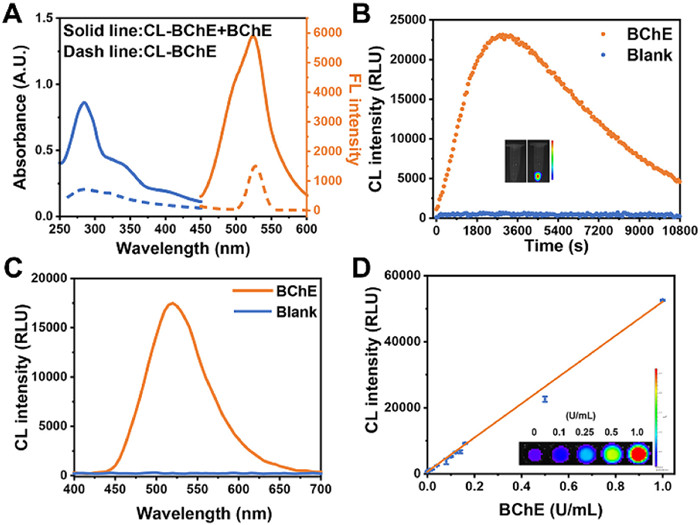
Haven demonstrated the response of the probe toward BChE, an investigation into the sensitivity of CL-BChE was further implemented. As shown in Fig. 1D, as the enzyme concentration increases, the chemiluminescence images captured with the IVIS system exhibited brighter signals. The quantification indicated that the chemiluminescence intensity of CL-BChE at 525 nm increased progressively with the concentration of BChE (0–1 U/mL), and a good linearity was found in the range of 0 to 1 U/mL (y = 681.76 + 51442.60x, R2 = 0.997). Of note, the limit of detection (LOD) was calculated as 6.25 × 10⁻³ U/mL (3 δ/k), suggesting the high sensitivity of CL-BChE to BChE. The results imply that CL-BChE holds satisfactory performance for the quantitative detection of BChE levels in cells as well as in biological samples.
Next, the verification toward the specificity of CL-BChE to BChE was considered. Cholinesterase inhibitor tacrine was co-incubated with BchE prior to responding [23]. As shown in Fig. S2 (Supporting information), the pretreatment of tacrine compromised significantly the chemiluminescence of probe CL-BChE. Further, the inhibition efficiency toward BChE versus the concentration of tacrine was also measured (IC50 = 32.88 nmol/L) (Fig. S1 in Supporting information), indicative of the competitive inhibition of tacrine on the enzymatic activation of the probe. These results demonstrate that the signals of CL-BChE were indeed triggered specifically by BChE. Moreover, the selectivity of probe CL-BChE for BChE against other biologically relevant analytes was also evaluated. As shown in Figs. S3 and S4 (Supporting information), no significant chemiluminescence was triggered by other interferents including enzymes such as acetylcholinesterase (AChE), tyrosinase (TYR), leucine aminopeptidase (LAP), glutamyl transpeptidase (GGT), β-galactosidase (β-Gal), alkaline phosphatase (ALP), glucose oxidase (GOD), β-glucuronidase (β-GN), amino acids such as cysteine (Cys), glutathione (GSH), homocysteine (Hcy), high concertation of cations such as Na+, K+, Ca2+, and Mg2+, and reactive bio-species such as H2O2, •OH, ONOO−, ClO−, NO2−. It should be noted that no obvious chemiluminescence was triggered in the presence of AChE, another cholinesterase in the same family as BChE. The above results demonstrated that probe CL-BChE can serve as a powerful tool for the specific detection of BChE in complex biological systems. Another requirement for application in living systems is a stable response behavior in the physiological pH range, therefore the pH effect on CL-BChE was studied. As shown in Fig. S5 (Supporting information), no chemiluminescence was observed under acidic conditions (pH < 6.0), which can be explained by the fact that the inability of the deprotonation leads to the forbidden access to CIEEL process. The abnormal chemiluminescence enhancement after raising the media pH above 10.0 can be ascribed to the non-specific hydrolysis of ester bonds under alkaline conditions. Nevertheless, the response of CL-BChE to BChE can proceed in a relatively steady state under physiological pH of 6.5 to 7.4, substantiating its applicability for detection and imaging in living cells, tissues, and animals.
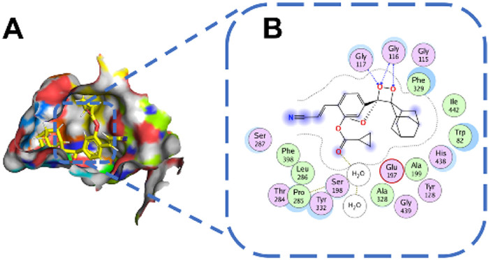
To further verify the potential response mechanism of CL-BChE to BChE, a high-performance liquid chromatography (HPLC) analysis was carried out, as shown in Fig. S6 (Supporting information). The chromatographic peaks with retention time at 7.15 min was corresponding to CL-BChE, which was diminished upon incubation with BChE at 37 ℃ for 5 h, indicating that the probe was hydrolyzed in the presence of BChE. Simultaneously, a new peak appeared at 3.66 min and intensified as the incubation time continues. To better understand the response mechanism of CL-BChE to BChE, molecular docking was implemented to estimate the binding interactions. As shown in Fig. 2A, the docking results showed that CL-BChE can smoothly access the active pocket of BChE. By analyzing the interactions between CL-BChE and key amino acid residues within the active site, it was observed that CL-BChE can form intermolecular hydrogen bonds with Gly116, Gly115, and Pro285 residues, and establish hydrophobic interactions with Gly117, Ala199, Ser198, and His438 residues (Fig. 2B). These amino acid residues align with those reported in the literature to be involved in the hydrolytic mechanism of BChE [24]. Similarly, molecular docking of CL-BChE and AChE was also implemented to verify the design strategy. However, as the results demonstrate (Fig. S7 in Supporting information), due to the smaller active pocket of AChE compared to BChE, CL-BChE could not effectively enter the enzyme’s active pocket and did not show strong interactions with key amino acid residues of AChE. Therefore, CL-BChE exhibits excellent selectivity towards BChE.
Inspired by the ideal optical response of CL-BChE in vitro, CL-BChE has found applications in pesticide residue detection [25]. Since the activity of BChE decreases upon exposure to pesticide residues, enzyme-biosensor systems can be served as a powerful tool for pesticide residue detection (Fig. S9 in Supporting information) [26]. Given the advantages of the excellent sensitivity and selectivity of CL-BChE, it may find opportunities for the detection of pesticide residue in real plant samples. Five model pesticides were adopted to assess the ability of CL-BChE to detect pesticides. Among these pesticides, trichlorfon, and metolcarb can bind to BChE and disrupt its normal function. The result indicates that CL-BChE can distinguish different types of pesticides with satisfying selectivity (Fig. S10 in Supporting information). To further verify the sensitivity, the inhibition curves of metolcarb and trichlorfon against BChE were measured by monitoring the chemiluminescence intensities of CL-BChE. As illustrated in Figs. 3A and B, the inhibition curves of the two pesticides fitted well to the hill equation, and the IC50 were calculated as 2.30334 µg/mL and 0.30788 µg/mL, respectively. These results show that CL-BChE can differentiate between different types of pesticides and detect the amount of pesticide residues.
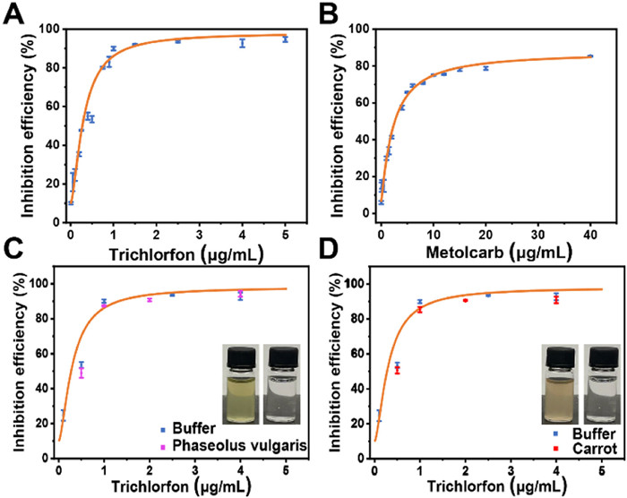
The anti-interference properties of CL-BChE against phytochromes were further verified. It was reported that the top six pesticide species detected in China were celery, apples, tomatoes, cucumbers, grapes, and Phaseolus vulgaris [27], so the three main phytochromes involved in these agricultural products, namely beta-carotene, chlorophyll, and anthocyanidin, were selected as representative interferents. The chemiluminescence signals were collected after vertexing the phytochromes (0.1 mmol/L) with CL-BChE response system (containing 0.5 U/mL BChE in PBS buffer). As expected, no obvious effect was detected by these phytochromes on the chemiluminescence from CL-BChE (Fig. S8 in Supporting information). The ability of CL-BChE for detecting pesticide residue in real vegetable samples was further verified. Phaseolus vulgaris, celery, cucumber and carrot were selected as were adopted as real vegetable model while trichlorfon was chosen as the pesticide model. The inhibition curves of BChE by pesticides in these vegetable samples were first investigated, as shown in Figs. 3C and D and Fig. S11 (Supporting information), despite variations in the color of the extraction solutions obtained from different vegetable samples, CL-BChE was still able to effectively detect pesticide residues in these extracts. The obtained inhibition curves showed no significant difference compared to those obtained in buffer solution. This demonstrates the capability of CL-BChE in detecting pesticide residues in real samples.
Inspired by the ideal optical response of CL-BChE in vitro, its capacity for tracking endogenous BChE in living cells was assessed. HepG2 cells were used as model cell lines for their high BChE expression [8]. The cytotoxicity of CL-BChE against HepG2 cells was evaluated with an MTT assay firstly, and the results demonstrated that CL-BChE exhibited no obvious cytotoxicity, suggesting its excellent biocompatibility (Fig. S12 in Supporting information). Next, HepG2 cells were incubated with CL-BChE (10 µmol/L) for 30 min and then the chemiluminescent images were collected with IVIS system under bioluminescence mode. As shown in Fig. S13 (Supporting information), a gradual increase in chemiluminescence intensity was observed with incubation time and reached the maximum at 35 min, substantiating that CL-BChE could be activated well by intracellular BChE. Subsequently, the endogenous BChE concentration-dependent chemiluminescence response was studied using black 96-well plates seeded with different numbers of cells. Upon staining with CL-BChE at 37 ℃ for 30 min, an increase in chemiluminescence intensity was observed with increasing cell numbers, as shown in Fig. 4A. While, in wells pre-incubated with cholinesterase inhibitors tacrine, the chemiluminescence intensity was significantly lower than other wells without inhibitors (Fig. 4B). Moreover, the chemiluminescence intensity shows a good linear relationship with cell numbers according to the fit results (Fig. S14 in Supporting information). These results suggest that CL-BChE is capable of real-time imaging of endogenous BChE activity in living cells, and additionally holds great promise for achieving in vivo BChE monitoring and assessment, considering the advantages of chemiluminescence in the in vivo imaging.

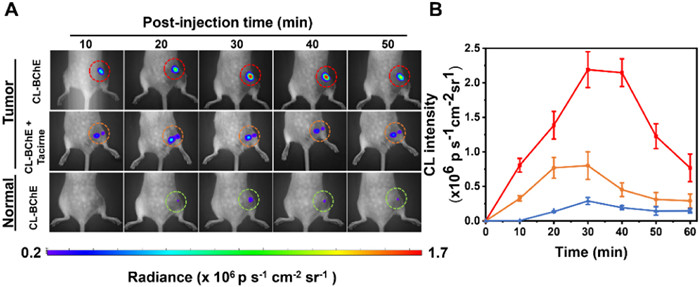
Animal models with BChE-overexpressed tumors were established by subcutaneous injection of HepG2 cells into the right back of each female BALB/c nude mouse. All animal experiments were performed according to the relevant ethical regulations of Central South University, and this study received approval from Central South University Experimental Animal Ethics Committee. Upon tumor formation, three of the tumor-bearing mice were randomly selected as a negative control group and injected intratumorally with the cholinesterase inhibitor tacrine 2 h prior to CL-BChE injection, while another three tumor-bearing mice were elected as experimental group and injected intratumorally with CL-BChE. In addition, three normal mice were injected with CL-BChE as a blank control group. After treatment with CL-BChE, chemiluminescence signals were collected from 0 to 50 min with an interval of 10 min. As depicted in Fig. 5A, a distinct chemiluminescence was observed at the tumor site in the experimental group and its intensity reached the maximum at 35 min. It is worth mentioning that the experimental group exhibited a 9-fold higher chemiluminescence enhancement than the blank control group (Fig. 5B). By contrast, no discernible chemiluminescence signals were detected in the negative control group, which is consistent with the inhibitory effect of tacrine revealed in vitro and in cells. All these results demonstrate that CL-BChE could be a promising tool for real-time monitoring of BChE activity in tumors and associated clinical settings.
In conclusion, a novel BChE-activatable chemiluminescent probe CL-BChE was well designed and successfully applied for BChE detection. When the cyclopropyl formate is removed by BChE, CL-BChE undergoes CIEEL process and emits bright chemiluminescence. The in vitro analysis demonstrated that CL-BChE exhibited a good response to BChE with high sensitivity and excellent selectivity. Further, the good biocompatibility allows the probe to be successfully applied for the detection of intracellular BChE in tumor cells. Owing to the absence of excitation light, the chemiluminescence modality allows the probe low background imaging of BChE in tumor-bearing animal models, unlike traditional BChE detection methods. Furthermore, given its excellent responsiveness to BChE, the potential of CL-BChE for detecting pesticide residues was revealed with great convenience. This work provides an innovative tool for the detection of BChE and, more importantly, a valuable imaging agent for revealing the role of BChE in associated pathology and pharmacology studies.
The authors declare that they have no known competing financial interests or personal relationships that could have appeared to influence the work reported in this paper.
Shuaige Bai: Data curation, Formal analysis, Investigation, Methodology, Writing – original draft, Writing – review & editing. Shuai Huang: Conceptualization, Formal analysis, Investigation, Methodology, Writing – original draft, Writing – review & editing. Ting Luo: Investigation, Writing – original draft. Bin Feng: Data curation, Formal analysis, Writing – original draft. Yanpeng Fang: Formal analysis, Investigation. Feiyi Chu: Data curation, Investigation, Writing – original draft. Jie Dong: Data curation, Resources, Writing – original draft. Wenbin Zeng: Conceptualization, Funding acquisition, Project administration, Resources, Supervision, Validation, Visualization, Writing – original draft, Writing – review & editing.
The authors gratefully acknowledge the financial support from the National Natural Science Foundation of China (Nos. 82272067, 81974386, and M-0696), Natural Science Foundation of Hunan Province (Nos. 2022JJ80052 and 2022JJ40656) and the Innovation Fund for Postgraduate Students of Central South University (No. 2023ZZTS0609).
Supplementary material associated with this article can be found, in the online version, at doi:
L. Santarpia, I. Grandone, F. Contaldo, et al., J. Cachexia. Sarcopeni. 4 (2013) 31–39. doi: 10.1007/s13539-012-0083-5
O.O. Ogunkeye, A.I. Roluga, Pathophysiology 13 (2006) 91–93. doi: 10.1016/j.pathophys.2006.01.002
O.O. Ogunkeye, E.K. Chuhwak, A.A.E. Otokwula, Pathophysiology 17 (2010) 29–32. doi: 10.1016/j.pathophys.2009.05.003
L. Rejc, V. Gómez-Vallejo, A. Joya, et al., Theranostics 11 (2021) 6542. doi: 10.7150/thno.54589
E. Podoly, D.E. Shalev, S. Shenhar-Tsarfaty, et al., J. Biol. Chem. 284 (2009) 17170–17179. doi: 10.1074/jbc.M109.004952
Q. Zhang, C. Fu, X. Guo, et al., ACS Sens. 6 (2021) 1138–1146. doi: 10.1021/acssensors.0c02398
G.L. Ellman, K.D. Courtney, V. Andres, et al., Biochem. Pharmacol. 7 (1961) 88–95. doi: 10.1016/0006-2952(61)90145-9
Y. Yang, L. Zhang, J. Wang, et al., Anal. Chem. 94 (2022) 13498–13506. doi: 10.1021/acs.analchem.2c02627
W. Zhang, J. Zhang, C. Qin, et al., Anal. Chim. Acta 1235 (2022) 340540. doi: 10.1016/j.aca.2022.340540
B. Feng, Y. Zhu, J. Wu, et al., Chin. Chem. Lett. 32 (2021) 3057–3060. doi: 10.1016/j.cclet.2021.03.074
M. Liu, S. Zhong, B. Feng, et al., Chin. Chem. Lett. 34 (2023) 107940. doi: 10.1016/j.cclet.2022.107940
J. Dong, J. Qian, K. Yu, et al., Research 6 (2023) 0075. doi: 10.34133/research.0075
M. Vacher, I. Fdez. Galván, B.W. Ding, et al., Chem. Rev. 118 (2018) 6927–6974. doi: 10.1021/acs.chemrev.7b00649
U. Haris, A.R. Lippert, ACS Sens. 8 (2023) 3–11. doi: 10.1021/acssensors.2c02371
F. McCapra, Acc. Chem. Res. 9 (1976) 201–208. doi: 10.1021/ar50102a001
A.P. Schaap, T.S. Chen, R.S. Handley, et al., Tetrahedron Lett. 28 (1987) 1155–1158. doi: 10.1016/S0040-4039(00)95313-9
A.P. Schaap, R.S. Handley, B.P. Giri, Tetrahedron Lett. 28 (1987) 935–938. doi: 10.1016/S0040-4039(00)95878-7
A.P. Schaap, M.D. Sandison, R.S. Handley, Tetrahedron Lett. 28 (1987) 1159–1162. doi: 10.1016/S0040-4039(00)95314-0
A.V. Trofimov, K. Mielke, R.F. Vasil’ev, et al., Photochem. Photobiol. 63 (1996) 463–467. doi: 10.1111/j.1751-1097.1996.tb03070.x
O. Green, T. Eilon, N. Hananya, et al., ACS Cent. Sci. 3 (2017) 349–358. doi: 10.1021/acscentsci.7b00058
Y. Gao, Y. Lin, T. Liu, et al., Chin. Chem. Lett. 30 (2019) 63–66. doi: 10.1016/j.cclet.2018.03.028
R. Blau, O. Shelef, D. Shabat, et al., Nat. Rev. Bioeng. 1 (2023) 648–664. doi: 10.1038/s44222-023-00074-0
J. Ding, R. Xiao, A. Bi, et al., Chin. Chem. Lett. 34 (2023) 108273. doi: 10.1016/j.cclet.2023.108273
C.G. Zhan, F. Zheng, D.W. Landry, J. Am. Chem. Soc. 125 (2003) 2462–2474. doi: 10.1021/ja020850+
F.P. Carvalho, Food Energy Secur. 6 (2017) 48–60. doi: 10.1002/fes3.108
Z. Wu, Z. Hao, Y. Chai, et al., Biosens. Bioelectron. 233 (2023) 115341. doi: 10.1016/j.bios.2023.115341
C. Li, H. Zhu, C. Li, et al., Food Chem. 354 (2021) 129552. doi: 10.1016/j.foodchem.2021.129552

Scheme 1 (A) Structure of BChE-specific chemiluminescent probe CL-BChE. (B, C) Schematic representation of the application of CL-BChE in CL-BChE tumor imaging and pesticide residue detection in real vegetable samples.

Figure 1 (A) Absorption (blue) and fluorescence emission (orange) spectra of CL-BChE (10 µmol/L) before (dash line) and after (solid line) incubation with BChE in PBS. (B) Chemiluminescence kinetic profiles of CL-BChE (10 µmol/L) in the absence or presence of BChE. (C) Chemiluminescence spectra of CL-BChE before (blue) and after (orange) incubation with BChE for 30 min. (D) Chemiluminescence intensities of CL-BChE (10 µmol/L) toward different concentrations of BChE (0 to 1.0 U/mL).

Figure 2 (A) Active site point surface of CL-BChE bound with BChE. (B) 2D ligand interaction plot of CL-BChE with the essential residues at the binding pocket of BChE.

Figure 3 Inhibition efficiency of different concentrations of metolcarb (A) and trichlorfon (B) to BChE (0.5 U/mL). Inhibition efficiency of different concentrations of trichlorfon to BChE (0.5 U/mL) in different vegetables extracts including Phaseolus vulgaris (C) and carrot (D).

Figure 4 (A) Chemiluminescence images of CL-BChE (10 µmol/L) upon incubation with different densities of HepG2 cells or cells pre-treated with tacrine (100 µmol/L) at 37 ℃ for 30 min. (B) Average chemiluminescence intensity of the different wells in A. Values are means ± SD (n = 3).

Figure 5 (A) Real-time chemiluminescence images of HepG2-tumor-bearing mice after intratumoral injection with CL-BChE (50 µmol/L, 100 µL), CL-BChE (50 µmol/L, 100 µL) with pre-injection of tacrine (500 µmol/L, 100 µL)) and normal mice injection with CL-BChE (50 µmol/L, 100 µL). The red, orange, and green dash circle represents the post-injection region. (B) Average chemiluminescence intensity of the circled section in A. Values are means ± SD (n = 3).

 扫一扫看文章

扫一扫关注我们
 DownLoad:
 下载:
下载: