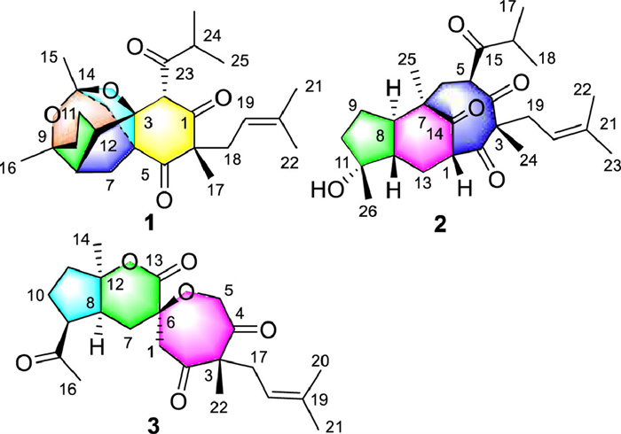
Figure 1.
Structures of compounds 1–3.
Hymoins A–C, three unusual polycyclic polyprenylated acylphloroglucinols with lipid-lowering activity from Hypericum monogynum
Chunmao Yuan , Yanrong Zeng , Lei Huang , Yu Mou , Jun Jin , Ping Yi , Yanmei Li , Xiaojiang Hao

Polycyclic polyprenylated acylphloroglucinols (PPAPs) are a special type of hybrid natural products, biosynthesized from acylphloroglucinol decorated with prenyl and other prenyl derivatives [1,2]. The structures of PPAPs are characterized by highly modified ring system formed between different types of prenyl and acylphloroglucinol core [1,2]. Their fantastic structures and excellent bioactivity engaged the interests of biologists and chemists [3-9]. It is noted that hyperforin (PPAP) is the main antidepressant active ingredient in GNC St. John's Wort Extract Capsules and Swisse Mood Tablets, which is widely used as dietary supplements in Europe and the United States [10,11]. PPAPs are widely discovered in the plants of the family Guttiferae, and more than 700 PPAPs have been identified up to now [1,2]. During our continue study for active PPAPs from the plants of Guttiferae [12-14], three unprecedented PPAPs (Fig. 1), hymoins A–C (1–3), were identified from the flowers of Hypericum monogynum.
To the best our knowledge, hymoin A represents the first intriguing 6/5/5/5/7 pentacyclic caged PPAP; hymoin B is characterized by an unprecedented rearranged 5/6/8 tricyclic ring system; hymoin C is reported to be the first rearranged PPAP with a fantastic spirocyclic 5/6/7 ring system. The plausible biosynthesis pathway was also supposed. Those compounds exhibited good lipid-lowering activity in oleic acid (OA)-induced HepG2 cells and the mechanistic study of 1 was also investigated.
Hymoin A (1) was isolated as colorless crystal and its molecular formula C26H36O5 was established by the positive high resolution electrospray ionization mass spectroscopy (HRESIMS) ([M + Na]+ m/z 451.2447; calcd. 451.2455), indicating nine indices of hydrogen deficiency (IHDs). The 1H and 13C nuclear magnetic resonance spectroscopy (NMR) spectra (Table S1 in Supporting information) combined with heteronuclear single quantum coherence (HSQC) spectrum exhibited the existence of 26 carbons, consisted of seven methyls, five methylenes, four aliphatic and one olefinic methines, and nine quaternary carbons (three carbonyls, δC 205.4, 207.6, and 211.4). The aforementioned functional moieties accounted for four IHDs, and the remaining five IHDs manifested 1 to be a pentacyclic ring system.
The planar structure of 1 could be established by comprehensive analysis of 2D NMR spectra (Fig. 2). Three segments in bold bonds were shown in Fig. 2 from 1H–1H heteronuclear singular quantum correlation (COSY) correlations. Obviously, HMBC correlations from H-2 to C-1, C-3, and C-4, from H2–7 to C-3, C-4, and C-5, and from H3–17 to C-1, C-5, C-6, and C-18, established ring A with a methyl at C-6. Furthermore, a 2-methylpropanoyl group and a prenyl group could be easily placed at C-2 and C-6, respectively (Fig. 2), verified from HMBC correlations of H-24 to C-2 and C-23, H3–22 to C-19 and C-20, and H2–18 to C-1, C-5, and C-6. What is more, the HMBC correlations from H-12 to C-3, C-4, C-7, and C-8, and from H3–16 to C-8, C-9, and C-10 could identify the fused rings B and C. Therefore, three rings A–C were assembled, as shown in Unit A. What's more, the linkages of C-4 and C-14 through C-13 could be assigned from the HMBC correlations from H3–15 to C-13 and C-14, and from H2–7 to C-4 and C-13, as shown in Unit B. Moreover, two unconnected oxygenated quaternary carbon [δC 92.8 (C-3); δC 85.9 (C-9)] and a typical ketal carbon [δC 106.4 (C-14)], coupled with two remaining IHDs implied that two O-bridges between C-3/C-14 and C-9/C-14 formed to assign rings D and E. Accordingly, this planar structure, featuring the intriguing 6/5/5/5/7 pentacyclic caged ring system, was defined.
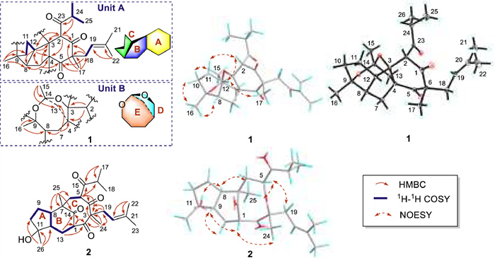
The relative configuration of 1 was deduced by nuclear overhauser effect spectroscopy (NOESY) experiment (Fig. 2), in which correlations of H-8/H3–16/H-10β and H-2/H-12/H3–17 implied that these groups were cofacial and randomly assigned as β-oriented. Inversely, the NOESY cross-peaks of H-10α/H3–15 disclosed the α-orientations of these protons. Herein, the relative configuration of 1 was determined in Fig. 2. Finally, the structure and absolute configuration of 1 was confirmed by single crystal X-ray diffraction with Cu Kα radiation in Fig. 2 (CCDC 2327212) [14].
The molecular formula of hymoin B (2), C26H38O5, with eight IHDs, was established by the positive HRESIMS ion at m/z 453.2608 [M + Na]+ (calcd. for C26H38O5, 453.2611). The 1H NMR data displayed the presence of seven methyls and one olefinic proton. The 13C NMR data together with heteronuclear single quantum coherence (HSQC) spectrum showed the existence of 26 carbon signals, including seven methyls, five methylenes, six methines (one olefinic carbon), and eight quaternary carbons (one olefinic and four carbonyl carbons). The above-mentioned functionalities accounted for five IHDs, and three additional rings were required to satisfy the remaining IHDs.
The 1H–1H COSY cross-peaks of H2–10/H2–9/H-8/H-12/H2–13/H-1, H-5/H2–6, and H2–19/H-20 revealed that 2 features three fragments in bonds (blue bold, Fig. 2). In the HMBC spectrum, 1H–13C long-range correlation signals from (Fig. 2) H3–26 to C-10, C-11, and C-12; from H2–13 to C-1 and C-14; and from H3–25 to C-6, C-7, C-8, and C-14, revealed the presence of the 1‑hydroxy-1,4-dimethyloctahydro-5H-inden-5-one motif (rings A and B). Moreover, the HMBC correlations from H3–24 to C-2, C-3, C-4, and C-19, and from H-5 to C-3 and C-4, together with the above established fragments generated an eight-membered ring C. Besides, a prenyl side chain was attached at C-3, which was verified through the HMBC corrections from H3–22 and H3–23 to C-20 (δC 117.3) and C-21 (δC 135.3) and from H2–19 to C-3 and C-4. A 2-methylpropanoyl group was placed at C-5 by the HMBC corrections of H3–17 and H3–18 to C-15 and C-16, and from H-5 to C-15 and C-16. Hence, this structure, named hymoin B, was defined.
The relative configuration of 2 was deduced by a NOESY experiment (Fig. 2). The correlations of H-9/H-1/H3–24 indicated that these groups were cofacial and assigned as β-oriented, whereas the correlations of 11-OH/H-8/H3–25/H-5/H-19a established the α-orientations of these protons. The absolute configuration of this compound was elucidated by quantum chemistry calculation. The calculated electronic circular dichroism (ECD) spectrum matched well with the experimental one (Fig. S2 in Supporting information) [14]. To the best of our knowledge, this compound was the first rearranged 5/6/8 tricyclic PPAP. The structure elucidation of compound 3 was shown in Supporting information.
A plausible biosynthesis pathway for compounds 1–3 was proposed (Scheme 1). Generally, the 2-methylpropanoyl was considered to be the initial precursor for these novel PPAPs [1], [2]. This could undergo different types of prenyltransferase, methyltransferase, and cyclization to get three critical intermediates, i, iv, and vi, which could generate different PPAP skeletons (1–3). As for compound 1, the intermediate i, underwent oxidative cleavage of a 11,14Δ double bond to get intermediate ii, followed by Adol reaction between C-12 and C-3, and reduction of a ketone of C-11 to obtain iii with a rare fused 5/5/6 ring system. This intermediate with two hydroxyl groups could undergo intramolecular etherification reaction to yield compound 1. What is more, The phloroglucinol meroterpene intermediate iv could be oxidated and changed to a radical intermediate v under light, followed by cyclization between C-6 and C-7 to get compound 2. In addition, the vital intermediate vi could undergo a series of oxidation and reduction to get intermediate vii, followed by Wagner-Meerwein rearrangement to get intermediate ix. Baeyr-Villiger oxidation occurred for this intermediate, followed by hydrolysis to get a polyhydroxy intermediate xi. Intermolecular etherification and esterification happened for intermediate xi to get compound 3 with the unusual spirocyclic 5/6/7 ring system.

Dysregulation of lipid metabolism contributed to the occurrence of non-alcoholic fatty liver disease (NAFLD) [12]. In the development of this disease, triglyceride (TG) is an important indicator of the accumulation of free fatty acids (FFAs) and lipid. Thus, the OA-induced HepG2 cell is applied as an anti-NAFLD model to screen the TG concentration of those isolates [12]. As a result, all isolates could inhibit the TG accumulation in OA-induced HepG2 cells without cytotoxicity in Fig. 3A and Fig. S31 (Supporting information) at three different concentrations (2, 4, and 8 µmol/L). Among them, compound 1 showed the best inhibitory effect on TG accumulation at the concentration of 4 µmol/L, better than the positive control atorvastatin (10 µmol/L). What is more, compound 1 also could prohibit the lipid droplet accumulation in OA-induced HepG2 cells using oil red O (ORO) staining in a concentration-dependent manner [15], better than the positive control (atorvastatin 10 µmol/L) (Fig. 3B and Fig. S31). Therefore, compound 1 was chosen for further mechanistic study.
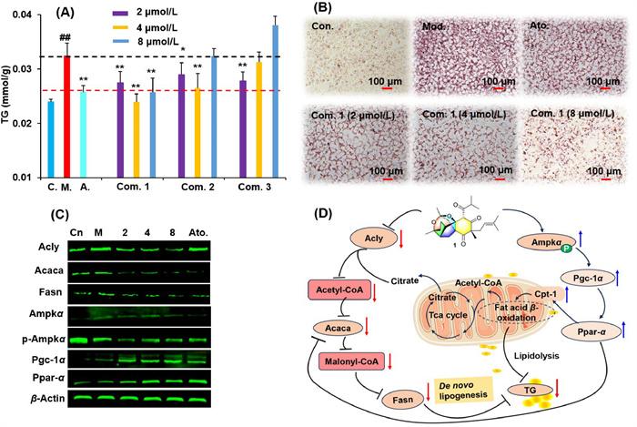
FFAs synthesis and metabolism play a vital role in the occurrence and development of NAFLD. Thus, a series of related proteins were investigated for compound 1. Three critical FFAs synthesis proteins [15,16], ATP citrate lyase (Acly), acetyl-CoA carboxylase (Acaca), and fatty acid synthase (Fasn) were checked by Western blot, and these proteins were significantly downregulated in OA-induced HepG2 cells treated with compound 1 for 24 h. Moreover, the AMP-activated protein kinase (Ampk) signal pathway is closely related to FFAs synthesis and TG metabolism, and thus, was applied for our investigation. As shown in Fig. 3C and Fig. S32 (Supporting information), the ratio of p-Ampkα/Ampkα was obviously downregulated by compound 1 in OA-induced HepG2 cells. Peroxisome proliferator-activated receptor-gamma coactivator (Pgc)−1α and the peroxisome proliferator activated receptor α (Ppar-α) are two vital FFAs metabolism proteins. Pgc-1α can bind to Ppar-α in mitochondrial and indirectly contribute to fatty acid (FA) transport and utilization, which are potential targets for the treatment of NAFLD. Interestingly, these two proteins could be upregulated by compound 1 (Fig. 3C and Fig. S32). In summary, compound 1 could exhibit excellent lipid-lowering activity in OA-induced HepG2 cells through inhibiting the proteins of FFAs synthesis and improving lipidolysis, as shown in Fig. 3D.
In conclusion, three unusual fantastic PPAPs, hymoins A–C (1–3), were isolated and identified from the flowers of Hypericum monogynum. Hymoin A (1) is discovered to be the first intriguing 6/5/5/5/7 pentacyclic caged PPAP. Hymoin B is characterized by an unprecedented rearranged 5/6/8 tricyclic ring system, while hymoin C features the first spirocyclic 5/6/7 ring system. Their possible biosynthetic pathways are proposed. The isolates could significantly suppress the TG concentration in OA-induced HepG2 cells at the concentrations of 2–8 µmol/L. Compound 1 not only could suppress the FFAs synthesis proteins, such as Acly, Acaca, and Fasn, but also could upregulate vital lipolysis proteins, Pgc-1α and Ppar-α. Compound 1, an unprecedented PPAP, was a potential lipid-lowering lead compound due to its excellent lipid-lowering activity.
The authors declare that they have no known competing financial interests or personal relationships that could have appeared to influence the work reported in this paper.
Chunmao Yuan: Conceptualization, Investigation, Writing – original draft, Writing – review & editing. Yanrong Zeng: Investigation, Methodology, Writing – original draft. Lei Huang: Conceptualization, Investigation, Methodology, Writing – review & editing. Yu Mou: Investigation, Methodology. Jun Jin: Formal analysis, Methodology. Ping Yi: Investigation, Methodology. Yanmei Li: Conceptualization, Software, Supervision. Xiaojiang Hao: Conceptualization, Methodology, Project administration, Supervision.
The work was financially supported by the National Natural Science Foundation of China (Nos. 32270413, 82060631, 82160808, and 82360035), the Science and Technology Department of Guizhou Province (Nos. QKHJC 2020–1Z076, QKHJC-ZK[2023]YB156, QKHJC-ZK[2021]YB569, and QKHPTRC [2020]5008), Excellent Young Talents Plan of Guizhou Medical University (2023, No. 106), the 13th batch of outstanding young scientific and technological talents in Guizhou Province (No. QKHPTRC [2021]5633), Guizhou Science and Technology Innovation Talent Team (No. QKHPTRC-CXTD[2022]007), and High-level Innovative Talents in Guizhou Province (Thousand Levels of Talent for Chunmao Yuan in 2018), the project of State Key Laboratory of Functions and Applications of Medicinal Plants, Guizhou Medical University (Nos. FAMP202102K and QJJ[2023]113) and Guizhou Provincial Engineering Research Center for Natural Drugs.
Supplementary material associated with this article can be found, in the online version, at doi:
X.W. Yang, R.B. Grossman, G. Xu, Chem. Rev. 118 (2018) 3508–3558. doi: 10.1021/acs.chemrev.7b00551
R. Ciochina, R.B. Grossman, Chem. Rev. 106 (2006) 3963–3986. doi: 10.1021/cr0500582
Y. Ji, B. Hong, I. Franzoni, et al., Angew. Chem. Int. Ed. 61(2022) e202116136.
Y.S. Ye, N.N. Jiang, X.W. Yang, G. Xu, Chin. Chem. Lett. 31 (2020) 2433–2436.
W.J. Lu, Y.Q. Zhang, Y.W. Li, et al., Chin. Chem. Lett. 33 (2022) 4121–4125.
L.J. Franov, J.D. Hart, G.A. Pullella, C.J. Sumby, J.H. George, Angew. Chem. Int. Ed. 61 (2022) e202200420.
K. Cottet, B. Xu, P. Coric, et al., J. Med. Chem. 59 (2016) 9560–9566. doi: 10.1021/acs.jmedchem.6b01182
S. Chen, X. Liu, C. Peng, et al., Cell Metab. 33 (2021) 565–580.
Z. Shi, J. Yin, Y. Xiao, et al., Chin. Chem. Lett. 35 (2024) 109458. doi: 10.1016/j.cclet.2023.109458
C.Y.W. Ang, L. Hu, T.M. Heinze, et al., J. Agr. Food Chem. 52 (2004) 6156–6164.
H.Y. Lou, F.W. Ma, P. Yi, et al., Arab. J. Chem. 18 (2022) 104057.
L. Huang, Z.Z. Zhang, Y.N. Li, et al., Org. Lett. 24 (2022) 5967–5971. doi: 10.1021/acs.orglett.2c02240
H.Y. Lou, Y.N. Li, P. Yi, et al., Org. Lett. 22 (2020) 6903–6906. doi: 10.1021/acs.orglett.0c02434
D.S. Tian, P. Yi, L. Xia, et al., Org. Lett. 18 (2016) 5904–5907. doi: 10.1021/acs.orglett.6b03004
L. Huang, Y.R. Zeng, Y.F. Li, et al., J. Funct. Foods 108 (2023) 105715.
G.F. Grabner, H. Xie, M. Schweiger, R. Zechner, Nat. Metab. 3 (2021) 1445–1465. doi: 10.1038/s42255-021-00493-6

Figure 2 The key 2D NMR correlations of compounds 1 and 2 and X-ray crystallographic structure of compound 1.

Figure 3 Effects of compounds 1–3 on lipid accumulation in OA-induced HepG2 cells and mechanistic study of 1. (A) Effects of three compounds 1–3 on the accumulation of TG in OA-induced HepG2 cells. (B) Lipid accumulation was observed for compound 1 with ORO staining under light microscopy. (C) OA-induced HepG2 cells were incubated with compound 1 (2, 4, and 8 µmol/L) for 24 h followed by Western blot for these proteins, Acly, Acaca, Fasn, Ampkα, p-Ampkα, Pgc-1α, and Ppar-α. C., Con., or Cn, DMSO group without adding OA; M. or Mod., DMSO group in OA-induced HepG2 cells; A. or Ato., positive control, atorvastatin (10 µmol/L). (D) Diagram of the pharmacological mechanism of compound 1. Data were obtained as the mean ± standard deviations (n = 3). ##P < 0.01 vs. the control group (C.); P < 0.05, **P < 0.01 vs. the OA group (M.).

 扫一扫看文章

扫一扫关注我们
 DownLoad:

 下载:
下载:
