National Engineering Research Center of Ophthalmology and Optometry, School of Ophthalmology and Optometry & Biomedical Engineering, Eye Hospital, Wenzhou Medical University, Wenzhou 325027, China
Received Date:
08 December 2023 Accepted Date:
03 February 2024 Revised Date:
10 January 2024 Available Online:
15 November 2024
Abstract:
The low drug bioavailability of eye drops challenges the therapy of ocular disorders with high efficacy. One of solutions is to extend the corneal retention and enhance the penetration of drug into cornea. Here we synthesize two fluorophore-conjugated peptide based analogs rich in positive charges (i.e., NBD-FFKK) and with a specific ligand (i.e., NBD-FFRGD), respectively, to visualize their performances in vitro and in vivo. The peptides both can self-assemble into supramolecular hydrogels with the microstructure of nanofibers. The in vitro experiments exhibit that two peptides are both uniformly distributed in cytoplasm, and the intracellular amount of peptide rich in positive charges is significantly larger than that of peptide with a specific ligand. The living corneal fluorescence shows that two peptides enter the corneal stroma within 15 min, and the peptide rich in positive charges is accumulated more extensively throughout the entire cornea, revealing that the supramolecular hydrogel eye drops penetrate the cornea more efficiently via electrostatic interaction than that via ligand-receptor interaction. This work, as a comparative study of supramolecular hydrogel eye drops on penetrating efficiency, indicates a possible direction for the design of eye drops with efficient corneal penetration.
Eye drops, as one of the most popular formulations, are mainly used for the local delivery of ophthalmic drugs [1-4]. However, one challenging issue in the topical instillation of eye drops is that tears flushing and drainage cause only a small portion of medication into the eyes, which means the low bioavailability of drug [5,6]. Besides increasing the frequency and dosage of administration, another strategy is to improve the bioavailability of drug via extending the corneal retention and enhancing the penetration of drug into cornea [7-9].
The peptide-based supramolecular hydrogel has emerged as a carrier for ophthalmic drug delivery [9-11]. Being similar to various hydrogels with outstanding bioactivities [12-15], the viscoelastic and thixotropic properties of peptide-based supramolecular hydrogel make them more suitable for use under shearing forces (e.g., physiological blinking), thereby drastically extending the corneal retention [16-19]. Moreover, according to the expression of proteins and enzymes in the cornea, the ingenious and rational design of peptides can achieve the active targeting effect, thereby effectively enhancing the penetration of drug into cornea. For example, there are some secretory mucins in the aqueous layer of tear films, which are also located in the epithelium as membrane-spanning mucins [20]. The expression of mucins inspires the design of positively charged lysine based peptide which increases the adhesion on corneal surface and the penetration into cornea via the electrostatic interaction with negative mucins [21]. The presence of integrin in corneal epithelial cells motivates the conjugation of tripeptide (arginine-glycine-aspartic acid, RGD) with drug which promotes the transcorneal permeability of drug via the ligand-receptor interaction [22,23]. The relationship of peptide-mediated pathway and penetrating efficiency after topical instillation of peptide-based supramolecular hydrogel eye drops, however, remains unexplored.
To visualize the penetrating efficiency of peptide-based supramolecular hydrogel eye drops, we synthesized two fluorophore-conjugated peptide based analogs rich in positive charges (i.e., NBD-FFKK) and with a specific ligand (i.e., NBD-FFRGD), respectively, and examined their performances in vitro and in vivo. Our results show that NBD-FFKK and NBD-FFRGD both self-assemble into supramolecular hydrogels with the microstructure of nanofibers. The intracellular amount of NBD-FFKK is significantly larger than that of NBD-FFRGD both in human corneal epithelial cells (HCEC) and human umbilical vein endothelial cells (HUVEC). After topical instillation of these supramolecular hydrogel eye drops, NBD-FFKK penetrates the cornea more efficiently than NBD-FFRGD, further revealing that supramolecular hydrogel eye drops via electrostatic interaction have the higher penetrating efficiency than that via ligand-receptor interaction. Besides establishing conjugation of positively charged peptides as a simple and effective way for enhancing the corneal penetration, the relationship of peptide-mediated pathway and penetrating efficiency may provide useful insights for designing peptide based supramolecular hydrogel eye drops with efficient corneal penetration.
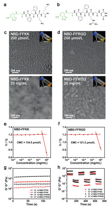
Figs. 1a and b show the structures of molecules as probes for fluorescent imaging of peptide-based supramolecular hydrogels. The synthetic peptides contain three critical features: (ⅰ) an environment-sensitive fluorophore (i.e., 4-nitro-2,1,3-benzoxadiazole, NBD), (ⅱ) a powerful self-assembled peptide motif (i.e., L-Phe-L-Phe, FF), and (ⅲ) a positively charged peptide (i.e., L-Lys-L-Lys, KK) for the electrostatic interaction with negatively charged mucins and a specific ligand based peptide (i.e., RGD) for the ligand-receptor interaction with integrin, respectively. After the straightforward synthesis using solid phase peptide synthesis, two analogs were purified by reverse-phase high performance liquid chromatography (RP-HPLC) and further determined with a purity of more than 95% by liquid chromatography-mass spectroscopy (LC-MS) (Figs. S1–S3 in Supporting information). As shown in the insets of Figs. 1c and d, NBD-FFKK and NBD-FFRGD formed transparent, yellow solutions at the concentration of 200 µmol/L. Transmission electron microscope (TEM) images showed that both solutions of NBD-FFKK and NBD-FFRGD produced a large number of nanoparticles and sparse nanofibers. After increasing the concentration to 20 mg/mL, NBD-FFKK and NBD-FFRGD both turned into stable hydrogels. As revealed by TEM, the hydrogel of NBD-FFKK consisted of abundant nanofibers and some nanoparticles, and the hydrogel of NBD-FFRGD encompassed much denser nanofibers and a little nanoparticles. The critical micellar concentration (CMC) of NBD-FFKK with a value of 134.5 µmol/L was quite close to that of NBD-FFRGD with a value of 121.2 µmol/L (Figs. 1e and f). The rheological property of two hydrogels was examined by a rheometer. As shown in Figs. 1g and h, the time sweeps of NBD-FFKK and NBD-FFRGD hydrogels showed that the storage modulus (G′) were larger than the loss modulus (G″) all the time at a strain of 1%, indicating the state of hydrogel. G′ of two hydrogels were both lower than G″ upon the strain was increased to 100%, suggesting the state of liquid. Moreover, when the strain was returned to 1%, NBD-FFKK and NBD-FFRGD both came back to the state of hydrogel, implying the feature of sol and gel transition by varying the shearing force.
Figure 1
Figure 1.
Molecular structures of (a) NBD-FFKK and (b) NBD-FFRGD. TEM images of (c) NBD-FFKK and (d) NBD-FFRGD at the concentration of 200 µmol/L and 20 mg/mL. Inset: corresponding optical images. Scale bar: 200 nm. CMC values of (e) NBD-FFKK and (f) NBD-FFRGD. (g) Time sweeps of NBD-FFKK (20 mg/mL) and NBD-FFRGD (20 mg/mL) hydrogels at a strain of 1% and a frequency of 1 Hz. (h) Step-strains (1% and 100%) of NBD-FFKK (20 mg/mL) and NBD-FFRGD (20 mg/mL) hydrogels at a frequency of 1 Hz.
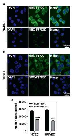
The positively charged materials may cause the certain cytotoxicity at high concentrations. After confirming that NBD-FFKK and NBD-FFRGD were innocuous to cells even at the concentration of 400 µmol/L (Fig. S4 in Supporting information), we incubated HCEC and HUVEC with peptides and compared the fluorescence using fluorescent confocal microscopy and flow cytometry, respectively (Fig. 2). From fluorescent confocal microscopy images, green fluorescent dots, belonging to the assemblies of NBD-FFKK and NBD-FFRGD, were well localized in the cytoplasm of HCEC, indicating the efficient uptake of peptides by cells. Moreover, NBD-FFKK exhibited a much brighter fluorescence than NBD-FFRGD, suggesting the more effective uptake of NBD-FFKK in the cellular environment. The same results were observed in HUVEC. In addition, the flow cytometry analysis also displayed that the mean fluorescence of NBD-FFKK was significantly stronger than that of NBD-FFRGD both in HCEC and HUVEC, which was consistent with the results of fluorescent confocal microscopy. These results imply that the penetrating efficiency of peptides via electrostatic interaction is likely higher than that via ligand-receptor interaction in the cellular environment.
Figure 2
Figure 2.
Fluorescent confocal microscopy images of (a) HCEC and (b) HUVEC with the treatment of NBD-FFKK and NBD-FFRGD at the concentration of 200 µmol/L for 2 h. Scale bar: 20 µm. (c) Flow cytometry analysis of HCEC and HUVEC with the treatment of NBD-FFKK and NBD-FFRGD at the concentration of 200 µmol/L for 2 h. ****P < 0.0001 vs. NBD-FFKK group (mean ± standard deviation, n = 3).
Fluorescein sodium was loaded in phosphate buffered solution (PBS), commercially available carbomer eye gel, NBD-FFKK hydrogel, and NBD-FFRGD hydrogel, respectively, to explore the precorneal retention (Fig. S5 in Supporting information). All animal experiments were approved by the Animal Care and Use Committee of Wenzhou Medical University (Approval No. xmsq2023–0925). The green fluorescein was only retained on the ocular surface until 10 min in PBS group, while the precorneal retention of fluorescein sodium loaded carbomer eye gel lasted for 30 min. Being similar to carbomer eye gel, the precorneal retention of NBD-FFRGD hydrogel was 30 min. Notably, NBD-FFKK hydrogel was adhesive on the ocular surface up to 60 min, confirming the enhanced adhesion on the corneal surface via the interaction with negative mucins.
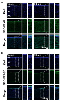
We further investigated the transcorneal behavior of peptides in living corneal tissues, to confirm the supposition about penetrating efficiency. As shown in Fig. 3 and Fig. S6 (Supporting information), upon single instillation of hydrogel eye drops at the concentration of 20 mg/mL, NBD-FFKK entered the cornea from epithelium to endodermis within a short time (i.e., 15 min). The fluorescence, belonging to NBD-FFKK, still retained in the entire cornea at 30 min, and the fluorescence intensity even became stronger. NBD-FFRGD was also able to penetrate the cornea within 15 min, and the fluorescence intensity, belonging to NBD-FFRGD, exhibited no statistical difference with that of NBD-FFKK at 15 min. However, NBD-FFRGD maintained the similar fluorescence intensity at 30 min with that at 15 min, suggesting that integrin in the epithelium was probably in a saturated state after the ligand-receptor interaction with RGD. These results indicate that two supramolecular hydrogel eye drops both show the effective penetration into cornea within a short time. Meanwhile, the penetrating efficiency of supramolecular hydrogel eye drops via electrostatic interaction was higher than that via ligand-receptor interaction with time going, which is also consistent with the in vitro results. Therefore, being similar to many drug delivery systems across barriers [24-35], the electrostatic interaction, being independent of receptors and not limited by the numbers of receptor molecules, may provide the higher throughput drug delivery on ocular surfaces.
Figure 3
Figure 3.In vivo corneal fluorescence distribution of (a) NBD-FFKK and (b) NBD-FFRGD after topical instillation at the concentration of 20 mg/mL. EP: Epithelium; ST: Stroma; ED: Endodermis. Scale bar: 100 µm.
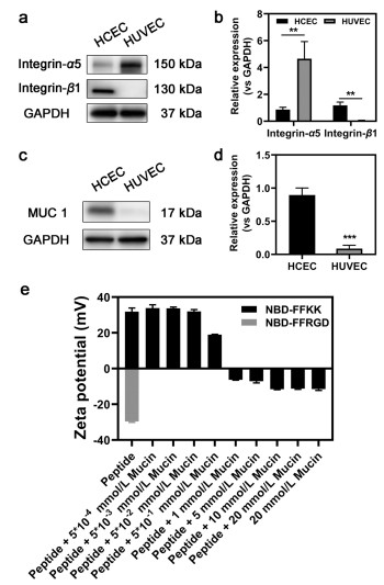
Western blot analysis further confirmed that the presence of integrin [36-38], as well as mucin 1 (MUC 1) which was a member of membrane-spanning mucins [39], effected the ligand-receptor interaction and electrostatic interaction, respectively (Figs. 4a–d). In addition, the variation of zeta potential recorded the interaction of positively charged NBD-FFKK and negatively charged mucins (Fig. 4e). The zeta potential value of NBD-FFKK was positive, while the zeta potential value of NBD-FFRGD and mucins were both negative. With the amount of mucins in NBD-FFKK increasing, the zeta potential value reduced, and exhibited the negative value when the concentration of mucins was more than the equivalent amount of NBD-FFKK, confirming the strong electrostatic interaction between NBD-FFKK and mucins.
Figure 4
Figure 4.
Western blot analysis (a) and quantification (b) of integrin-α5 and integrin-β1 expression in HCEC and HUVEC. Western blot analysis (c) and quantification (d) of MUC 1 expression in HCEC and HUVEC. Glyceraldehyde-3-phosphate dehydrogenase (GAPDH) was used as loading control. **P < 0.01, ***P < 0.001 vs. HCEC group (n = 3). (e) Zeta potential variation of NBD-FFKK and NBD-FFRGD at the concentration of 1 mmol/L with different concentrations of mucins (n = 3). Data are represented as mean ± standard deviation.
In summary, by comparing the cellular uptake in vitro and corneal distribution in vivo of positively charged peptide and specific ligand-based peptide, this work demonstrates that the supramolecular hydrogel eye drops effectively penetrate the cornea after topical instillation via electrostatic interaction or ligand-receptor interaction, which sets the stage for their ophthalmological application in clinic. For example, by conjugating tissue-specific targeting peptides with therapeutic agents to generate hydrogels, it might be an effective way to achieve the long-term and targeted treatment of ocular disorders with lower side effects to other tissues. Compared to the ligand-receptor interaction, although the electrostatic interaction shows low affinity and lack of specificity, it enhances the penetrating efficiency of eye drops into cornea because of abundant mucins on the surface of eyes. Conjugating positively charged peptides with therapeutic agents provides a high-throughput drug delivery capacity via electrostatic interaction, which might improve the efficiency of drug delivery systems for eye diseases. Besides underscoring the ingenious design of peptide-based supramolecular hydrogel as eye drops for drug delivery across corneal barriers, this work suggests that the conjugation of positively charged peptides to drugs may be a powerful way to achieve the high penetrating efficiency into eyes.
Declaration of competing interest
The authors declare that they have no known competing financial interests or personal relationships that could have appeared to influence the work reported in this paper.
Acknowledgments
This work was supported by National Natural Science Foundation of China (Nos. 82102215 and 82372129) and Wenzhou Medical University Scientific Research Fund (No. KYQD20210602).
Supplementary materials
Supplementary material associated with this article can be found, in the online version, at doi:10.1016/j.cclet.2024.109629.
[1]
B.M. Davis, E.M. Normando, L. Guo, et al., Small 10 (2014) 1575–1584. doi: 10.1002/smll.201303433
Figure 1
Molecular structures of (a) NBD-FFKK and (b) NBD-FFRGD. TEM images of (c) NBD-FFKK and (d) NBD-FFRGD at the concentration of 200 µmol/L and 20 mg/mL. Inset: corresponding optical images. Scale bar: 200 nm. CMC values of (e) NBD-FFKK and (f) NBD-FFRGD. (g) Time sweeps of NBD-FFKK (20 mg/mL) and NBD-FFRGD (20 mg/mL) hydrogels at a strain of 1% and a frequency of 1 Hz. (h) Step-strains (1% and 100%) of NBD-FFKK (20 mg/mL) and NBD-FFRGD (20 mg/mL) hydrogels at a frequency of 1 Hz.
Figure 2
Fluorescent confocal microscopy images of (a) HCEC and (b) HUVEC with the treatment of NBD-FFKK and NBD-FFRGD at the concentration of 200 µmol/L for 2 h. Scale bar: 20 µm. (c) Flow cytometry analysis of HCEC and HUVEC with the treatment of NBD-FFKK and NBD-FFRGD at the concentration of 200 µmol/L for 2 h. ****P < 0.0001 vs. NBD-FFKK group (mean ± standard deviation, n = 3).
Figure 3In vivo corneal fluorescence distribution of (a) NBD-FFKK and (b) NBD-FFRGD after topical instillation at the concentration of 20 mg/mL. EP: Epithelium; ST: Stroma; ED: Endodermis. Scale bar: 100 µm.
Figure 4
Western blot analysis (a) and quantification (b) of integrin-α5 and integrin-β1 expression in HCEC and HUVEC. Western blot analysis (c) and quantification (d) of MUC 1 expression in HCEC and HUVEC. Glyceraldehyde-3-phosphate dehydrogenase (GAPDH) was used as loading control. **P < 0.01, ***P < 0.001 vs. HCEC group (n = 3). (e) Zeta potential variation of NBD-FFKK and NBD-FFRGD at the concentration of 1 mmol/L with different concentrations of mucins (n = 3). Data are represented as mean ± standard deviation.

 DownLoad:

 下载:
下载:


