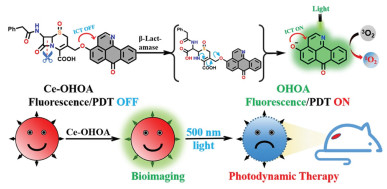
Scheme 1.
Illustration of Ce-OHOA for selective imaging and photodynamic antibacterial.

β-Lactam antibiotics are antibiotics that contain a β-lactam structure in their molecules [1]. This class of antibiotics is the most widely used and has several advantages, including strong bactericidal activity, low toxicity, wide indications, and good clinical efficacy [2,3]. However, the widespread use of these antibiotics inevitably leads to the emergence of drug-resistant bacteria, resulting in reduced therapeutic efficacy and other issues [4,5]. β-Lactamase, also known as cephalosporinase or penicillinase (E.C.3.5.2.6), is a zinc ion-dependent amide hydrolase that degrades penicillin antibiotics, such as penicillin G [6,7]. The destruction of β-lactam antibiotics' structure by bacteria, due to the high expression of β-lactamase, is considered one of the most common mechanisms of bacterial resistance. Therefore, the development of diagnostic and therapeutic reagents that target β-lactamase is of great significance in addressing bacterial resistance [8,9].
Antimicrobial photodynamic therapy (aPDT) is a novel of antimicrobial method that utilizes a light source and photosensitizer (PS) to eradicate bacteria [10]. The principle of this technology is that PSs are activated by specific wavelengths of light to produce reactive oxygen species (ROS) molecules. These molecules rapidly oxidize bacterial lipids and proteins, leading to the destruction of the bacterial membrane structure and initiating cell death [11]. PS shows high broad-spectrum bactericidal activity and can effectively kill gram-positive, gram-positive bacteria or drug-resistant bacteria [12]. In aPDT, the PS is the key factor. A number of small molecule antimicrobial PSs have been developed, and many of them demonstrate strong bacterial inhibition at the nanomolar level [13-18]. However, most PS does not possess targeting properties, as a result, they will inevitably cause damage to the surrounding normal cells while targeting the bacteria [19]. The emergence of activatable PSs effectively solves the targeting issue associated with conventional PSs. Before being activated by a specific biological microenvironment, the fluorescence and photodynamic effects of activatable PS are weak. After activation, the fluorescence and photodynamic effects of the PS are enhanced [20-22]. At present, most of the activatable PSs are primarily used in antitumor research, with fewer reports on their use in antibacterial applications [23-31]. Therefore, the development of activatable PSs for precise anti-drug-resistant bacteria therapy is crucial.
Several near-infrared activatable antimicrobial PSs have been reported to offer significant advantages in terms of wavelength and depth of penetration. However, the core of the PSs used in these studies has low single-linear oxygen (1O2) yields [32,33]. This necessitates higher concentrations and light doses for antimicrobial activity. 4-Hydroxyl-oxoisoaporphine (OHOA), which was first reported by Xu et al., stands out due to its high fluorescence quantum yield (0.72 in phosphate buffer saline (PBS)), high 1O2 yield (0.06), and low cytotoxicity [34]. Despite its excellent performance as a PS, OHOA has not been previously designed to be an activatable PS. In this study, we synthesized an activated PS called Ce-OHOA by connecting the OHOA and cephalosporin structures using ether bonds. Ce-OHOA exhibits a low fluorescence quantum yield and a 1O2 yield. However, when it undergoes hydrolysis by β-lactamase, OHOA with high fluorescence quantum yields and high 1O2 yields is generated. Experimental results in vitro and in vivo showed that Ce-OHOA could not only be used for fluorescence imaging of methicillin-resistant Staphylococcus aureus (MRSA) with high expression of β-lactamase enzymes, but also exhibited excellent photodynamic antimicrobial effects both in vivo and in vitro (Scheme 1).
OHOA was synthesized using previously reported literature methods [34]. The probe Ce-OHOA consisted of OHOA and Ce-Cl, and the complete synthesis procedure of Ce-OHOA is shown in Scheme S1 (Supporting information). The structure of Ce-OHOA and its intermediates were found to be unstable on silica gel and could not be effectively separated using conventional column chromatography to obtain pure products. Therefore, the products obtained during the synthesis step were purified using pulping. Ce-OHOA was characterized by 1H nuclear magnetic resonance (NMR), 13C NMR, and high resolution mass spectroscopy (HRMS) analyses (Figs. S1–S4 in Supporting information). The basic photophysical properties of Ce-OHOA and OHOA in PBS buffer are listed in Table S1 (Supporting information).
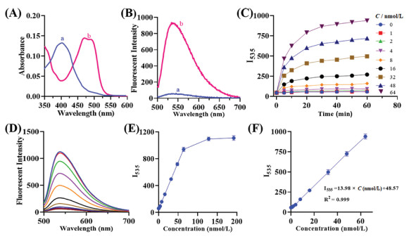
Fig. 1A shows the absorption spectra of Ce-OHOA (blue) and Ce-OHOA + β-lactamase (red). The maximum absorption position of Ce-OHOA shifted from 410 nm to 476 nm before and after reacting with β-lactamase. In addition, the fluorescence emission spectrum of Ce-OHOA, when reacted with β-lactamase, exhibited a significant enhancement under 480 nm light excitation. The maximum emission intensity (I535) was observed at 535 nm (Fig. 1B). We next measured the appropriate pH and temperature for the reaction of Ce-OHOA with β-lactamase, as shown in Fig. S5A (Supporting information). The fluorescence intensity of Ce-OHOA in different pH environments before Ce-OHOA did not interact with the enzyme was consistently low. The fluorescence of Ce-OHOA after interaction with β-lactamase, varied significantly in different pH environments, with the highest intensity observed at pH 7.4. In the temperature-dependent experiments (Fig. S5B in Supporting information), the fluorescence intensity of Ce-OHOA remained low at 25–40 ℃ before interacting with β-lactamase. After interacting with β-lactamase, the fluorescence intensity of Ce-OHOA was the strongest at 37 ℃. Therefore, we chose to study the reaction of Ce-OHOA with β-lactamase at 37 ℃ and pH 7.4. The fluorescence intensity of Ce-OHOA, which is time-dependent, shows a rapid increase with different amounts of β-lactamase (Figs. 1C–E). However, after approximately 60 min, the rate of increase in fluorescence intensity decreases. In addition, there was a good linear relationship between the intensity and β-lactamase concentration within the range of 0–64 nmol/L. A limit of detection (LOD) of 0.83 nmol/L was determined (Fig. 1F). To specifically detect the Ce-OHOA interacting by β-lactamase, the fluorescence intensity at 535 nm was measured. The selectivity of Ce-OHOA was tested, and the results in Fig. S6 (Supporting information) show that Ce-OHOA is not interfered with by other analytes. In order to investigate the recognition mechanism of Ce-OHOA with β-lactamase, Ce-OHOA was co-incubated with β-lactamase (64 nmol/L) for 60 min and the products after the reaction were monitored by high performance liquid chromatography (HPLC). At the end of the reaction, a new peak was observed at 2.29 min, which had a retention time (Rt) consistent with that of OHOA (Rt = 2.29 min). In contrast, the peak of Ce-OHOA at 4.72 min decreased in size (Fig. S7 in Supporting information). Mass spectrometry showed that the molecular weight of the peak with retention time of 2.29 min was consistent with OHOA (Fig. S8 in Supporting information) [35]. HPLC analysis showed that Ce-OHOA could be hydrolyzed by β-lactamase to produce OHOA.
In order to gain a clearer understanding of the interaction between Ce-OHOA and β-lactamase probes, we employed molecular docking software. As shown in Fig. S9A (Supporting information), the carboxyl group and the carbonyl group of the amide bond in the structure of Ce-OHOA form multiple sets of hydrogen bonds with Ala237, Ser70, Ser130, Ser235, and Ser244 of β-lactamase to achieve precise recognition. The distance between the hydroxyl group of Ser70, which plays a catalytic role in the β-lactamase, and the carbonyl carbon atom of the Ce-OHOA's lactam ring is only 0.32 nm. This close proximity ensures that the hydrolysis of the amide bond proceeds normally. In order to investigate the luminescence mechanism of Ce-OHOA after hydrolysis by β-lactamase, we utilized the calculated molecular orbital energies of Ce-OHOA and OHOA. As shown in Fig. S9B (Supporting information), the energy level difference between the highest occupied molecular orbital and the lowest unoccupied molecular orbital of Ce-OHOA is 3.38 eV, which is higher than that of OHOA (2.83 eV). Therefore, Ce-OHOA requires a higher energy of light for excitation. When Ce-OHOA is irradiated with 500 nm light, it does not undergo intramolecular charge transfer (ICT) due to insufficient light energy, resulting in weak luminescence [36]. When 500 nm light is used to irradiate OHOA, the ICT occurs easily and it exhibits strong luminescence.
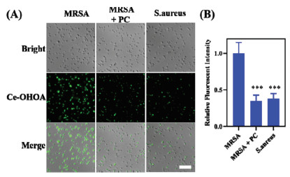
Subsequently, time-dependent MRSA imaging experiments were conducted. As shown in Fig. S10 (Supporting information), a weak green fluorescence emission appeared after 20 min of incubation of Ce-OHOA with MRSA, reaching its brightest at 60 min. Therefore, 60 min was chosen as the optimal time for bacterial fluorescence imaging. As shown in Fig. 2, the fluorescence signal of MRSA was significantly weakened after treating with potassium clavulanate (PC) compared to the untreated group. The same weakening of the fluorescence signal was also observed in S. aureus, which expresses a lower level of β-lactamase. The above results suggest that the fluorescent signal in bacteria is generated through the specific recognition of Ce-OHOA by β-lactamase.
In order to minimize the false damage of photosensitizers to normal tissues, Ce-OHOA was designed as an activatable type. The release of 1O2 was detected by monitoring the absorption at 400 nm using the commercial dye ABDA. The results showed that at 500 nm light emitting diode (LED), Ce-OHOA hardly produced 1O2 without β-lactamase activation (Fig. S11A in Supporting information). After treatment with β-lactamase, the absorption of ABDA at 400 nm was reduced by approximately 57% (Fig. S11B in Supporting information). This demonstrates that Ce-OHOA can release large amounts of rapidly 1O2 for antimicrobial use only after selective activation by β-lactamase.
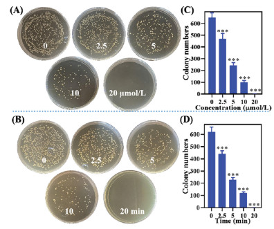
We then investigated the PDT effect of Ce-OHOA on MRSA. We conducted a series of experiments to investigate the concentration-dependent and light dose-dependent photoinactivation effects of Ce-OHOA against MRSA. In the concentration-dependent group, different concentrations of Ce-OHOA were cultured with MRSA for 60 min, then irradiated with or without LED. In the non-LED group (Fig. S12 in Supporting information), the survival of MRSA decreased with increasing Ce-OHOA concentration, but even at concentrations as high as 100 µmol/L, the survival rate of MRSA survival remained above 60%. In the LED group (Fig. 3), the survival rate of MRSA gradually decreased as the concentrations of Ce-OHOA increased. When the concentration of Ce-OHOA was only 2.5 µmol/L, the survival rate of MRSA decreased significantly to 74.6%. However, when the concentration of Ce-OHOA was increased to 20 µmol/L, the survival rate dropped to only 0.1% (Figs. 3A and C). In the light dose-dependent group (Figs. 3B and D), the survival rate of MRSA decreased significantly and gradually with the increase in light dose. The survival rate of MRSA significantly decreased to 72.1% at 2.5 min, and only 0.1% at 20 min. These results indicate that Ce-OHOA has a strong bactericidal effect on MRSA strains that exhibit high expression of β-lactamase under light conditions, and its antibacterial concentration and light dose are obviously lower than those reported activatable photosensitizer (Table S2 in Supporting information).
Diamidino-2-phenylindole (DAPI) and propidium iodide (PI) co-staining was used to study the mechanism of bacterial death. As depicted in Fig. S13 (Supporting information), when the MRSA were incubated with Ce-OHOA without light irradiation, only blue fluorescence (DAPI) was observed in the bacteria. However, under the light condition, both blue fluorescence (DAPI) and red fluorescence (PI) was observed in the bacteria. DAPI stains bacteria with intact cell membranes, while PI only stains bacteria with damaged cell membranes [14]. This suggesting that the bacteria incubated with the Ce-OHOA experienced damage to their cell membranes of under light conditions, potentially leading to their death.
The favorable in vitro antimicrobial effect of Ce-OHOA prompted us to further investigate its effect in vivo using aPDT. We chose a rat wound model to study the antimicrobial potential of Ce-OHOA. To assess the safety of Ce-OHOA at the infection site in rats, we initially examined the photo/dark toxicity of Ce-OHOA on RS1 cells (rat skin fibroblast cell line). As demonstrated in Fig. S14 (Supporting information), Ce-OHOA showed good biocompatibility with RS1 cells, with cell survival remained above 85% even under light conditions when the concentration was below 40 µmol/L.
After demonstrating the favorable biocompatibility of Ce-OHOA with normal skin, we conducted additional photodynamic antimicrobial validation using a rat wound infection model (Adherence to the ethical standards set by the Animal Ethics Committee at Wenzhou Medical University was ensured, approval number: Wydw7019–0134). As depicted in Figs. 4A and B, the rate of wound healing was slower in the saline, saline + LED, and Ce-OHOA groups when compared to the Ce-OHOA + LED group. On day 10, the infected wound form the Ce-OHOA + LED group only slight marks, while all other control groups showed varying degrees of unrepaired wounds. Under the irradiation of the 500 nm LED, the rats did not show any recurrence of the wound or signs of septic infection. Moreover, the body weight of rat fluctuated within the normal range during the treatment (Fig. 4C), indicating that Ce-OHOA accelerates the healing of infected wounds through antibacterial photodynamic therapy.
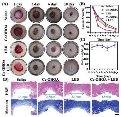
The therapeutic efficacy was further examined by performing hematoxylin-eosin staining (H&E) staining and Masson staining on day 10 after infection. As illustrated in Fig. 4D, H&E histological analysis showed that Ce-OHOA plus irradiation resulted in shorter mean length of granulation gap and the formation of thicker epithelial tissue with improved histological architecture compared to the other three groups. Masson staining showed an organized arrangement of collagen. These results strongly indicate that Ce-OHOA plus LED irradiation has a significant advantage in suppressing MRSA infections and promoting wound healing.
In this paper, we developed an activatable photosensitizer (Ce-OHOA) against MRSA with high expression of β-lactamase. Ce-OHOA not only shows good sensitivity and high anti-interference ability to β-lactamase in aqueous solution, but also can image the MRSA with high expression of β-lactamase in vitro. In addition, in vitro and in vivo antibacterial experiments, Ce-OHOA showed a strong antibacterial effect at low light dose (12 J/cm2) and low concentration (10 µmol/L), and contributed to the wound healing of MRSA-infected animal models. Overall, Ce-OHOA is outstanding in fluorescence detection and photodynamic antibacterial function. Such rationally designed activatable photosensitizer will provide a new idea for the study of diagnostic and therapeutic reagents for drug-resistant bacteria.
The authors declare that they have no known competing financial interests or personal relationships that could have appeared to influence the work reported in this paper.
This work was financially supported by the National Natural Science Foundation of China (No. 21977081), Zhejiang Provincial Natural Science of Foundation of China (No. LQ21H190006).
Supplementary material associated with this article can be found, in the online version, at doi:
E. Minaldi, E.J. Phillips, A. Norton, Clin. Rev. Allergy Immunol. 62 (2022) 449–462.
C.L. Tooke, P. Hinchliffe, E.C. Bragginton, et al., J. Mol. Biol. 431 (2019) 3472–3500. doi: 10.1016/j.jmb.2019.04.002
L.M. Lima, B.N.M. da Silva, G. Barbosa, et al., Eur. J. Med. Chem. 15 (2020) 112829.
M.L.M. Salverda, J.A.G.M. De Visser, M. Barlow, FEMS Microbiol. Rev. 34 (2010) 1015–1036. doi: 10.1111/j.1574-6976.2010.00222.x
J. Li, W. Sun, Z. Yang, G. Gao, et al., ACS Appl. Mater. Interfaces 12 (2020) 54378–54386. doi: 10.1021/acsami.0c15244
E. Dellus-Gur, M. Elias, E. Caselli, F. Prati, et al., J. Mol. Biol. 427 (2015) 2396–2409. doi: 10.1016/j.jmb.2015.05.011
M.F. Mojica, M. -A. Rossi, A.J. Vila, et al., Lancet Infect. Dis. 22 (2022) 28–34. doi: 10.1016/S1473-3099(21)00777-5
Z. Yi, X. Xu, X. Meng, et al., Chin. Chem. Lett. 34 (2023) 108238. doi: 10.1016/j.cclet.2023.108238
H. Zhang, C. He, L. Shen, et al., Chin. Chem. Lett. 34 (2023) 108160. doi: 10.1016/j.cclet.2023.108160
S.P. Songca, Y. Adjei, Int. J. Mol. Sci. 23 (2022) 3209. doi: 10.3390/ijms23063209
F.F. Sperandio, Y.Y. Huang, M.R. Hamblin, Drug Disc. 8 (2013) 108–120. doi: 10.2174/1574891X113089990012
H.R. Jia, Y.X. Zhu, Z. Chen, F.G. Wu, ACS Appl. Mater. Interfaces 9 (2017) 15943–15951. doi: 10.1021/acsami.7b02562
N. Vn, Z. Z, T. Bz, Y. J, Chem. Soc. Rev. 51 (2022) 3324–3340. doi: 10.1039/D1CS00647A
G. Lin, M. Hu, R. Zhang, et al., J. Med. Chem. 64 (2021) 18143–18157. doi: 10.1021/acs.jmedchem.1c01643
E. Caruso, S. Banfi, P. Barbieri, et al., J. Photochem. Photobiol. B 114 (2012) 44–51. doi: 10.1016/j.jphotobiol.2012.05.007
F. Hu, S. Xu, B. Liu, Adv. Mater. 30 (2018) e1801350. doi: 10.1002/adma.201801350
E. Zhao, Y. Chen, H. Wang, et al., ACS Appl. Mater. Interfaces 7 (2015) 7180–7188. doi: 10.1021/am509142k
S. Wang, W. Wu, P. Manghnani, et al., ACS Nano 13 (2019) 3095–3105. doi: 10.1021/acsnano.8b08398
E. Caruso, S. Ferrara, P. Ferruti, et al., Lasers Med. Sci. 33 (2018) 1401–1407. doi: 10.1007/s10103-017-2345-0
Y. Wen, N. Jing, M. Zhang, et al., Adv. Sci. 10 (2023) e2206681. doi: 10.1002/advs.202206681
X. Li, F. Huo, Y. Zhang, F. Cheng, et al., J. Mater. Chem. B 10 (2022) 5504–5519. doi: 10.1039/d2tb00922f
X. He, Z. Zheng, F. Zhang, et al., ACS Appl. Bio Mater. 3 (2020) 7886–7897. doi: 10.1021/acsabm.0c01029
Z. Li, Q. Feng, J. Hou, et al., Bioorg. Chem. 143 (2023) 107021.
B. Peng, G. Chen, Y. Li, et al., Anal. Chem. 94 (2022) 11159–11167. doi: 10.1021/acs.analchem.2c01189
H.W. Liu, X.X. Hu, K. Li, et al., Chem. Sci. 8 (2017) 7689–7695. doi: 10.1039/C7SC03454G
J. Huang, C. Zhang, X. Wang, et al., Angew. Chem. Int. Ed. 62 (2023) e202303982. doi: 10.1002/anie.202303982
J. Miao, Y. Huo, G. Yao, et al., Angew. Chem. Int. Ed. 61 (2022) e202201815. doi: 10.1002/anie.202201815
Y. Luo, Z. Zeng, T. Shan, et al., Theranostics 12 (2022) 3610–3627. doi: 10.7150/thno.70308
X. Zhou, H. Li, C. Shi, et al., Biomaterials 253 (2020) 120089. doi: 10.1016/j.biomaterials.2020.120089
Q. Zhao, G. Qing, J. Yu, et al., Chin. Chem. Lett. 35 (2024) 108535. doi: 10.1016/j.cclet.2023.108535
Y. Chen, X. Zhao, T. Xiong, et al., Sci. China Chem. 64 (2021) 808–816. doi: 10.1007/s11426-020-9947-4
C. Zhao, W. Sun, B. Tan, et al., Sens. Actuat. B: Chem. 382 (2023) 133502. doi: 10.1016/j.snb.2023.133502
Y. Xu, H. Chen, S. Xu, et al., ACS Sens. 7 (2022) 1361–1371. doi: 10.1021/acssensors.1c02485
Q. Xu, Y. Ji, M. Chen, et al., Photochem. Photobiol. Sci. 20 (2021) 501–512. doi: 10.1007/s43630-021-00030-0
G. Cheng, J. Xing, Z. Pi, et al., Chin. Chem. Lett. 30 (2019) 656–659. doi: 10.1016/j.cclet.2018.12.003
C. Yan, Z. Guo, W. Chi, et al., Nat. Commun. 12 (2021) 3869. doi: 10.1038/s41467-021-24187-5

Scheme 1 Illustration of Ce-OHOA for selective imaging and photodynamic antibacterial.

Figure 1 (A) Absorption and (B) fluorescence emission spectra of Ce-OHOA (10 µmol/L) before (a) and after (b) reaction with β-lactamase (64 nmol/L) at 25 ℃ for 30 min in PBS (pH 7.4). λex = 480 nm. (C) Plots of fluorescence intensity I535. The reaction time of Ce-OHOA (10 µmol/L) with varied concentrations of β-lactamase (0–64 nmol/L). λex = 480 nm. (D) Fluorescence emission spectra of Ce-OHOA (10 µmol/L) with 0–192 nmol/L β-lactamase. (E) Fluorescence intensity I535 of Ce-OHOA (10 µmol/L) with 0–192 nmol/L β-lactamase. (F) Linear fitting curve of fluorescence intensity I535 toward the concentration of β-lactamase (0–64 nmol/L). λex = 480 nm.

Figure 2 (A) Confocal fluorescence images of MRSA, MRSA+PC and S. aureus (non-expressing β-lactamase) incubated with Ce-OHOA (10 µmol/L). Scale bar: 20 µm. (B) Relative pixel intensity of the corresponding fluorescence images in pane A. ***P < 0.001 vs. MRSA group. Error bars: mean ± standard deviation (SD) (n = 10).

Figure 3 Digital images of clonogenic assay performed in plates, with clones produced by MRSA under different concentrations (A) or different illumination times (B). (C, D) Colony numbers are counted using Image J. All the data are presented as mean ± SD (n = 3). ***P < 0.001 vs. control group.

Figure 4 In vivo antibacterial evaluation of Ce-OHOA for MRSA-infected wounds of Rat. (A) Successive photographs of wounds without and with aPDT treatment. Inside diameter of stainless-steel ring = 16 mm. (B) Time-dependent lesion area. (C) Body weight change of rats. (D) H&E staining and Masson staining images of the sectioned tissues of wounds without and with aPDT treatment on day 10 post-infection. Scale bar: 1.0 mm. Error bars: mean ± SD (n = 4).

 扫一扫看文章

扫一扫关注我们
 DownLoad:
 下载:
下载: