Figure 1.
Schematic diagram of sensing mechanism of the three-dimensional spectral array. Reprinted with permission [26]. Copyright 2021, Elsevier.
Multivariate chemical analysis: From sensors to sensor arrays
Xueling Yu , Lixing Fu , Tong Wang , Zhixin Liu , Na Niu , Ligang Chen

The sensor is an information acquisition and processing device that can convert specific input information into readable output signals according to certain rules [1,2]. The sensor is mainly composed of the receptor (sensing element) and the transducer [3]. The receptor is the sensing material used to accept the stimulus from the analyte. The transducer is a signal conversion system that converts input signals from the receptor into a measurable signal. From the perspective of analyte type, sensors can be divided into physical sensors and chemical sensors. Physical sensors can sense physical information such as light, sound, temperature, and pressure. Chemical sensors are used to obtain chemical information (substance composition, concentration, and identity). This review mainly discusses the chemical sensor.
With the development of sensing technology, chemical sensors have adopted a variety of analytical strategies to track the subtle changes in the interaction of the analyte with the sensing elements. According to the different signal transduction mechanisms, chemical sensors can be divided into electrochemical, mass, and optical sensors [4]. At present, chemical sensors have been used in environmental analysis [5,6], medical detection [7,8], food safety [9,10], and other fields. The preparation of chemical sensors with excellent performance has been an important task. A sensor with good performance requires excellent sensitivity, selectivity, stability, and reversibility. However, it is difficult for a single sensor to have all the functions at once. For example, the more sensitive a sensor is, the less inherently stable it usually is. And when there is interference, the accuracy of detection results is difficult to evaluate. Therefore, there are many limitations to using a single signal characteristic of a single sensor to analyze an analyte.
Sensor arrays are called "electronic noses" or "electronic tongues". Humans can recognize substances through the cross-reactivity of different receptor cells in the olfactory or taste organs with different molecules [11,12]. Similarly, several different sensors form a sensor array, which also has the characteristic of cross-reactivity [3]. Namely, each sensing element has different degrees of response to the different analytes (usually a series of molecules with similar structure or chemical properties) in the system. Different sensing elements also have different degrees of response to the same analyte. These cross-reactive sensing elements avoid their specificity to a single target [13]. Compared with conventional sensors, sensor arrays can not only identify single targets but also distinguish multi-component mixed samples. Therefore, array sensing is of great significance for the analysis of complex systems and multi-component samples. The cross-reaction of chemical sensor arrays leads to the generation of multi-dimensional signals. It is necessary to analyze multi-dimensional data or data patterns with appropriate pattern recognition methods [14,15].
This review primarily summarizes the sensing materials for array design and shows their applications in food monitoring, medical diagnosis, and environmental monitoring. We also discuss several important pattern recognition methods for processing high-dimensional data. Finally, based on the analysis of the limitations of current array technology, the direction of development of sensor arrays is predicted. We hope that this review will guide the further development of chemical sensor arrays and promote their practical applications in sensing.
The core of a chemical sensor array is the design of the sensing elements. Sensing elements should provide a differential response to analytes and good signal transduction performance. Therefore, the synthesis and modification of sensing materials become the key steps in array design. At present, many functional and highly active sensing materials are used in array design. Among them, metal-organic frameworks, graphene, quantum dots, rare earth up-conversion luminescent materials, molecularly imprinted polymers, and precious metal nanomaterials with good stability and surface modification have received much attention in this field.
Metal–organic frameworks (MOFs) are new crystalline porous materials composed of organic connectors and metal centers [16,17]. The three main parts of MOFs are topological structures, inorganic metal centers, and organic ligands. The permanent porosity of MOF provides a large specific surface area and potential active sites for target sensing (such as coordinated unsaturated open metal sites and terminated functional groups) [18].
The low mechanical strength, poor stability, and single function of single MOF materials limit their application. Therefore, composite MOFs prepared by combining carbon materials, nanoparticles, and organic molecules have been widely studied [19,20]. MOF-based composites also show significant strengths and potential in chemical sensing. By modifying electrode surfaces with catalytically active MOF-based composites, the sensitivity, selectivity, and interference resistance of electrochemical sensors can be improved [21]. Wang et al. [22] combined Cu-MOFs with polyvinylpyrrolidone (PVP) by a one-pot solvothermal method to obtain three-dimensional (3D) nitrogen-doped large-medium-microporous carbon composites (N/Cu-HPC). The introduction of PVP gives N/Cu-HPC excellent hydrophilicity to prevent clustering. The higher electronegative nitrogen species significantly improve electrocatalytic performance. The large-medium-microporous structure improves the binding and charge transmission between the neonicotinoid molecules and the active site. Interestingly, Zhou et al. [23] fabricated an Mn-PCN-222/ITO electrode by liquid deposition of Mn-PCN-222 (manganese metalloporphyrin) film on conductive indium tin oxide (ITO) glass using a fast and simple modular assembly method. The metal porphyrin unit as the catalytic center and the porous Mn-PCN-222 structure can be efficiently combined. The sensor has the advantages of a high density at the active site, a large specific surface area, and a sensitive response. It has also demonstrated multifunctional voltammetry sensing for a variety of analytes (nitroaromatic hydrocarbons, phenolic resins, quinone-hydroquinone, heavy metal ions, biological species, and azo dyes).
MOFs are an excellent platform for building array sensing, as they can regulate the topology, and porosity (pore size and geometry) through rational selection of ligands and metal nodes, and thus influence specific responses to external stimuli. Sensor arrays based on different MOFs materials have been used to detect various analytes. Chen et al. [24] prepared three zirconium porphyrin-emitting metal–organic frameworks (PCNs) with different topologies and constructed a sensor array (Fig. S1 in Supporting information). The sensing principle is the static fluorescence quenching of PCNs during adsorption with perfluoroalkyl substances (PFASs). Different PFAS showed different fluorescence response signals. Combined with pattern recognition methods, the PCN array could successfully distinguish six different PFASs in water. This array is a mechanical combination of different sensor elements, and most of the sensor arrays reported are based on this form. However, this form increases the complexity of array technology in terms of experimental manipulation and data processing.
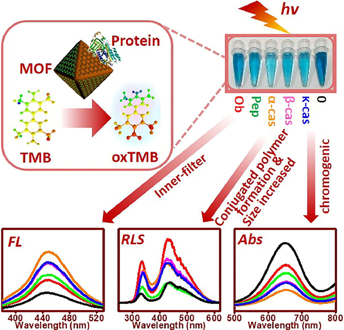
Another form of the array is the integration of multiple signals onto a single element. This form reduces the complexity of the array. Wang et al. [25] combined three luminescent ligands onto a UiO-66 framework to synthesize a white-emitting MOF for distinguishing berberine homologs. The MOF generates a distinctive three-channel fluorescent signal pattern for each analyte. Therefore, only one measurement of the fluorescence spectrum can be used for rapid detection of the substance. The arrays successfully identified five structurally similar berberine homologues at concentrations as low as 2 µmol/L, and in combination with chemical titration, this method can be used for the analysis of the samples at unknown concentrations. In addition, different signal types (such as fluorescence, phosphorescence, resonance light scattering, and absorption) can also be integrated into a single sensing element. Lin et al. [26] prepared an array sensing platform based on MOF/3, 3′, 5, 5′-tetramethylbenzidine. Phosphoproteins were adsorbed by MOF, and the absorbance, fluorescence, and resonance light scattering signals were altered. The sensing mechanism is shown in Fig. 1. The three-dimensional spectral array sensing system can identify five typical phosphoproteins (with detection limits as low as 5 nmol/L and classification accuracy of 100%). At the same time, the array-sensing platform can distinguish abnormally phosphorylated cancer cells from normal cells, validating its utility for diagnosing phosphorylation-related diseases. Compared to conventional array sensing, a single MOF-based sensor array ensures sufficient data information while significantly reducing the complexity and time of analytical operations.
Graphene is a carbon material composed of a two-dimensional honeycomb lattice of sp2 hybrid monolayer carbon atoms [27,28]. Graphene has excellent electronic, optical, thermal, and mechanical properties, giving it the triple role of structuring probe molecules, signal transmission, and signal amplification. The size, number of layers, shape, and chemical groups of graphene can affect significantly the performance of the sensor, making graphene an ideal material for array sensing. Differences in graphene functionalization parameters, differences in local morphology, and chemical inhomogeneities can lead to different responses of the sensing elements in the array to the analyte [29].
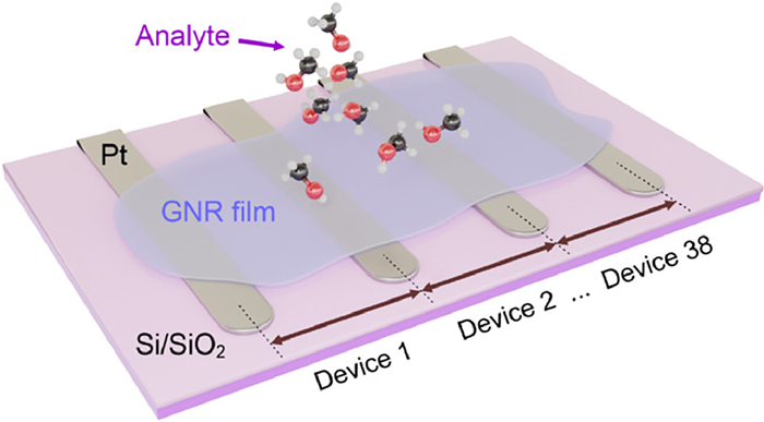
Graphene has a short history, having only been isolated for the first time in 2004 [30]. Mechanical stripping is the original synthesis method for graphene, which produces graphene with the best structural integrity. However, the technology cannot control the number of layers and the low yield. Therefore, it is limited to scientific research. In recent years, chemical vapor deposition (CVD) has provided an effective method for the controllable preparation of large graphene sheets. For example, Xia et al. [31] synthesized a high-quality van der Waals methylammonium halide lead/graphene heterostructure by the two-step CVD method for constructing high-performance image sensors. In practice, the semi-metallic nature of graphene itself limits its application, while graphene nanoribbons (GNRs) have attracted attention due to their open band gap properties [32]. Shekhirev et al. [33] designed an innovative scheme for preparing GNRs based on CVD using Si/SiO2 containing 39 Pt electrodes as a planar substrate. After GNR film is grown, Pt electrodes are bridged, and adjacent Pt electrodes form sensor devices. These sensor devices can independently detect the measured substance, thus forming a sensor array consisting of 38 sensor units. The morphology of the GNR domain (size, orientation, microscopic crack, etc.) results in the difference between each sensing unit. The array is shown in Fig. 2. The interaction of the measured molecule with the GNR sensor leads to a reduction in resistance. The array can effectively distinguish different chemical classes of analytes, including alcohols such as methanol, ethanol, and isopropanol, and amines such as n-butylamine, diethylamine, and triethylamine.
Graphene can also be chemically modified to produce graphene derivatives, including graphene oxide (GO) [34,35] and reduced graphene oxide (rGO) [36]. In contrast to the original graphene, the GO matrix plane contains epoxy and carbon radicals with carboxyl and hydroxyl groups at its edges. The inherent properties of graphene enable GO to be partially reduced to form rGO. GO, rGO and raw graphene are often collectively referred to as graphene-related materials. In terms of chemical sensors, graphene materials show great potential. Graphene and GO have been used to construct different types of chemical sensors based on various sensing mechanisms, mainly optical and electrochemical sensors.
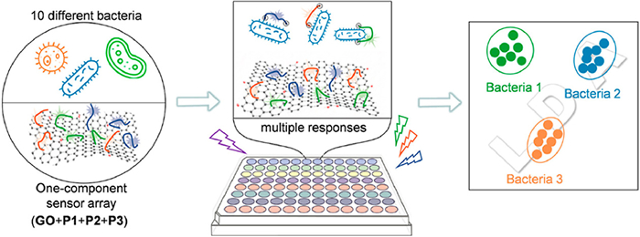
Graphene and GO are universal quenchers that can effectively quench the fluorescence of fluorescent dye molecules, quantum dots, and up-conversion nanomaterials through fluorescence energy resonance transfer and non-radiative dipole interaction [37]. Wang et al. [38] designed a single-component multichannel sensor array formed by electrostatic complexation of GO with three different polyethylene imines (PEIs). Three different fluorophores were used for the fluorescent labeling of PEIs. Graphene oxide can interact electrostatically with the three PFIs and cause fluorescence quenching in the attached fluorophores. Different bacteria compete with GO to bind PEIs and make the fluorophore produce different fluorescence signals. The array can identify different species and concentrations of bacteria, as well as different proportions of mixed bacteria. The sensing and recognition processes of the array for bacteria are shown in Fig. 3. The array can identify bacteria at different concentrations (OD600 = 0.025–1) and mixed bacteria. Its application to urine has also shown tremendous identification capability (OD600 = 0.125, 94% accuracy). Constructing sensor arrays with multiple signal outputs with a minimum number of sensing elements saves significant cost and time and offers a strong method for the diagnosis of clinical bacterial infection.
The graphene-modified electrode not only has excellent electrical conductivity but can also enrich the detection molecules due to its large specific surface area, and the sensor can feedback to larger signal strength. To date, one of the most promising applications of graphene electrochemical sensors is the gas sensor [39,40]. The honeycomb structure of the individual carbon layers exposes all the carbon atoms to the environment, thus maximizing the contact area between the gas and the graphene. It makes up for the shortcomings of other gas-sensitive materials. Most graphene-based gas sensors work on the principle of their change in electrical conductivity, which is caused by the adsorption of gas molecules on the surface of graphene. Chen et al. [41] constructed an electronic nose based on Go-Mx+ (M = Co, Fe, Cu, and Ce) sensing elements by using GO in combination with different types of metal ions (Mx+). Under the action of polyvalent Mx+, a cross-linked GO network was formed, and then the hydrazine vapor reduced GO to rGO in situ. The porous rGO-M film could be evenly coated on the polyethylene terephthalate substrate, thus forming the sensing element. The electron nose successfully distinguished four types of exhaled breath biomarkers, including acetone, isoprene, ammonia, and hydrothion at sub-ppm concentrations. In addition, the electronic nose was able to accurately differentiate between the healthy group and lung cancer patients in clinical studies (95.8% sensitivity and 96.0% specificity). This suggests that graphene-based sensor arrays have great potential for use in non-invasive medical diagnostics.
Quantum dots (QDs) are a kind of inorganic semiconductor and zero-dimensional nanomaterial, with unique optical and electromagnetic properties and good surface modifiability. QDs can modulate energy states and charge interactions by changing their composition and structure. Due to the quantum domain effect, closed electron carriers are formed inside the QDs, and the discrete electronic energy states and optical leap energies of QDs can be precisely modulated, which allows QDs to be used often as optical tags in chemical sensing. In addition, QDs can be assembled into semiconductor materials by manipulating chemical surfaces using capped molecules with different physical and chemical properties, increasing electrical conductivity, and converting chemical stimuli into electrical signals. As a result, quantum dots have been widely used for optical and electrochemical sensing [42].
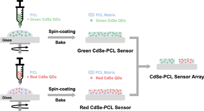
QDs are usually divided into two categories: metal-based (M-based) and carbon-based (C-based) materials [43]. M-based QDs mainly refer to the QDs formed by the combination of the elements of the groups IIIA and VA, IIB and VIA, and IVA and VIA, such as CdSe QDs, CdTe QDs, and ZnS QDs. With size-tunable electrical and optical properties, M-based QDs have attracted widespread interest in the development of luminescent materials for array sensing. Aznar-Gadea et al. [44] embedded green CdSe QDs (emitting at 560 nm) and red CdSe QDs (emitting at 597 nm) into polycaprolactone (PCL) to constitute a sensor array for detecting explosive markers. The mechanical properties and stability of QDs sensors can be improved by embedding QDs in PCL. Based on the differential variation in fluorescence intensity caused by different molecules adsorbed on the surface of the quantum dots, the array can identify three explosive markers. Response times for the two sensing elements were as short as 30 s for the analytes. Detection limits for 3-nitrotoluene, 4-nitrotoluene 2,3-dimethyl-2,3-dinitrobutane were 0.055, 2.7, and 0.7 ng, respectively. The design concept of the array is shown in Fig. 4.
In practical applications, QDs are usually doped to improve performance, that is, ions or atoms of appropriate elements are incorporated into the main lattice of the QDs. Ions or atoms doped into the main lattice of the QDs will result in new electron energy levels and new electron-hole complex centers, thus changing the number of emission centers in the host QDs. The doped QDs can not only retain the optical advantages of the main QDs but also effectively avoid the self-quenching phenomenon. Jiao et al. [45] synthesized four kinds of Mn-doped ZnS (Mn–ZnS) QDs modified with n-acetylcysteine, citric acid, mercaptopropionic acid, and triammonium n-dithiocarboxyamino-diacetate, respectively. They are designed as electronic tongues to distinguish heavy metal ions (copper, mercury, silver, and cadmium) from complex samples. Since the fluorescence quenching effects of different ions on QDs are disparate, the array sensor can recognize different metal ions in unknown mixtures. Based on the luminescence characteristics of Mn–ZnS, Wu et al. [46] designed Mn–ZnS QDs as a single probe sensor array by using the three-channel optical signals of fluorescence, phosphorescence, and resonance light scattering of Mn–ZnS QDs, successfully realizing the recognition and differentiation of eight proteins.
Carbon quantum dots (CDs) have been the focus of research in recent years due to their low toxicity, easy availability, and excellent water solubility [47]. CDs are a kind of quasi-spherical carbon nanoparticle with particle sizes less than 10 nm, mainly composed of C, H, and O elements [48]. Because the surface of CDs contains rich functional groups such as hydroxyl, carboxyl, and carbonyl, they are well dispersed in water and easy to functionalize. The fluorescence of exposed CDs is very weak or even absent. To improve the quantum yield, water solubility, and stability, CDs can be doped with N, S, P, and B atoms. Xu et al. [49] designed a two-channel fluorescence sensor array based on the internal filter effect (IFE) using quinaldine red and cetylpyridinium chloride-quinidine as raw materials for the detection of tetracycline, oxytetracycline, doxycycline, and metacycline. The IFE-based approach does not require complex modifications to CDs. Chen et al. [50] developed a two-channel (fluorescence and absorbance channels) sensor array consisting of four polyvinyl alcohol (PVA) composite CDs films, which can classify a variety of polycyclic aromatic hydrocarbons (PAHs) by the IFE mechanism. The preparation flow chart is shown in Fig. S2 (Supporting information). After pattern recognition algorithms, the array can identify 16 PAHs with 100% accuracy at concentrations as low as 57 nmol/L. In addition, the sensor array showed excellent immunity to interference in complex environments and rapid identification and quantification of PAHs in real environmental samples (soil and lakebed sludge).
In addition, CDs prepared from biomass can be doped with elements from the biomass itself, which simplifies the modification step. For example, fluorescent CDs have been produced using fruit peels [51]. And our research group [52] synthesized bone CDs of three animals (pig, cow, and sheep) by the hydrothermal method and formed arrays. The array distinguished between these five ions and their binary and ternary mixtures with 100% accuracy. Simultaneously, metal ions in environmental water were also successfully distinguished. The array was successfully used for the identification of heavy metal ions in aqueous environments. The bones contain large amounts of Ca, N, and S elements, which allow these atoms to be doped into the CDs. Bones can increase the quantum yield of CDs compared to the peel as a precursor.
According to the different relative positions between the excitation band and the emission band, luminescent materials can be divided into two types: Stokes luminescent materials and anti-Stokes luminescent materials [53,54]. Most luminescent materials (such as organic dyes and quantum dots) generally obey Stokes' law, which states that the wavelength of emitted light should always be longer than that of excited light. However, in some cases, the wavelength of the emitted light may be shorter than that of the excited light. The photon energy of the emitted light is greater than that of the excited light, a phenomenon known as "anti-Stokes luminescence" or "up-conversion luminescence" [55]. Most of the trivalent ions of rare earth elements can realize the up-conversion process [56]. Rare earth up-conversion nanomaterials are generally composed of matrix, sensitizer, and activator [54]. The matrix material itself does not emit light, but it has a great influence on the luminescence performance of rare earth up-conversion materials. The matrix material with low phonon energy and matching with a rare-earth doped ion lattice can improve the up-conversion efficiency [56]. Activator refers to the ion used for luminescence in the up-conversion luminescence system. The activator will emit dazzling fluorescence when it is excited by excited light or receives the energy provided by the sensitizer. The sensitizer is the bridge between the excitation light and the activator. Sensitizer particles, also known as activation ions, absorb excitation energy and then transfer the absorbed energy to luminescent ions. After absorbing energy, the activated ions transition from the ground state to the excited state and then undergo an up-conversion luminescence process.
Chemical sensing is one of the most important applications of up-conversion nanomaterials, which can control the signal of up-conversion optical sensors by changing the substrate material and ion doping [57,58]. In addition, the main mechanisms of up-conversion luminescence include excited state absorption, energy transfer, and photon avalanche. Achieving the high brightness and sensitivity of up-conversion materials relies on discovering the most efficient energy transfer pathways through material design. External stimuli can be used to achieve chemical sensing by controllably interfering with these energy transfer processes [58]. NaYF4 is one of the most ideal up-conversion matrix materials. Abbasi-Moayed et al. [59] design an emerging multichannel sensor array based on NaYF4 doped lanthanide (Yb, Er, and Tm) up-conversion nanoparticles (UCNPs). The energy levels of the lanthanide ions provide ideal emission bands with cross-reaction, and these emission bands are used as sensing elements to recognize four neurotransmitters (NTs): dopamine, norepinephrine, levodopa, and serotonin. Under alkaline conditions, the oxidation products of these NTs can quench the fluorescence emission of UCNPs to different degrees. NTs can be distinguished very accurately from the fingerprint multi-channel emission curve. The design provides an innovative insight into the use of up-conversion luminescent materials to distinguish various chemical features.
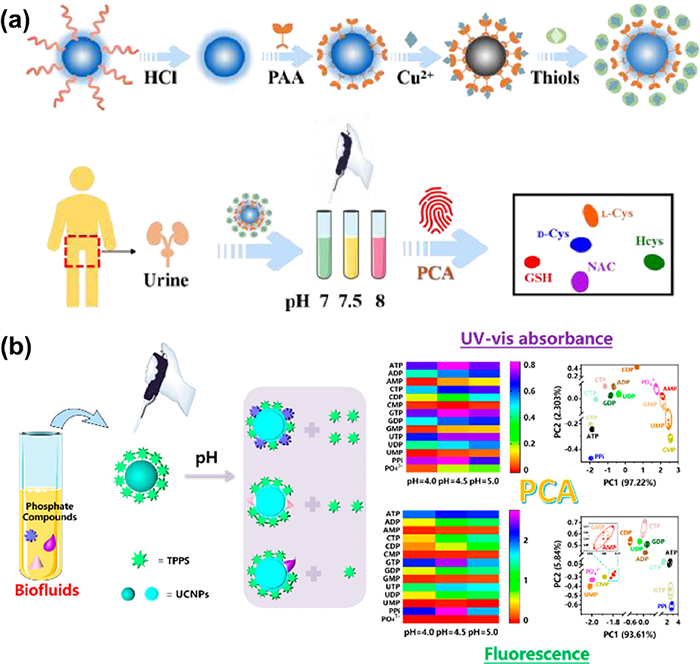
Another effective way to simplify sensing elements is to modulate the properties of the material by changing external conditions (such as temperature, solvent, and pH). pH is one of the most important external conditions affecting the fundamental properties of the material [60]. As shown in Fig. 5a, Wang et al. [61] designed a fluorescent sensor array of pH-regulated single-sensor components. The oleic acid-coated lanthanide UCNPs (Ln-UCNPs) with the core-shell structure were synthesized by solvothermal method, and oleic acid-free Ln-UCNPs were obtained after acid treatment. Ln-UCNPs without ligands have good water dispersion and surface modifiability. The desired function can be achieved by specific surface modification of the Ln-UCNPs. The fluorescent probes (PAA/Ln-UCNPs) were obtained by modifying the Ln-UCNPs with polyacrylic acid (PAA). Cu2+ can quench the fluorescence of PAA/UCNPs by complexing PAA. The fluorescence response of PAA/Ln-UCNPs to Cu2+ differs at different pH values, thus forming a PAA/Ln-UCNPs-Cu2+ sensor array at three pH values. Based on the coordination of Cu2+ with sulfhydryl groups, the arrays were successfully used for the identification of five thiols as well as the chiral enantiomers of cysteine. By analyzing human urine, this method provides a new protocol for monitoring homocysteine in humans. Yan et al. [62] designed a pH-regulated dual-mode optical sensor array using tetraphenylporphyrin tetrasulfonic acid hydrate (TPPS) modified above ligand-free Ln-UCNPs. As shown in Fig. 5b, the constructed organic/inorganic hybrids (TPPS/Ln-UCNPs) can exhibit different optical properties in terms of UV–vis absorption and fluorescence through pH adjustment. The phosphate groups in the phosphate compounds can be firmly bound to the surface of Ln-UCNPs with lanthanide ions via Lewis acid/base interactions. Due to the different affinities of phosphate compounds with Ln-UCNPs, different photoresponse patterns are presented. The sensor array successfully identified 14 phosphate compounds and their hybrids.
Molecularly imprinted polymers (MIPs) are synthesized by molecular imprinting technology (MIT) with specific recognition functions for specific target molecules (template molecules) [63]. MIT is a special technology that simulates the mechanism of antigen and antibody recognition [64,65]. During the synthesis of MIPs, imprinted cavities that complement the size, shape, and functional group of the template molecule are generated. These cavities can use a "lock and key" mechanism to selectively bind template molecules. MIPs have high affinity and selectivity for template molecules, but may still cross-react with other compounds, especially compounds with similar chemical properties to the template molecules, so MIPs can also be designed as sensor arrays. By using different templates in the imprinting process, a range of polymers with different binding selectivity can be quickly prepared [66].
MIPs have a certain electrochemical inertia and their conductivity and electrocatalytic activity are relatively poor. This severely affects the responsiveness and detection sensitivity of electrochemical sensors that rely on electron transfer signals [67]. Although MIPs electrical performance is poor, the combination with chemical sensors significantly improves the selectivity and sensitivity of the sensors. Therefore, new synthesis, polymerization, and immobilization routes are being explored to improve the overall performance of electrochemical sensors. Wang et al. [68] synthesized a voltammetric sensor array integrated with three polypyrrole MIPs-based sensors by the electropolymerization method for paracetamol, ascorbic acid, and uric quantitative detection. Electropolymerization is easy to operate and reproducible, which is beneficial for the direct integration of a voltammetry sensor array.
At present, MIPs optical sensors generally use visible light, fluorescence, or chemiluminescence as signal sources, with fluorescence or chemiluminescence groups as functional monomers connected to the skeleton of MIPs. The combination of molecular imprinting technology with chemiluminescence and fluorescence detection technology can achieve the purpose of sensitive and specific detection. MIPs fluorescence sensor combines molecular imprinting technology with fluorescence detection technology. Based on the specific recognition performance of MIPs, the recognition signal is converted into a fluorescence signal, and the signal response of MIPs material to the target analyte is enhanced. Our research group [69] synthesized CDs by the hydrothermal method using walnut shells as carbon sources. As shown in Fig. 6, using metronidazole and tinidazole as template molecules, the sol-gel method was used to form a molecularly imprinted layer on the surface of CDs. The imprinted molecular cavities combined with metronidazole, tinidazole, and their structural analogues, caused varying levels of fluorescence quenching in MIPs. Based on this principle, The array can successfully recognize these 5-nitroimidazoles over the broad range (20–5000 nmol/L). The array was 100% accurate in identifying 5-nitroimidazoles in both distilled water and real water samples.

In recent years, the combination of photonic crystals and molecular imprinting technology has prepared a new responsive photonic crystal material, namely molecularly imprinted photonic crystal (MIPC), which has also been gradually used in optical sensing. Through molecularly imprinted technology, the target molecule is introduced into the gap of the photonic crystal to form a rigid structure, and then the target molecule is removed to form the molecularly imprinted photonic crystal structure. MIPC is a kind of crystal material with a periodic dielectric structure on the optical scale that can be designed and prepared artificially. Lin et al. [70] prepared a four-channel MIPC-based sensor array to identify diverse sulfonamides (SA). Three of the sensing elements were obtained by preparing sulfaguanidine, sulfamethazine, or sulfathiazole as template molecules. The fourth obtained was obtained in the absence of a template molecule. The sensing process of the array is shown in Fig. S3 (Supporting information). The array can distinguish six individual SAs or mixtures of them at three concentrations (10−4 mol/L, 10−6 mol/L, and 10−8 mol/L). In addition, the array can be used for SAs recognition of fish samples with an accuracy of 90.9%.
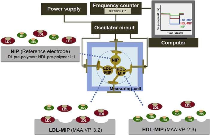
Mass sensitive sensors based on MIPs have also been developed rapidly. There are three kinds of molecularly imprinted quality sensors: acoustic surface sensors, quartz crystal microbalance (QCM) sensors, and cantilever beam chemical sensors. At present, molecular imprinting technology is often used in conjunction with quartz QCM sensors. The QCM sensor consists of a thin quartz crystal with electrodes on both sides, and its surface can be loaded with MIPs to adsorb the target molecule to increase the crystal mass. This influences the variation of crystal resonance frequency to provide real-time measurement. Chunta et al. [71] designed a three-electrode MIPs-QCM sensor array for the detection of two atherosclerosis biomarkers (low-density lipoprotein (LDL) and high-density lipoprotein (HDL)). The array consists of two MIPs (LDL-MIP, HDL-MIP) and a reference non-imprinted polymer (NIP). As shown in Fig. 7, two MIPs contain monomeric mixtures of methacrylic acid (MAA) and n-vinylpyrrolidone in different proportions with selective recognition properties for LDL and HDL. NIP was used as a reference electrode. The sensing signal (frequency as a function of time) is recorded using a frequency counter. The array shows the ability to differentiate lipoproteins, which opens the way for MIPs-QCM multi-sensing arrays.
Precious metal nanomaterials are new materials containing precious metals (Au, Ag, Pt, Pd, Ru, pH, Os, and Ir) in the size range of 0.1–100 nm developed and prepared by nanotechnology [72]. Different sizes of precious metal nanomaterials have different optical properties, including plasmon resonance, surface Raman enhancement, and fluorescence [73]. The plasmon resonance and luminescence properties of precious metal nanomaterials are highly dependent on the surface state, the environment, and the structural properties of the surface-modified molecules. Thus, by simply adjusting the surface chemical state, complemented by specific molecular modifications, the light absorption and emission of precious metal nanomaterials can be purposefully modulated. In this way, new colorimetric and optical sensors based on noble metal nanomaterials have been developed. For example, in fluorescence sensing, Zhang et al. [74] prepared a fluorescent sensor array of individual gold nanoclusters (AuNC) to detect heavy metal ions (HMIs). Fluorescent AuNC was prepared by the reduction of the gold precursor HAuCl4 by 2-mercapto-1-methylimidazole (MMI) in the presence of polyvinylpyrrolidone (PVP). As shown in Fig. S4 (Supporting information), the fluorescence emission of the probe (PVP/MMI-AuNC) was regulated by pH and exhibited yellow and red fluorescence at pH 12.0 and 6.0, respectively. Thus, dual-channel sensing with a single probe can be achieved by just altering the pH of the medium.
The excellent localized surface plasmon resonance properties of precious metal nanoparticles make them advantageous for application in colorimetric detection. Most detection principles are based on color or spectral changes caused by the aggregation of precious metal nanoparticles. This strategy has the advantages of short timescales and ease of operation [75]. For example, Fahimi-Kashanide et al. [76] developed a colorimetric sensor array based on the aggregation of citrate-coated gold nanoparticles (AuNPs) capable of the detection and identification of five organophosphorus pesticides in the concentration range of 120–400 ng/mL. However, the aggregation reaction is susceptible to environmental factors, which limits the application of precious metal nanomaterials. One way to avoid this limitation is to change the shape of the nanomaterials. Triangular-shaped silver nanoparticles (TSNPs) are anisotropic plasmonic nanoparticles with highly modulated surface plasmon resonance absorption. Therefore, TSNPs are a good colorimetric sensing element. Keshavarzi et al. [77] designed a time-dimensionally responsive TSNPs colorimetric sensor array (Fig. 8) to distinguish halide ions. Halide anions (I−, Br−, and Cl−) can etch the cusps of TSNPs, producing hemispherical TSNPs to varying degrees. The structural change of TSNPs to spherical nanoparticles results in a significant blue shift in the plasmon resonance band of TSNPs. The color change of TSNPs etched at different reaction time intervals was recorded to produce a response pattern for each ion, which successfully distinguished the three halide ions. The array also successfully classified binary and ternary mixtures of three halogen ions with 100% specificity and 100% sensitivity. In addition, the array was able to accurately determine the three halide ions in real water samples, demonstrating its potential for application in real samples. These results show that the chrono-type colorimetric sensor is a simple and effective sensor, which will provide a novel idea for the development of chemical sensor arrays with a single sensing element.

In addition to nanomaterials composed of a single precious metal element, they also include alloys and core–shell structured precious metal nanomaterials. Nanomaterials composed of two or more metals are called alloys. The alloys usually exhibit better properties than single-component noble metal nanomaterials through regulation. Precious metal core–shell nanomaterials (M@N) are a class of nanomaterials with a core and a shell composed of two metal elements. Core–shell structured noble metal nanomaterials have tunable optical properties, as well as superior chemical and thermal stability. For example, Qiang et al. [78] synthesized a colorimetric sensor array based on Cu@AuNPs modified by non-specific DNA strands (15A, 15C and 15T). Different DNAs and detection proteins interact differently with each other, which results in different steric hindrances. It also affects the distance of iodide ions to the surface of Cu@AuNPs, leading to significant changes in absorption response and color. Fig. S5 (Supporting information) shows its sensing mechanism. The sensor array successfully distinguished between eight proteins at a concentration of 20 nmol/L in pure water buffer and real serum samples with 100% accuracy.
Chemical sensor arrays are multidimensional. The increase in dimension will multiply the amount of data, which brings difficulties to function approximation, model fitting, information extraction, and calculation. These data usually cannot be processed using simple methods such as linear regression. Generally, multidimensional data in array analysis is processed by multivariate statistical analysis based on pattern recognition. We will focus here on three of the most common pattern recognition methods: hierarchical cluster analysis (HCA), principal component analysis (PCA), and linear discriminant analysis (LDA). The three pattern recognition methods are specifically described in Text S1 (Supporting information).
Food quality and safety are related to human life quality and health. The supervision of the food industry is becoming more and more important. The chemical composition of food is complex, and there are some highly similar substances. For a long time, it has been a difficult problem to develop a technology to detect food contaminants, raw material sources, freshness, adulteration, and other aspects. The chemical sensor array has been regarded as a good solution to these problems. In addition, the chemical sensor array can predict the storage conditions and storage time of food and evaluate its quality. With the development of chemical sensing and chemometrics, electronic nose and electronic tongue have been widely used in meat products, wine, aquatic products, vegetables, fruits, food, condiments, and other fields [79–82]. Table S1 (Supporting information) lists several applications of chemical sensor arrays in food monitoring.
Food traceability is one of the keys to ensuring food safety. The research and development of food traceability technology have been widely concerned. Array sensing technology is widely used in food traceability research due to its good specificity and comprehensiveness in acquiring information. Wang et al. [83] constructed a voltammetry electronic tongue based on three nano-composite modified electrodes and applied it in the identification of rice wines from different regions. In addition, the sensor array is very effective in distinguishing the geographical origin and variety of honey [84,85], wine [86], tea [87,88], and grain [89,90] using multivariate analysis techniques.
Freshness is an important indicator of the quality of food. People often judge the quality of food comprehensively through smell, vision, and taste. However, sensory evaluation is too subjective and easily affected by external environmental factors, so the accuracy and repeatability of results are not credible enough. Sensor array technology can provide an objective evaluation. In the storage process of meat and eggs, proteins, fats, and carbohydrates contained in them are easily decomposed by microorganisms and enzymes, resulting in freshness reduction, spoilage, and even the generation of harmful substances [91]. The volatile components changed significantly. Deng et al. [92] designed an electronic nose to assess the shelf life of eggs. The electronic nose consists of four QCMs modified by different functional materials as sensing units, which can enhance cross-sensitivity. The electronic nose can obtain information about egg samples by measuring the change in resonance frequency caused by adsorbed egg volatile gases (ammonia, amine, sulfide, aldehyde, ketone, alcohol, and other compounds). QCM technology provides a nondestructive and sensitive method for evaluating the quality of eggs with different shelf lives and provides an alternative strategy for evaluating the freshness of eggs. At present, in addition to the detection of volatile gases, another commonly used indicator to evaluate the freshness of food is the content of bioamines (BAs). BAs levels are very low in fresh foods. However, BAs concentrations may rise significantly during certain stages of storage and processing of protein-rich meat and fish. Orouji et al. [93] designed a multicolor sensor array consisting of two types of AuNRs and AuNSs and used it to detect biogenic amines (BAs) in meat and fish. In the presence of BAs, silver ions metalize AuNRs and AuNSs to form Au@Ag core–shell nanoparticles. The changes in AuNSs and AuNRs resulted in a blue shift of plasma bands and significant color changes in the solution, which verified the potential of the array in the field measurement.
At present, there are a few illegal traders for profiteering, adulteration in food, even toxic and harmful substances, damaging consumer rights and interests, and harming people's health. Therefore, people pay attention to the problems of food authenticity assessment and food adulteration identification methods. For example, Zhang et al. [94] designed a new fluorescence sensor array composed of perylene probes induced by different cationic polymers that can distinguish five main milk proteins at nanomolar concentration and be successfully used for qualitative identification and quantitative detection of adulterated milk. Pan et al. [95] designed a colorimetric sensor array for the identification of ginseng. The sensor array used phenylboronic acid as an acceptor to analyze the composition of ginseng diols to identify ginseng. The array successfully classified 40 samples from three types of ginsengs based on both pH change and indicators displacement assay. The sensor array can be used for the rapid identification of ginseng grades and species.
The development of rapid, effective, and accurate medical diagnosis methods has been the focus of medical research in the past decades. A chemical sensor array has become a promising disease diagnosis method with its advantages of non-invasiveness and high diagnostic accuracy [96]. Chemical sensor arrays can associate specific biomarkers, such as proteins, amino acids, and biological small molecules, with the patient's disease and detect physiological changes by measuring exhaled breath, blood, serum, urine, and other body fluids (sweat, tears, and saliva) [97]. At present, chemical sensor arrays have made some important achievements in medical analysis. Table S2 (Supporting information) lists several applications of chemical sensor arrays in medical diagnosis.
The electronic tongue can detect all kinds of body fluids, and the test results of body fluids can be linked to pathological changes in the body [98–101]. Electronic tongues are already being used to diagnose diseases as diverse as cancer, neurodegenerative diseases such as Alzheimer's disease (AD) and Parkinson's disease (PD), and metabolic diseases such as diabetes. Neurodegenerative diseases are accompanied by damage to the neural network, and these pathological changes are irreversible. Therefore, it is important to use certain tools for the early diagnosis of these diseases. Li et al. [102] established a fluorescence sensor array with CDs doped with two different oxidants (ammonium persulfate and hydrogen peroxide) as sensing elements. The array can recognize and distinguish four typical amyloid-β1-42, α-synuclein, islet amyloid polypeptide, insulin, and two common serum proteins (bovine serum albumin and immunoglobulin) with an accuracy of 100%. Amyloid-β1-42 and α-synuclein are considered markers of AD and PD, respectively, so this array has proven its potential value in the diagnosis of neurodegenerative diseases. Kim et al. [103] constructed a sensor array based on densely packed carbon nanotubes specifically for AD. This array was successfully used in clinical diagnosis to distinguish AD patients from healthy controls with an average sensitivity of 90.0%, selectivity of 90.0%, and average accuracy of 88.6%.
Through the analysis of volatile organic compounds (VOCs) in exhaled breath, the gas exchange between air and blood can be detected to judge various physiological characteristics. Various electronic noses have been used in the analysis of exhaled air and have made remarkable progress in the early diagnosis of lung cancer and various respiratory diseases. In addition to pulmonary and respiratory diseases, various other diseases also have unique exhaled characteristics. Li et al. [104] designed a QCM-based virtual sensor array with multiple harmonics and used it for the diagnosis of diabetic ketosis. The multi-dimensional response of the array was analyzed using machine learning algorithms such as PCA, and artificial neural network (ANN), which were able to accurately identify different VOCs and mixtures and predict acetone concentration in a complex background with 90.6% accuracy. The array analysis verified the ability to identify exhaled air in "diabetic ketosis" with 95% accuracy.
With the rapid development of globalization, industrialization, and urbanization, excessive emissions of pollutants (heavy metal ions, pesticides, antibiotics, explosives, VOCs, toxic gases, toxic small molecules, radioactive ions, etc.) have seriously affected the ecological balance and indirectly threatened biological health [105,106]. These factors may lead to fatal and irreversible damage if the pollutant emissions are not controlled. Therefore, environmental pollutant detection is an indispensable fundamental part and necessary condition for environmental pollution management. Environmental systems are highly complex. Environmental contaminant detection has to deal with more changing environments and more complex and unpredictable components than detection in medical and food fields. So traditional techniques for detecting single factors have many limitations for environmental monitoring, while chemical sensor arrays can effectively identify environmental data and can describe the interrelationships between various environmental contaminants. Table S3 (Supporting information) briefly introduces several applications of chemical sensor arrays in environmental monitoring. Currently, exploring novel pollutant sensor arrays with environmental resistance and versatility is a daunting task.
Contaminants with high solubility and mobility can easily diffuse into water and soil. For example, heavy metal ions and organic matter are discharged into the environment, entering the human body through the food chain or direct contact, and accumulate in the body causing cancer, neuronal degeneration, blood, and urinary system damage. Xu et al. [107] constructed a multi-emission fluorescent sensor array based on CDs and a novel lanthanide complex, which successfully identified seven heavy metal ions with detection limits as low as 0.05 µmol/L. The array was also effective in identifying metal ions in real samples (lake water and soil) with an accuracy of 93.3% and 100%, respectively.
Polluting gases such as NOx, SO2, and CO2 can cause serious environmental problems such as photochemical smog, acid rain, and ozone layer depletion. Long-term exposure to these gases can also lead to health risks, such as chest congestion, asthma, or headaches. Therefore, it is important to monitor airborne pollutants. Moon et al. [108] designed a fully integrated chemoresistance sensor array for the detection of NOx in roadways. The array can be used to assess the spatial and temporal variation of NOx and can be an important tool for air quality detection. Li et al. [109] designed a colorimetric sensor array of gold nanoparticles based on a needle-printing method. The array can quantitatively identify 15 photochemical pollutants related to air quality monitoring and predict the 24-hour air quality of indoor office environment.
In this review, we provide a comprehensive review of the development and status of chemical sensor arrays. Array sensing technology has become an important tool in analytical testing nowadays by its high sensitivity, good accuracy, and low detection limits. Chemical sensing has witnessed the development of a diversity of sensing materials. Arrays based on various types of sensing materials have proven their usefulness in distinguishing similar analytes and mixtures. The use of array technology to detect complex analyses inevitably generates large data sets, and chemometrics provides a viable avenue for data processing and statistical analysis. With the development of chemical sensor array technology, complex compounds, biomolecules, and actual samples can be accurately identified. Sensor array technology will lay a solid foundation for the progress of human civilization and sustainable development.
Although chemical sensor arrays have been applied in many fields, many opportunities and challenges remain.
Chemical sensor arrays mainly provide a composite response to mixtures and are more suitable for the overall qualitative analysis of mixtures. The main limitation of sensor arrays is that they are difficult to provide a complete quantitative analysis of each component in a mixture. Quantitative analysis is based on finding the linear relationships hidden in the database. However, the limitations of current data analysis capabilities have affected the development of array sensing technology in this field. Currently, most research is focused on specific algorithms for specific arrays and experimental data that do not make full use of the feature information contained in the sensed signal. In order to further improve the performance of existing algorithms, a systematic comparison of various data processing methods based on the same data set is necessary. Drawing on results from other research areas and introducing new data processing methods into this research area. We expect revolutionary approaches to data processing for sensor arrays.
Arrays based on multiple sensing elements are still the mainstream of design. The design, optimization, and implementation of multi-sensor elements are time-consuming and expensive. Therefore, one direction of development for arrays is novel designs based on multiple signals to maximize the sensing characteristics of sensing elements. Opposing reaction modes (such as on and off reactions) or environmental factors (such as temperature, solvent, and pH) can be incorporated into the sensing system to simplify the preparation of sensing elements. Furthermore, exploring novel sensing signals for building arrays, such as combining ultra-sensitive surface-enhanced Raman scattering signals with array-based strategies, could be seen as a future direction.
The detection results of the array are dependent on the database constructed. This requires the selection of specific combinations of components to simulate most possibilities. As a result, any small variation or disturbance to the species can lead to large deviations from the simulated sample. This leads to the fact that despite the achievements of array sensing in controlled laboratory environments, its application in realistic, complex scenarios still has some difficulties to overcome. The system must be able to match an unknown sample to the response of a set of training analyses and effectively eliminate possible disturbances. This requires exploring new probes that are resistant to interference and suitable for multi-component sensing, and ensuring that the responses of analytes and arrays are reproducible and stable over long periods. In addition, the combination of digital imaging devices (such as scanners and cameras) with paper or plain plastic film-based sensor arrays will facilitate rapid detection and in situ analysis of arrays.
The authors declare that they have no known competing financial interests or personal relationships that could have appeared to influence the work reported in this paper.
This project was funded by Natural Science Foundation of Heilongjiang Province (No. LH2022B004), Fundamental Research Funds for the Central Universities (No. 2572022DJ01), 111 Project (No. B20088), Heilongjiang Touyan Innovation Team Program (Tree Genetics and Breeding Innovation Team).
Supplementary material associated with this article can be found, in the online version, at doi:
D.A.C. Sedgwick, P.T.D. James, ChemistryOpen 7 (2018) 215–216. doi: 10.1002/open.201800027
I.L.J. Celine, R.G. Ana, C.F. Ana, et al., TrAC: Trend. Anal. Chem. 91 (2017) 53–66. doi: 10.1016/j.trac.2017.04.003
J.P. Anzenbacher, P. Lubal, P. Buček, et al., Chem. Soc. Rev. 39 (2010) 3954–3979. doi: 10.1039/b926220m
J.R. Askim, M. Mahmoudi, K.S. Suslick, Chem. Soc. Rev. 42 (2013) 8649–8682. doi: 10.1039/c3cs60179j
K.B.R. Teodoro, F.L. Migliorini, M.H.M. Facure, et al., Carbohydr. Polym. 207 (2019) 747–754. doi: 10.1016/j.carbpol.2018.12.022
M. Li, X. Wang, Y. Zhu, et al., Chin. Chem. Lett. 34 (2023) 107299. doi: 10.1016/j.cclet.2022.03.022
X.R. Liu, M.P. Qian, H.L. Qi, et al., J. Anal. Test. 4 (2020) 114–121. doi: 10.1007/s41664-020-00130-3
W. Wojciech, D. Tomasz, G. Jacek, et al., Curr. Med. Chem. 26 (2017) 197–215.
Y.X. Li, H.Y. Qin, C. Hu, et al., J. Anal. Test. 6 (2022) 431–440. doi: 10.1007/s41664-022-00235-x
Z. Zhang, Y. Lou, C. Guo, et al., Trends Food Sci. Technol. 118 (2021) 569–588. doi: 10.1016/j.tifs.2021.10.024
K. Sanggon, B. Jacob, A.B. Faraj, et al., Front. Chem. 9 (2021) 629329. doi: 10.3389/fchem.2021.629329
K.J. Albert, N.S. Lewis, C.L. Schauer, et al., Chem. Rev. 100 (2001) 2595–2626.
A. Hierlemann, R. Gutierrez-Osuna, Chem. Rev. 108 (2008) 563–613. doi: 10.1021/cr068116m
R.A. Jon, M. Morteza, S.S. Kenneth, Chem. Soc. Rev. 42 (2013) 8649–8682. doi: 10.1039/c3cs60179j
P. Anzenbacher, P. Lubal, P. Bucek, et al., Chem. Soc. Rev. 39 (2010) 3954–3979. doi: 10.1039/b926220m
C. Zhu, R.E. Gerald, J. Huang, IEEE Sens. J. 21 (2021) 19647–19661. doi: 10.1109/JSEN.2021.3094092
D. Zhao, Y. Cui, Y. Yang, et al., CrystEngComm 18 (2016) 3746–3759. doi: 10.1039/C6CE00545D
H. Yuan, N. Li, W. Fan, et al., Adv. Sci. 9 (2022) 2104374. doi: 10.1002/advs.202104374
M. Huangfu, M. Wang, C. Lin, et al., Dalton Trans. 50 (2021) 3429–3449. doi: 10.1039/D0DT04276E
P. Tong, J. Liang, X. Jiang, et al., Crit. Rev. Anal. Chem. 50 (2020) 376–392. doi: 10.1080/10408347.2019.1642732
S. Liu, C. Lai, X. Liu, et al., Coord. Chem. Rev. 424 (2020) 213520. doi: 10.1016/j.ccr.2020.213520
Q. Wang, H. Zhangsun, Y. Zhao, et al., J. Hazard. Mater. 411 (2021) 125122. doi: 10.1016/j.jhazmat.2021.125122
Z. Zhou, S. Mukherjee, S. Hou, et al., Angew. Chem. Int. Ed. 60 (2021) 20551–20557. doi: 10.1002/anie.202107860
B. Chen, Z. Yang, X. Qu, et al., ACS Appl. Mater. Interfaces 13 (2021) 47706–47716. doi: 10.1021/acsami.1c15528
Q. Wang, Q. Liu, X.M. Du, et al., J. Mater. Chem. C 8 (2020) 1433–1439. doi: 10.1039/C9TC05180E
X. Lin, E. Shuang, X. Chen, J. Colloid Interface Sci. 602 (2021) 513–519. doi: 10.1016/j.jcis.2021.06.025
Z.J. Li, Q.F. Xia, Rev. Anal. Chem. 31 (2012) 57–81.
D.G. Papageorgiou, I.A. Kinloch, R.J. Young, Prog. Mater. Sci. 90 (2017) 75–127. doi: 10.1016/j.pmatsci.2017.07.004
M.K. Rabchinskii, V.V. Sysoev, A.S. Varezhnikov, et al., ACS Appl. Mater. Interfaces 15 (2023) 28370–28386. doi: 10.1021/acsami.3c02833
K.S. Novoselov, A.K. Geim, S.V. Morozov, et al., Science 306 (2004) 666–669. doi: 10.1126/science.1102896
K. Xia, W. Wu, M. Zhu, et al., Sci. Bull. 65 (2020) 343–349. doi: 10.1016/j.scib.2019.12.015
L. Cai, W. He, X. Xue, et al., Natl. Sci. Rev. 8 (2021) nwaa298. doi: 10.1093/nsr/nwaa298
M. Shekhirev, A. Lipatov, A. Torres, et al., ACS Appl. Mater. Interfaces 12 (2020) 7392–7402. doi: 10.1021/acsami.9b13946
A.D. da Silva, W.J. Paschoalino, J.P.V. Damasceno, et al., ChemElectroChem 7 (2020) 4508–4525. doi: 10.1002/celc.202001168
H. Huang, H. Shi, P. Das, et al., Adv. Funct. Mater. 30 (2020) 1909035. doi: 10.1002/adfm.201909035
R. Tarcan, O. Todor-Boer, I. Petrovai, et al., J. Mater. Chem. C 8 (2020) 1198–1224. doi: 10.1039/C9TC04916A
H. Zhang, H. Zhang, A. Aldalbahi, et al., Biosens. Bioelectron. 89 (2017) 96–106. doi: 10.1016/j.bios.2016.07.030
H. Wang, L. Zhou, J. Qin, et al., Anal. Chem. 94 (2022) 10291–10298. doi: 10.1021/acs.analchem.2c02236
K. Xu, C. Fu, Z. Gao, et al., Instrum. Sci. Technol. 46 (2018) 115–145. doi: 10.1080/10739149.2017.1340896
C. Wang, Y. Wang, Z. Yang, et al., Ceram. Int. 47 (2021) 16367–16384. doi: 10.1016/j.ceramint.2021.02.144
Q. Chen, Z. Chen, D. Liu, et al., ACS Appl. Mater. Interfaces 12 (2020) 17713–17724. doi: 10.1021/acsami.0c00720
F.P. García de Arquer, D.V. Talapin, V.I. Klimov, et al., Science 373 (2021) eaaz8541. doi: 10.1126/science.aaz8541
A. Khojastehnezhad, F. Taghavi, E. Yaghoobi, et al., Talanta 235 (2021) 122753. doi: 10.1016/j.talanta.2021.122753
E. Aznar-Gadea, P.J. Rodriguez-Canto, S.A. Sánchez, et al., ACS Appl. Nano Mater. 5 (2022) 6717–6725. doi: 10.1021/acsanm.2c00743
Z. Jiao, P. Zhang, H. Chen, et al., Sens. Actuators B: Chem. 295 (2019) 110–116. doi: 10.1016/j.snb.2019.05.059
P. Wu, L.N. Miao, H.F. Wang, et al., Angew. Chem. Int. Ed. 50 (2011) 8118–8121. doi: 10.1002/anie.201101882
B.K. Walther, C.Z. Dinu, D.M. Guldi, et al., Mater. Today 39 (2020) 23–46. doi: 10.1016/j.mattod.2020.04.008
P. Devi, P. Rajput, A. Thakur, et al., TrAC: Trend. Anal. Chem. 114 (2019) 171–195. doi: 10.1016/j.trac.2019.03.003
Z. Xu, Z. Wang, M. Liu, et al., Spectrochim. Acta A 232 (2020) 118147. . doi: 10.1016/j.saa.2020.118147
L. Chen, X. Tian, Y. Li, et al., J. Hazard. Mater. 424 (2022) 127694. doi: 10.1016/j.jhazmat.2021.127694
W. Du, X. Xu, H. Hao, et al., Sci. China Chem. 58 (2015) 863–870. doi: 10.1007/s11426-014-5256-y
L. Fu, T. Liu, F. Yang, et al., J. Photochem. Photobiol. A 424 (2022) 113638. doi: 10.1016/j.jphotochem.2021.113638
J.F.C. Loo, Y.H. Chien, F. Yin, et al., Coord. Chem. Rev. 400 (2019) 213042. doi: 10.1016/j.ccr.2019.213042
M.K. Tsang, G. Bai, J. Hao, Chem. Soc. Rev. 44 (2015) 1585–1607. doi: 10.1039/C4CS00171K
M. Safdar, A. Ghazy, M. Lastusaari, et al., J. Mater. Chem. C 8 (2020) 6946–6965. doi: 10.1039/D0TC01216E
L.D. Sun, H. Dong, P.Z. Zhang, et al., Annu. Rev. Phys. Chem. 66 (2015) 619–642. doi: 10.1146/annurev-physchem-040214-121344
A. Jouyban, E. Rahimpour, Talanta 220 (2020) 121383. doi: 10.1016/j.talanta.2020.121383
G. Lin, D. Jin, ACS Sens. 6 (2021) 4272–4282. doi: 10.1021/acssensors.1c02101
S. Abbasi-Moayed, A. Bigdeli, M.R. Hormozi-Nezhad, ACS Appl. Mater. Interfaces 12 (2020) 52976–52982. doi: 10.1021/acsami.0c17200
T. Li, X. Zhu, X. Hai, et al., ACS Sens. 8 (2023) 994–1016. doi: 10.1021/acssensors.2c02596
Q.Q. Wang, R. Hu, Z.Q. Fang, et al., Chin. Chem. Lett. 33 (2022) 3782–3786. doi: 10.1016/j.cclet.2021.11.012
Q. Yan, X.Y. Ding, Z.H. Chen, et al., Anal. Chem. 90 (2018) 10536–10542. doi: 10.1021/acs.analchem.8b02603
L. Chen, X. Wang, W. Lu, et al., Chem. Soc. Rev. 45 (2016) 2137–2211. doi: 10.1039/C6CS00061D
O.S. Ahmad, T.S. Bedwell, C. Esen, et al., Trends Biotechnol. 37 (2019) 294–309. doi: 10.1016/j.tibtech.2018.08.009
J.J. BelBruno, Chem. Rev. 119 (2019) 94–119. doi: 10.1021/acs.chemrev.8b00171
K.D. Shimizu, C.J. Stephenson, Curr. Opin. Chem. Biol. 14 (2010) 743–750. doi: 10.1016/j.cbpa.2010.07.007
A. Herrera-Chacón, X. Cetó, M. del Valle, Anal. Bioanal. Chem. 413 (2021) 6117–6140. doi: 10.1007/s00216-021-03313-8
M. Wang, X. Cetó, M. del Valle, Biosens. Bioelectron. 198 (2022) 113807. doi: 10.1016/j.bios.2021.113807
Q. Sun, L. Fu, C. Yin, et al., Sens. Actuators B: Chem. 373 (2022) 132716. doi: 10.1016/j.snb.2022.132716
Z.Z. Lin, L. Li, G.Y. Fu, et al., Anal. Chim. Acta 1101 (2020) 32–40. doi: 10.1016/j.aca.2019.12.032
S. Chunta, R. Suedee, S. Singsanan, et al., Sens. Actuators B: Chem. 298 (2019) 126828. doi: 10.1016/j.snb.2019.126828
J. Sun, Y. Lu, L. He, et al., TrAC: Trend. Anal. Chem. 122 (2020) 115754. doi: 10.1016/j.trac.2019.115754
Z. Yuan, C.C. Hu, H.T. Chang, et al., Analyst 141 (2016) 1611–1626. doi: 10.1039/C5AN02651B
X.P. Zhang, K.Y. Huang, S.B. He, et al., J. Hazard. Mater. 405 (2021) 124259. doi: 10.1016/j.jhazmat.2020.124259
A. Thomas, U.K.G. Kumer, Spectrochim. Acta Part A 188 (2018) 113–119. doi: 10.1016/j.saa.2017.06.040
N. Fahimi-Kashani, M.R. Hormozi-Nezhad, Anal. Chem. 88 (2016) 8099–8106. doi: 10.1021/acs.analchem.6b01616
P. Keshavarzi, S. Abbasi-Moayed, M. Khodabakhsh, et al., Talanta 259 (2023) 124528. doi: 10.1016/j.talanta.2023.124528
H. Qiang, X. Wei, Q. Liu, et al., ACS Sustain. Chem. Eng. 6 (2018) 15720–15726. doi: 10.1021/acssuschemeng.8b04235
M. Peris, L. Escuder-Gilabert, Trends Food Sci. Technol. 58 (2016) 40–54. doi: 10.1016/j.tifs.2016.10.014
A. Villalonga, A. Sánchez, B. Mayol, et al., Curr. Opin. Food Sci. 43 (2022) 18–26. doi: 10.1016/j.cofs.2021.09.006
C. Dincer, R. Bruch, E. Costa-Rama, et al., Adv. Mater. 31 (2019) 1806739. doi: 10.1002/adma.201806739
T. Yang, T.V. Duncan, Nat. Nanotechnol. 16 (2021) 251–265. doi: 10.1038/s41565-021-00867-7
J. Wang, L. Zhu, W. Zhang, et al., Anal. Chim. Acta 1050 (2019) 60–70. doi: 10.1016/j.aca.2018.11.016
L. Sobrino-Gregorio, F. Tanleque-Alberto, R. Bataller, et al., J. Sci. Food Agric. 100 (2020) 212–217. doi: 10.1002/jsfa.10022
N. El Alami El Hassani, K. Tahri, E. Llobet, et al., Food Chem. 243 (2018) 36–42. doi: 10.1016/j.foodchem.2017.09.067
M.J. Moehring, P.D. Harrington, Crit. Rev. Anal. Chem. 52 (2021) 1–12.
M. Shuai, C. Peng, H. Niu, et al., Food Chem. 374 (2022) 131713. doi: 10.1016/j.foodchem.2021.131713
L. Cheng, Y. Wang, J. Zhang, et al., LWT-Food Sci. Technol. 150 (2021) 111875. doi: 10.1016/j.lwt.2021.111875
M. Arslan, M. Zareef, H.E. Tahir, et al., Food Control 132 (2022) 108513. doi: 10.1016/j.foodcont.2021.108513
H.Y. Liu, S.A. Wadood, Y. Xia, et al., Crit. Rev. Food Sci. Nutr. 63 (2021) 1–24.
H. Singh, G. Singh, N. Kaur, et al., Biosens. Bioelectron. 196 (2022) 113687. doi: 10.1016/j.bios.2021.113687
F. Deng, W. Chen, J. Wang, et al., Sens. Actuators B: Chem. 265 (2018) 394–402. doi: 10.1016/j.snb.2018.03.010
A. Orouji, F. Ghasemi, A. Bigdeli, et al., ACS Appl. Mater. Interfaces 13 (2021) 20865–20874. doi: 10.1021/acsami.1c03183
L. Zhang, J. Hou, H. Zhou, et al., Food Chem. 343 (2021) 128492. doi: 10.1016/j.foodchem.2020.128492
Y. Pan, M. Yang, M. Zhang, et al., Sens. Actuators B: Chem. 294 (2019) 48–54. doi: 10.1016/j.snb.2019.05.029
J.E. Fitzgerald, E.T.H. Bui, N.M. Simon, et al., Trends Biotechnol. 35 (2017) 33–42. doi: 10.1016/j.tibtech.2016.08.005
M. Tabata, Y. Miyahara, Sens. Actuators B: Chem. 352 (2022) 131033. doi: 10.1016/j.snb.2021.131033
V. Mani, T. Beduk, W. Khushaim, et al., TrAC: Trend. Anal. Chem. 135 (2021) 116164. doi: 10.1016/j.trac.2020.116164
S. Veeralingam, S. Badhulika, Nanoscale 12 (2020) 15336–15347. doi: 10.1039/D0NR03427D
M. Colombo, S.J. McGurnaghan, L.A.K. Blackbourn, et al., Diabetologia 63 (2020) 788–798. doi: 10.1007/s00125-019-05081-8
K. Vanarsa, S. Soomro, T. Zhang, et al., Ann. Rheum. Dis. 79 (2020) 1349. doi: 10.1136/annrheumdis-2019-216312
Q.Y. Li, L. Ma, L. Li, et al., Chem. Eng. J. 430 (2022) 132696. doi: 10.1016/j.cej.2021.132696
K. Kim, M.J. Kim, D.W. Kim, et al., Nat. Commun. 11 (2020) 119. doi: 10.1038/s41467-019-13901-z
D. Li, Z. Xie, M. Qu, et al., ACS Appl. Mater. Interfaces 13 (2021) 47043–47051. doi: 10.1021/acsami.1c13046
G.L. Yang, X.L. Jiang, H. Xu, et al., Small 17 (2021) 2005327. doi: 10.1002/smll.202005327
S. Tajik, H. Beitollahi, F. Garkani Nejad, et al., J. Mater. Chem. A 9 (2021) 8195–8220. doi: 10.1039/D0TA08344E
Z. Xu, J. Chen, Y. Liu, et al., Chem. Eng. J. 441 (2022) 135690. doi: 10.1016/j.cej.2022.135690
H.G. Moon, Y. Jung, B. Shin, et al., Adv. Sci. 7 (2020) 2002014. doi: 10.1002/advs.202002014
Z. Li, R. Zhang, X. Lu, et al., Anal. Chem. 93 (2021) 13990–13997. doi: 10.1021/acs.analchem.1c03457

Figure 1 Schematic diagram of sensing mechanism of the three-dimensional spectral array. Reprinted with permission [26]. Copyright 2021, Elsevier.

Figure 2 Schematic diagram of graphene nanoribbon and Pt electrode forming an array of 38 sensing units. Reprinted with permission [33]. Copyright 2020, American Chemical Society.

Figure 3 Schematic diagram of PEI-GO composite single-component multi-channel sensor array for bacteria detection. Reprinted with permission [38]. Copyright 2022, American Chemical Society.

Figure 4 Schematic diagram of the synthesis of CdSe-PCL sensor array. Reprinted with permission [44]. Copyright 2022, American Chemical Society.

Figure 5 (a) The design and application of pH-regulated PAA/UCNPs-Cu2+ fluorescent sensor arrays. Reprinted with permission [61]. Copyright 2022, Elsevier. (b) The pH-regulated dual-mode optical sensor array for phosphoric acid compound recognition. Reprinted with permission [62]. Copyright 2018, American Chemical Society.

Figure 6 Schematic diagram of synthesis and recognition of molecularly imprinted polymer array. Reprinted with permission [69]. Copyright 2022, Elsevier.

Figure 7 Schematic of a three-electrode MIPs-QCM sensor array for lipoprotein detection. Reprinted with permission [71]. Copyright 2019, Elsevier.

 扫一扫看文章

扫一扫关注我们
 DownLoad:
 下载:
下载:

 下载:
下载: