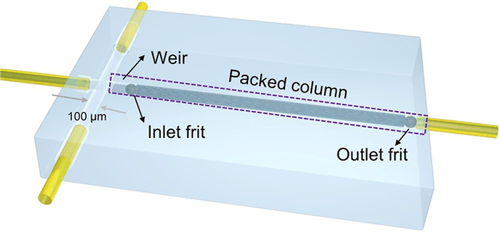
Figure 1.
Schematic diagram of a microchip.
Nano-sized stationary phase packings retained by single-particle frit for microchip liquid chromatography
Wangyan Hu , Ke Li , Xiangnan Dou , Ning Li , Xiayan Wang

Due to the advantages of requiring ultra-small sample volumes and high separation efficiency, there is a growing interest in transferring conventional liquid chromatography (LC) onto miniaturized LC systems [1,2]. Capillary and microchip formats are the two primary formats for miniaturized LC systems [3–6]. Microchip liquid chromatography (microchip-LC) is gaining popularity due to its zero-dead volume connection and ease of integration [7,8]. The chromatographic column is the core component of a microchip-LC, which has a significant impact on separation performance. Although open tubular columns and monolithic columns were initially developed for use in microchip-LC systems due to their ease of preparation, their low phase ratio and susceptibility to swelling have limited their widespread use in the microchip format [9,10]. Packed columns remain the preferred stationary phase for microchip-LC due to their high sample capacity and improved separation efficiency [11,12]. The use of nano-sized particle-packed columns on microchips is anticipated to increase column efficiency and reduce analysis time due to the improved separation performance resulting from a decrease in term A (eddy diffusion) of the van Deemter equation [13,14]. However, there are two major challenges in preparing nano-sized particle-packed columns on a microchip: the difficulty of retaining particles in the microchannels, and the packing technique required to achieve a uniform and dense packing of the nano-sized particles in the microchannels.
Several studies have reported methods for blocking particles in microchip channels using weirs [15,16], porous polymeric frits [17–19], and porous single-particle frits [20,21]. The fabrication of "weir" structures on microchips is challenging because it requires nanoscale manufacturing techniques to create barriers that are smaller than the particle size, in order to block the movement of nanoparticles. While monolithic column frits may have a lower tolerance for organic solvents, porous single-particle silica frits exhibit excellent permeability and rigidity as an alternative option.
There are relatively few reports on the preparation of nano-sized particles packed columns on microfluidic chips. Wirth et al. [22] were the first to prepare a nano-sized particles packed column using self-assembled colloidal silica beads. Reverse phase chromatographic separation of three dyes was successfully achieved using this method. Other researchers have also reported the construction of packed columns on microfluidic chips using nano-sized colloidal silica beads for size separation of biomolecules such as proteins [23,24], amino acids [25], and DNA [26]. These studies have a common feature of utilizing electric drives. However, electric drives have significant shortcomings when applied to chromatographic separations, including the inability to generate stable mobile phase gradients [27] and being easily affected by pH, ionic strength, and organic phase ratio in the mobile phase [28,29]. To our knowledge, the use of pressure-driven nano-sized particle-packed columns for microchip liquid chromatography is rarely reported. Pressure-driven chromatographic separation has higher requirements on the microchip platform, which needs to be made of glass or quartz with good pressure resistance due to the high backpressure generated by packing nano-sized particles [30]. Filling and immobilizing nano-sized packing on glass microchips is currently an issue that needs to be addressed [31,32].
In this work, we constructed a packed column on a glass microchip by filling it with nano-sized C18-functionalized silica particles. A surface tension-based single-particle picking technique was established to insert porous single-particle frit into glass microchannels. Additionally, we developed a slurry filling method that utilizes air pressure to inject nano-sized packings into the microchannel. Pressure-driven microchip-LC based on a packed column filled with nano-sized silica particles was developed and applied for the separation of four polycyclic aromatic hydrocarbons (PAHs). The on-chip packed column with 550 nm size particles showed excellent performance with a plate number of 106/m.
The glass microchip was prepared using wet etching and thermal bonding techniques, as described in our previous research [18]. The glass microchip was designed to include a sample injection channel, mobile phase capillary connecting channel and separation channel (Fig. 1). The sample injection channel was cross-sectional with a diameter of 100 µm. The separation channel was 5 mm in length with a diameter of 100 µm. The sample and mobile phase were introduced to the microchip via a capillary connecting channel with a diameter of 380 µm. The weir and frit were combined to retain packing particles. The single silica particle frit has good mechanical strength and porous structure, which can not only withstand the high pressure during the separation process, but also facilitates good penetration of the mobile phase and sample. However, the frit particle size (~90 µm) is slightly smaller than the inner diameter of the separation channel, which will cause the movement of the column bed under high pressure. The weir can serve to block the inlet frit. The combination of the weir and the porous silica single-particle can firmly retain the slurry of nano-sized packings in the microfluidic channel. A restricted weir (100 µm in length, 50 µm in height) was located 500 µm away from the injection channel and used to fix the single-particle frit.
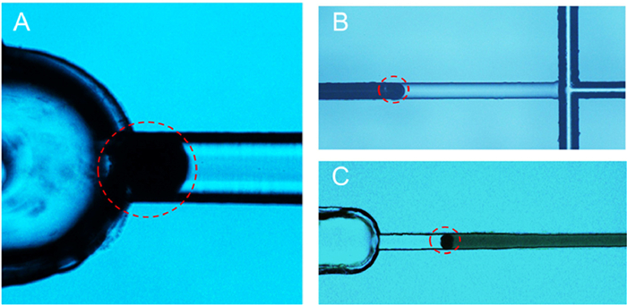
The microchip-LC packed column was prepared by combining the weir and the porous silica single-particle as frit to retain the filling particles. Placing a single particle into a rigid glass microchannel is a challenging task that needs to be addressed. The single-particle frit was fabricated using the single-particle picking technique. Due to the effect of surface tension, a single silica particle is encapsulated within a water droplet. The droplet containing the single particle is then drawn into a capillary via capillary action. The precise positioning of single particles can be achieved using a microscope and a three-dimensional adjustment stage, which facilitates the picking of a single particle using a capillary tube. The picking device is shown in Fig. S1 (Supporting information). A single particle measuring 90 µm was picked up and transferred into a capillary. It was then drawn into the interface between the separation channel and the end capillary channel (Fig. 2A), and pushed towards the entrance of the separation channel, where it functioned as the inlet frit (Fig. 2B). The outlet frit is introduced into the chip channel using the same method and placed at the end of the packed column (Fig. 2C).
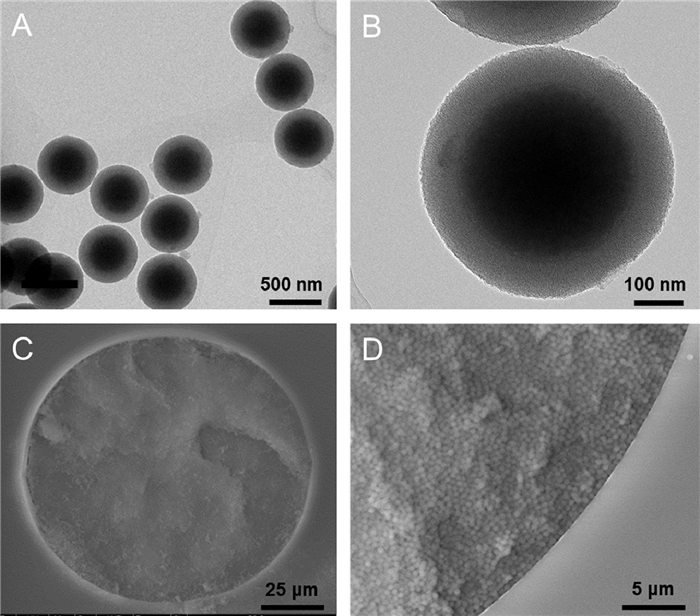
The microcolumn was packed with C18-functionalized silica particles of nano size. As shown in Figs. 3A and B, the TEM image revealed that they possess a spherical structure, with an average particle size of 550 nm. The separation channel was filled by 550 nm of C18-functionalized silica particles by the slurry-packing method. The slurry of 10 mg/mL 550 nm C18-functionalized silica particles was packed into the microchannel by applying nitrogen pressure on the slurry liquid surface (Fig. S2 in Supporting information). Then, the column bed was compacted by the high pressure of 3000 psi from the chromatographic pump. The uniformity and compactness of the packed column significantly affect the performance of chromatographic separation. For example, the high uniformity of the packed column results in a reduction of the A-term (eddy diffusion) in the Van Deemter equation. Hence, the effect of nitrogen pressure ranging from 50 psi to 200 psi on the packing of microcolumn is investigated (Fig. S3 in Supporting information). The results indicate that the nitrogen pressure can have an impact on the uniformity of the packed column bed. At very high pressures, such as 200 psi, the microcolumn bed can crack due to excessive nitrogen pressure. On the other hand, low pressure of 50 psi leads to uneven compaction of the column bed and requires more time for packing. The pressure of 100 psi has been shown to provide a good packing density and uniformity. Additionally, SEM images confirm that the packed column bed is well connected to the inner wall of the microchip channel (Figs. 3C and D).
The chromatographic performance of a microcolumn packed with nano-sized silica particles was evaluated by separating a mixture of four polycyclic aromatic hydrocarbons (PAHs): Naphthalene (NAP), Fluorene (FLU), Anthracene (ANT) and Pyrene (PYR). In reversed-phase chromatography, the organic phase content in the mobile phase is critical for effective separation. Therefore, the impact of the organic phase content on the separation of PAHs using the nano-sized silica particles packed microcolumn was investigated (Fig. 4A). When the acetonitrile content is at 60%, severe peak overlap occurs, resulting in only three peaks being observed. However, baseline separation of the four PAHs has been achieved with a 50% acetonitrile content in the mobile phase. When the acetonitrile content is further reduced to 40%, although all four PAHs can be separated completely, there is an increase in peak broadening due to the low organic phase content. Therefore, an acetonitrile content of 50% was selected as the mobile phase for the eluent.
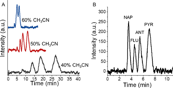
The microchip-LC system was assembled with a nanoflow pump operating at nL/min, a glass microchip, a freeze-thaw valve, and a UV detector. The separation of a mixture of four PAHs was achieved using a packed column filled with C18-functionalized silica particles of 550 nm on a glass microchip. The sample injection was controlled by the freeze-thaw valve through its "off/on" state. Using the optimized chromatographic separation conditions, all four PAHs were completely separated within 8 min using the developed microchip-LC system (Fig. 4B). The separation performance is summarized in Table S1 (Supporting information). The theoretical plate number of four PAHs were NNAP = 111,857 plates/m, NFLU = 145,408 plates/m, NANT = 160,582 plates/m and NPYR = 102,682 plates/m. The resolutions between adjacent components of four PAHs were RNAP-FLU = 1.56, RFLU-ANT = 1.51, and RANT-PYR = 1.60.
The repeatability and stability of the microcolumn packed with nano-sized silica particles were evaluated by separating four PAHs standards. The results, presented in Table 1, show that repeatability was assessed by evaluating intraday and interday precision of retention time. The intraday repeatability ranged from 1.01% to 2.23% (RSD, n = 10) and the interday repeatability was between 1.04% and 1.34% (RSD, over 3 days). Furthermore, the stability of the microcolumn packed with nano-sized silica particles was investigated, and the RSD of the peak asymmetry factor ranged from 1.45% to 2.01%. These findings indicate that the microcolumn packed with nano-sized silica particles has stable performance.
 DownLoad:
CSV
DownLoad:
CSV

|
To evaluate the validity of the microchip-LC method based on the microcolumn packed with nano-sized silica particles, various analytical parameters such as linearity, the limit of detection (LOD), and the limit of quantification (LOQ) were investigated and presented in Table S2 (Supporting information). Linear calibration curves were generated by plotting the peak area versus the concentrations of each PAH standard (Fig. S4 in Supporting information). The calibration curves were linear within the range of 10.0–40.0 µg/mL for naphthalene, 2.0–8.0 µg/mL for fluorene, 0.5–2.0 µg/mL for anthracene, and 4.0–16.0 µg/mL for pyrene. LOD and LOQ were determined as three times and ten times the signal-to-noise ratio, respectively. The repeatability of the method was evaluated by analyzing six replicates of the standards over three days (n = 6 × 3). The RSDs of intraday analysis ranged from 3.55% to 4.48%, while the RSDs of interday analysis ranged from 3.60% to 4.86%.
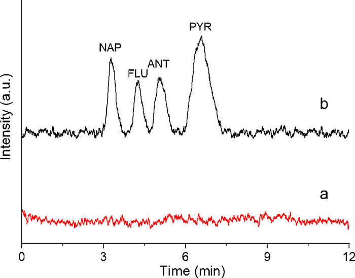
The applicability of the proposed method was verified by applying the microchip-LC to analyze PAHs in practical samples of river water. The chromatogram obtained from the direct analysis of river water showed no PAHs present (Fig. 5). Therefore, spiking experiments were conducted by adding 10 µg/mL of NAP, 2 µg/mL of FLU, 0.5 µg/mL of ANT, and 4 µg/mL of PYR to the river water. The spiked river water was successfully analyzed with a recovery of 98.6%.
In conclusion, this study demonstrates the successful packing of nano-sized silica particles into a miniaturized glass microchip channel using a combination of weir-based blockage and single-particle frit. The results show that this approach leads to excellent separation performance in the resulting packed column. With a short separation column of only 5 mm, rapid separation of PAHs was achieved within just 8 min, with more than 106 plates number. These findings highlight the potential of utilizing nano-sized packings for microchip chromatographic separation. The advantages of integration, fast analysis and low sample consumption of microchip-LC will have broad application prospects in the detection of precious and ultra-small volume biological samples and be more deeply applied in the field of portable detection.
The authors declare that they have no known competing financial interests or personal relationships that could have appeared to influence the work reported in this paper.
This work was supported by the National Natural Science Foundation of China (No. 21936001) and the Beijing Outstanding Young Scientist Program (No. BJJWZYJH01201910005017).
Supplementary material associated with this article can be found, in the online version, at doi:
C. Aydoğan, B. Beltekin, H. Aslan, et al., J. Chromatogr. Open 2 (2022) 100066. doi: 10.1016/j.jcoa.2022.100066
L. Ma, N. Li, J. Wang, et al., Trends Anal. Chem. 160 (2023) 116951. doi: 10.1016/j.trac.2023.116951
J.S. da Silva Burato, J.V. Basolli Borsatto, F.M. Lanças, Talanta 253 (2023) 124106. doi: 10.1016/j.talanta.2022.124106
W.M. Zhang, Z.S. Han, Y.Q. Liang, et al., Chin. Chem. Lett 32 (2021) 2183–2186. doi: 10.1016/j.cclet.2020.12.007
P.C. Chen, W.Z. Zhang, W.R. Chen, et al., Sens. Actuat. B 350 (2022) 130888. doi: 10.1016/j.snb.2021.130888
T.C. Silva, M. Eppink, M. Ottens, J. Chromatogr. A 1681 (2022) 463451. doi: 10.1016/j.chroma.2022.463451
Y. Deng, L. Qiao, N. Gasilova, X.X. Zhang, H.H. Girault, Chin. Chem. Lett. 27 (2016) 85–87. doi: 10.1016/j.cclet.2015.09.017
G.L. Gauthier, R. Grimm, Drug Discov. Today: Technol. 3 (2006) 59–66. doi: 10.1016/j.ddtec.2006.03.013
M.A. Ahmed, B.M.B. Felisilda, J.P. Quirino, Anal. Chim. Acta 1088 (2019) 20–34. doi: 10.1016/j.aca.2019.08.016
Y.J. Ma, M. Li, H. Yu, R.S. Li, Chin. Chem. Lett. 24 (2013) 1067–1069. doi: 10.1016/j.cclet.2013.06.029
K.K. Unger, R. Skudas, M.M. Schulte, J. Chromatogr. A 1184 (2008) 393–415. doi: 10.1016/j.chroma.2007.11.118
C. Bignardi, L. Elviri, A. Penna, M. Careri, A. Mangia, J. Chromatogr. A 1217 (2010) 7579–7585. doi: 10.1016/j.chroma.2010.10.037
G. Desmet, S. Eeltink, Anal. Chem. 85 (2013) 543–556. doi: 10.1021/ac303317c
Y. Wang, F. Ai, S.C. Ng, T.T. Tan, J. Chromatogr. A 1228 (2012) 99–109. doi: 10.1016/j.chroma.2011.08.085
S. Thurmann, A. Dittmar, D. Belder, J. Chromatogr. A 1340 (2014) 59–67. doi: 10.1016/j.chroma.2014.03.009
R.F. Gerhardt, A.J. Peretzki, S.K. Piendl, D. Belder, Anal. Chem. 89 (2017) 13030–13037. doi: 10.1021/acs.analchem.7b04331
S. Thurmann, L. Mauritz, C. Heck, D. Belder, J. Chromatogr. A 1370 (2014) 33–39. doi: 10.1016/j.chroma.2014.10.008
A.J. Peretzki, D. Belder, J. Chromatogr. A 1612 (2020) 460653. doi: 10.1016/j.chroma.2019.460653
S. Thurmann, C. Lotter, J.J. Heiland, B. Chankvetadze, D. Belder, Anal. Chem. 87 (2015) 5568–5576. doi: 10.1021/acs.analchem.5b00210
K. Li, W. Hu, Y. Zhou, et al., Talanta 215 (2020) 120896. doi: 10.1016/j.talanta.2020.120896
B. Zhang, E.T. Bergstrom, D.M. Goodall, P. Myers, Anal. Chem. 79 (2007) 9229–9233. doi: 10.1021/ac0713297
S. Zheng, E. Ross, M.A. Legg, M.J. Wirth, J. Am. Chem. Soc. 128 (2006) 9016–9017. doi: 10.1021/ja062676l
B. Wei, D.S. Malkin, M.J. Wirth, Anal. Chem. 82 (2010) 10216–10221. doi: 10.1021/ac102438w
N. Shaabani, A.B. Jemere, D.J. Harrison, Electrophoresis 37 (2016) 2602–2609. doi: 10.1002/elps.201600224
J. Park, D. Lee, W. Kim, et al., Anal. Chem. 79 (2007) 3214–3219. doi: 10.1021/ac061714g
Y. Zeng, D.J. Harrison, Anal. Chem. 79 (2007) 2289–2295. doi: 10.1021/ac061931h
C.E. Nazario, M.R. Silva, M.S. Franco, F.M. Lancas, J. Chromatogr. A 1421 (2015) 18–37. doi: 10.1016/j.chroma.2015.08.051
K.D. Bartle, R.A. Carney, A. Cavazza, et al., J. Chromatogr. A 892 (2000) 279–290. doi: 10.1016/S0021-9673(00)00150-3
D. Dutta, J.M. Ramsey, Lab Chip 11 (2011) 3081–3088. doi: 10.1039/c1lc20329k
J.P. Kutter, J. Chromatogr. A 1221 (2012) 72–82. doi: 10.1016/j.chroma.2011.10.044
J.P. Grinias, R.T. Kennedy, Trends Anal. Chem. 81 (2016) 110–117. doi: 10.1016/j.trac.2015.08.002
A. Nagy, A. Gaspar, J. Chromatogr. A 1304 (2013) 251–256. doi: 10.1016/j.chroma.2013.06.065

Figure 2 Microscope images of a single-particle frit for the microchip-packed column. (A) A single particle at the interface between the separation channel and the end capillary channel. (B) A single particle as an inlet frit. (C) A single particle as an outlet frit.

Figure 3 Characterization of separation column packed by nano-sized silica nanoparticles. (A) TEM image of C18-functionalized nano-sized silica particle. (B) The enlarged TEM image of silica nanoparticle. (C) SEM image of a cross-section view of separation column on microchip. (D) The enlarged SEM image of the edge of the section of the packed column.

Figure 4 The microchip liquid chromatographic separation of four PAHs. (A) The optimization of chromatographic separation of different organic phase concentrations. Conditions: the mixed standard of 3 µg/mL NAP, 2 µg/mL FLU, 0.5 µg/mL ANT and 4 µg/mL PYR. (B) The chromatogram of a mixture of four PAHs standard under optimum condition. Conditions: the mixed standard of 10 µg/mL NAP, 2 µg/mL FLU, 0.5 µg/mL ANT and 4 µg/mL PYR; Mobile phase: 50% acetonitrile (v/v); Flow rate: 1000 nL/min; UV detection wavelength: 254 nm.

Figure 5 The chromatogram of four PAHs of river water (a) and river water spiked with standard (b) using microchip-LC system. Sample: river water and river water spiked with standard samples of 10 µg/mL NAP, 2 µg/mL FLU, 0.5 µg/mL ANT and 4 µg/mL PYR; Mobile phase: 50% acetonitrile (v/v); Flow rate: 1000 nL/min; UV detection wavelength: 254 nm.
Table 1. Relative standard deviation (RSD) values for the retention time and peak asymmetry factor of the four PAHs.
|
|
 下载: 导出CSV

 扫一扫看文章

扫一扫关注我们

 下载:
下载: