Citation:
Qiu-Yi Duan, Fu-Gen Wu. Exploring three-dimensional biological tissues with new chemical labels[J]. Chinese Chemical Letters,
2023, 34(12): 108625.
doi:
10.1016/j.cclet.2023.108625
Exploring three-dimensional biological tissues with new chemical labels
English
Exploring three-dimensional biological tissues with new chemical labels
Received Date:
27 February 2023 Accepted Date:
29 May 2023 Revised Date:
31 March 2023 Available Online:
15 December 2023
Abstract:
Three-dimensional (3D) histology has exhibited tremendous potential in fundamental research and clinical disease grading, but compatible labeling techniques are still lacking. Recently in Science Advances, Pac et al. report a new histological technique termed 3DNFC, which realizes 3D fluorescence imaging of thick tissues via citrate-based in situ fluorophore formation.
Contemporary disease diagnosis often relies on the histological analysis of surgically excised or biopsied specimens [1]. However, traditional two-dimensional (2D) histological workflow yields 2D slides of the samples, bringing with it huge limitations [1,2], such as undersampling-induced limited reliability, insufficient accuracy and reproductivity, and the time-wasting and destructive workflow. Therefore, the concept of three-dimensional (3D) histology emerges as demanded by the times. Conventional nonfluorescent histological stains (e.g., hematoxylin and eosin (H&E)) lack the compatibility with the volumetric imaging, so fluorescence staining and imaging play an indispensable role in 3D histology. In the early cases, the 3D histology relies on the fluorescence imaging of proteins expressed from transgenes [3,4], such as green fluorescent protein, but the transgenic manipulation steps are time-consuming and only suitable for research goals rather than clinical applications [5]. The use of fluorescently labeled antibodies avoids the genetic manipulation, but possesses other disadvantages, such as the inability to provide general pathological information, high cost, and slow thick tissue penetration [6]. Autofluorescence of biological samples has also been used for fluorescence imaging, but its requirement of a strong excitation laser also seriously hampers its application together with other fluorescent labels [7]. By contrast, small-molecule fluorescent stains present better tissue penetration ability and relatively stronger fluorescence intensity [8-10], but the affinity-based small-molecule fluorescent stains may lead to the production of uneven staining results in relatively thick tissues, and fluorescent stains with reactive functional groups are also relatively expensive and their nonspecific binding may generate false-positive readouts [7]. To sum up, in 3D histology, the fluorescence staining and imaging of the thick tissues await technological innovation.
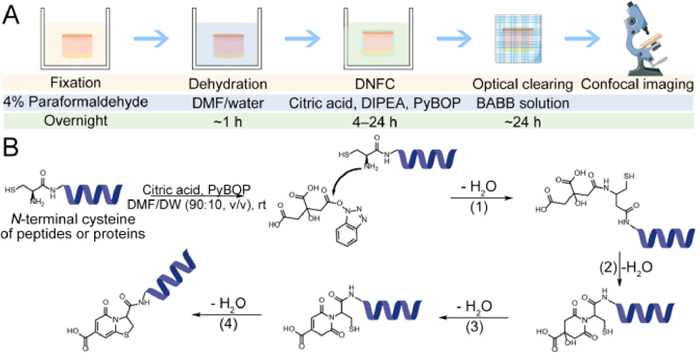
Recently in Science Advances, Pac et al. [11] reported a 3D histological technique termed "3D tissue imaging through de novo formation of citrate-based fluorophores" (3DNFC), which is the integration of DNFC ("de novo formation of citrate-based fluorophores") staining and 3DISCO ("3D imaging of solvent-cleared organs") techniques, and represents a novel approach to utilize in situ fluorophore formation for 3D fluorescence imaging. In the DNFC process, the nonfluorescent citrate is utilized to from a blue fluorophore with N-terminal cysteine (Cys) in the tissue. Since the N-terminal Cys moieties are distributed heterogeneously within the tissue and cells, the general and detailed structures of the specimen may exhibit different labeling levels, which can be detected via fluorescence contrast imaging. The general steps of 3DNFC include tissue fixation, dehydration with N, N-dimethylformamide (DMF), fluorophore formation via DNFC, tissue clearing via 3DISCO, and confocal imaging and 3D reconstruction (Fig. 1A).
Figure 1
Figure 1.
Procedure and mechanism of 3DNFC. (A) Scheme illustrating the treatment, material, and duration in every step in 3DNFC. (B) Scheme showing the proposed mechanism of the fluorophore formation from N-terminal cysteine of peptides or proteins and citric acid in the presence of PyBOP. Reproduced with permission [11]. Copyright 2022, The Authors, some rights reserved; exclusive licensee American Association for the Advancement of Science. No claim to original U.S. Government Works.
Based on the finding that Cys can react with citrate to form 5-oxo-2, 3-dihydro-5H-[1,3]thiazolo[3, 2-a]pyridine-3,7-dicarboxylic acid (TPA), a strong blue fluorophore [12], the authors developed the DNFC technique for in situ fluorophore formation in tissues. During the DNFC process, one carboxylic group of citrate is first activated by benzotriazole-1-yloxytripyrrolidinophosphonium hexafluorophosphate (PyBOP), and then the primary amine group of N-terminal Cys on the peptides or proteins in the tissue attacks the activated carboxylic group to yield an amide bond, followed by imide formation, dehydration, and intramolecular condensation, eventually forming the TPA structure (Fig. 1B). The 3DISCO technique in the 3DNFC is an organic solvent-based tissue clearing method. Herein, benzyl alcohol-benzyl benzoate (BABB) is used for optical clearing, thus reducing the light scattering in the thick tissue specimens and enabling their morphological features and detailed structures to be observed nondestructively with a confocal microscope.
Consequently, the combination of DNFC and 3DISCO techniques in 3DNFC not only successfully exhibited the detailed characteristic structures of the kidney, lung, liver, tongue, esophagus, intestine, brain, and ovary in the 2D mouse specimens, but also realized volumetric imaging of whole mouse lung lobe and mouse spleen tissue, providing the spatial and morphological information of the samples in a 3D manner. In addition, the authors also demonstrated the feasibility of 3DNFC technique in 3D imaging as well as quantitative diagnostic evaluation of pathological tissues in the nonalcoholic fatty liver disease (NAFLD) model and human-derived breast cancer specimens, and these applications of this technique successfully indicated the development of NAFLD in the mouse model and the distribution of the cancer cells and vascular channels at different depths in the breast cancer specimen, respectively.
In conclusion, such a staining technique not only utilizes a cheap and nonfluorescent hydrophilic small molecule, citrate, enhancing the staining penetration depth and avoiding the nonspecific interactions-induced false-positive readouts, but also realizes in situ fluorophore formation under a mild reaction condition with the aid of PyBOP, which avoids the damage to the specimen and allows its broad compatibility with other labeling methods. This technique has provided a practical tool for precise 3D reconstruction, spatial analysis, and pathological diagnosis, presenting its promising potential in playing the same important role in 3D histology as H & E staining plays in 2D histology.
However, there are still some issues requiring further investigation, which we also think of as the challenges and opportunities existing in the 3D histology. First, reducing biomolecule loss or damage during the 3DNFC process also represents an important direction that needs further improvement. For instance, since the dehydration and optical clearing steps use organic solutions, the lipid components in the samples are defective. Although the 3DNFC technique can sketch out the lipid vacuoles, it cannot directly and clearly lighten the lipid droplets (if necessary). Similarly, harsh treatment conditions may also cause damage to RNAs [13], which is another challenge to overcome. Second, since 3DNFC is a technique that can present the general characteristics of the specimen while some other staining techniques may provide the distribution and abundance of specific biomolecules, the combination of 3DNFC and other staining techniques can generate more accurate and detailed information. Although the authors have successfully applied 3DNFC together with the staining by propidium iodide (PI) and specific fluorescent antibodies, the compatibility between 3DNFC and other staining techniques such as organic dye staining, immunofluorescence staining, and fluorescence in situ hybridization (FISH) labeling of nucleic acids may need further investigation. For example, questions like whether the 3DISCO process can lead to better antibody penetration and whether permeabilization of the tissue with detergents is needed for achieving a better costaining effect are worth concern. Third, whether the time of the whole 3DNFC can be shortened by optimizing the clearing and staining conditions or by improving the scanning technology (since the problem of long scanning time still exists in 3DISCO) to match the clinical requirement under urgent circumstances is a key question in the clinical application of 3DNFC. Finally, the integration of 3DNFC and the advanced fluorescence microscopy, multiscale imaging data processing technique, and artificial intelligence-based data analysis method can also be a meaningful direction for 3D histology and benefit the application of 3D histology in fundamental research and clinical disease grading.
Declaration of competing interest
The authors declare that they have no known competing financial interests or personal relationships that could have appeared to influence the work reported in this paper.
[1]
J.T.C. Liu, A.K. Glaser, K. Bera, et al., Nat. Biomed. Eng. 5 (2021) 203–218. doi: 10.1038/s41551-020-00681-x
[2]
A. Ertürk, K. Becker, N. Jährling, et al., Nat. Protoc. 7 (2012) 1983–1995. doi: 10.1038/nprot.2012.119
[3]
A. Ertürk, C.P. Mauch, F. Hellal, et al., Nat. Med. 18 (2012) 166–171. doi: 10.1038/nm.2600
Figure 1
Procedure and mechanism of 3DNFC. (A) Scheme illustrating the treatment, material, and duration in every step in 3DNFC. (B) Scheme showing the proposed mechanism of the fluorophore formation from N-terminal cysteine of peptides or proteins and citric acid in the presence of PyBOP. Reproduced with permission [11]. Copyright 2022, The Authors, some rights reserved; exclusive licensee American Association for the Advancement of Science. No claim to original U.S. Government Works.

 DownLoad:

 下载:
下载:


