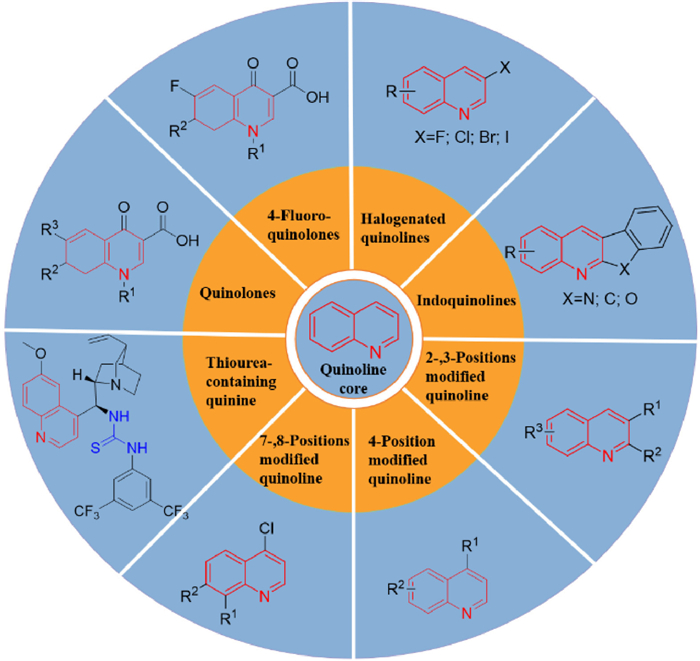
Figure 1.
Structures of quinoline analogues related to anti-MRSA activities.
Quinoline-based anti-MRSA agents: Current development, structure-activity relationships, and mechanisms
Hong Yao , Liping Cui , Hang Liu , Xueyu Li , Lin Shen , Ruige Yang , Shangshang Qin , Yong Guo

Antibiotics are primarily used to treat a variety of bacterial infections, but due to overuse and misuse of antibiotics, bacteria have become resistant to currently approved marketed drugs [1,2]. The spread of antimicrobial resistance (AMR) has made once treatable diseases deadly again, and bacterial infections have become a widespread global health risk [3-6]. Staphylococcus aureus (S. aureus) is a round, Gram-positive bacteria that causes a variety of diseases ranging from skin and soft tissue infections to severe pneumonia, sepsis, and meningitis. Currently, more than 60% of S. aureus isolates are methicillin-resistant S. aureus (MRSA) [7-12]. Methicillin resistance in S. aureus strains is caused by the in vivo gene encoding PBP2a, which has an ultra-low β-lactam affinity and is capable of cell wall synthesis at normally lethal β-lactam concentrations to complete the bacterial reproductive growth, hence MRSA is also known as a superbug [13,14]. And as the last line of defense, vancomycin, MRSA has also developed resistance to it. Therefore, there is an urgent need to develop new antimicrobials with anti-MRSA potency [15,16].
Among the heterocyclic derivatives containing nitrogen atoms, quinolines are used in drug design and discovery due to their wide range of biological activities [17,18]. Quinolines have been reported to have anticancer [19], antimalarial [20], antihypertensive [21], anti-inflammatory and analgesic [22,23], antibacterial [24], anti-human immunodeficiency virus (HIV) [25], antitubercular [26], anti-cardiovascular activities [27], and inhibition of tyrosine kinase [28] properties. At present, the drugs such as ofloxacin, norfloxacin, ciprofloxacin, and sparfloxacin, which have been approved for marketing to treat various bacterial infections, all contain a quinoline backbone in their structures [29,30]. Although there are some reviews on the biological activity of quinolines, a review on the antibacterial activity of quinolines against MRSA has not been reported. Herein, we reviewed the structure–activity relationships (SARs) and different mechanisms of potential antibacterial effects of quinoline analogues (Fig. 1) against MRSA strains in recent two decades to provide insights into the development of new potent quinoline-based antimicrobial agents.
4-Quinolones such as nemonoxacin [31,32] and delafloxacin [33] play a key role in the clinical anti-MRSA infection treatment. However, MRSA has developed resistance to 4-quinolones [34], so it is becoming increasingly urgent to improve the efficacy of 4-quinolones. Modifications of the C-7 position of 4-quinolones are thought to be beneficial for improving antibacterial potency, solubility, and safety [35,36].
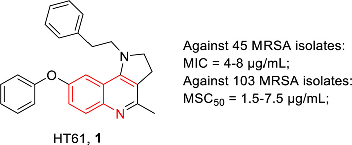
HT-61 (1, Fig. 2), is a quinolone derivative with a molecular weight of about 400 Daltons that is particularly active against S. aureus, including MRSA [37,38]. Compound 1 not only acted on reproductive bacteria with minimum inhibitory concentrations (MICs) higher than those of marketed antibiotics but also displayed significant efficacy against non-reproductive bacteria. The MICs of 1 against 45 MRSA isolates were 4–8 µg/mL and its minimum stationary phase-cidal concentration 50 (MSC50) values against 103 MRSA isolates were 1.5–7.5 µg/mL. Compound 1 was significantly more potent than daptomycin, which required 50 µg/mL to completely kill fixed-stage MRSA. Additionally, compound 1 can target bacterial cell membranes and act on the plasma membrane of bacterial cells by disrupting the membrane potential of S. aureus, resulting in the release of cellular contents. Moreover, compound 1 in combination with several antibiotics against clinical MRSA strains was also investigated, and the results showed that compound 1 had significant synergistic activity when it combined with gentamicin, neomycin, or chlorhexidine.
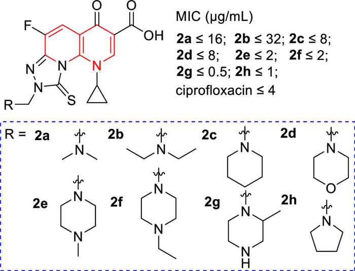
A few tricyclic fluoroquinolones, [1,2,4]triazolo[3,4-h][1,8] naphthyridine-8-one-7-carboxylic acid derivatives 2a–h (Fig. 3), whose C-8 positions contained a functional Mannich base part, were evaluated for their in vitro antibacterial activities against MRSA [39]. The results showed that the Mannich base derived from the cycloaliphatic amine donors (2d–h) exhibited strong antibacterial activity against MRSA, with MIC values of 0.5–2 µg/mL, and its potency was 2–8 times stronger than that of ciprofloxacin. Among them, especially compound 2g had the best antibacterial activity with an MIC value of 0.5 µg/mL, which was 8 times higher than that of ciprofloxacin (Fig. 3).
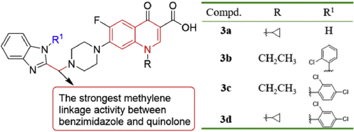
The new benzimidazole quinolones drugs displayed promising antibacterial action [40]. Antibacterial evaluation in vitro showed that most of the title benzimidazole quinolones exhibited good antibacterial activity against the tested strains, especially against MRSA, even better than the reference drugs [40]. For example, methylene-bridge linkers, halogenated benzyl derivatives, NH2CH2 linkers and benzimidazole complexes directly linked to quinolone nucleus displayed good anti-MRSA activity, with MIC values of 0.125–0.5 µg/mL. They were more active than chloramphenicol (MIC = 16 µg/mL), norfloxacin (MIC = 8 µg/mL), ciprofloxacin (MIC = 2 µg/mL), and clinafloxacin (MIC = 1 µg/mL). The anti-MRSA activity of methylene-linked compounds 3a, 3b, 3c, and 3d (Fig. 4, MIC = 0.125 µg/mL) was 8–64 times higher than that of norfloxacin, ciprofloxacin, and clinafloxacin. Compound 3d showed rapid bactericidal activity, and the number of viable bacteria decreased by more than 3 logs (CFU/mL) within an hour at a concentration of 6× MIC. Moreover, 3d not only can inhibit the formation of biofilm but also can disperse the formed bacterial biofilm, while exhibiting low toxicity to mammalian cells and not inducing bacterial resistance. The results of SARs indicated that when the benzimidazole and quinolone nucleus were linked by a methylene linkage, there was a significant effect on the enhancement of antibacterial activity (Fig. 4).
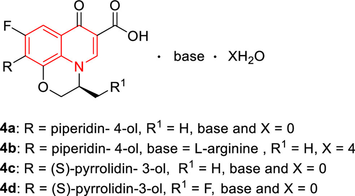
In 2015, some novel quinolone derivatives based on levofloxacin as the core were synthesized by Huang et al. [41]. The antibacterial activity of 4a–d (Fig. 5, MIC = 1 µg/mL) was 4 times greater than that of moxifloxacin (MIC = 4 µg/mL), 32 times stronger than that of levofloxacin (MIC = 32 µg/mL) and as isoenergetic as vancomycin (MIC = 1 µg/mL). Compound 4b had better solubility (5.33 mg/mL) and lower toxicity than levofloxacin. In addition, the lethal dose 50 (LD50) of 4b was 1402 mg/kg, while the LD50 of levofloxacin was 358 mg/kg.
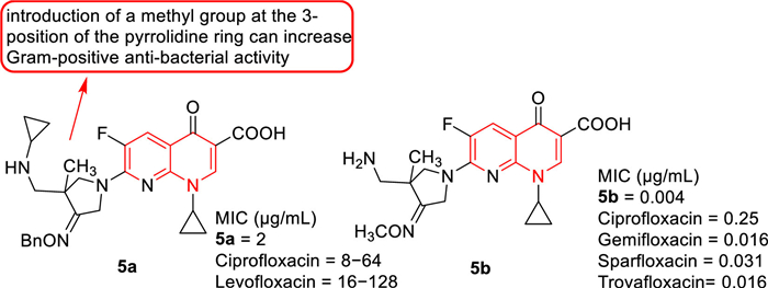
Zhang et al. [42] reported the development of fluoroquinolone derivatives containing 3-alkoxyimino-4-(cyclopropylanimo)methylpyrrolidine moiety and evaluated their biological activities. Among these fluoroquinolone derivatives, compound 5a (Fig. 6) exhibited the strongest antibacterial activity against MRSA with an MIC value of 2 µg/mL, 4–64 times higher than ciprofloxacin and levofloxacin (MIC: 8–128 µg/mL) against MRSA. The SARs indicated that the introduction of methyl at 3-position of the pyrrolidine ring increased antibacterial activity against Gram-positive bacteria, which was consistent with the results reported by Yun et al. [43]. The compounds from the two research groups had the same parent nuclear structure, and compound 5b synthesized by Yun et al. [43] displayed good in vivo efficacy against MRSA in a mouse infection model (Fig. 6).
Cui et al. [44] inserted the triazole ethanol moiety into the N-1 position of quinolones to change the different substituents on the benzene ring of quinolones, and prepared a new class of quinolones triazoles. Most of these new quinolone triazoles can effectively inhibit MRSA growth (MIC = 0.5-16 µg/mL), which were more effective than clinical drug chloramphenicol (MIC = 16 µg/mL). Compounds 6a, 6b, and 6c (Fig. 7) with MIC values of 1 µg/mL were 8 and 16 times more active than norfloxacin (MIC = 8 µg/mL) and chloramphenicol (MIC = 16 µg/mL), respectively. In particular, 7-trifluoromethyl intermediate 6d (Fig. 7, MIC = 0.5 µg/mL), as a new potential anti-MRSA candidate, exhibited 16 and 32 times higher antibacterial activity than the standard drugs norfloxacin and chloramphenicol, respectively. In addition, Cui's [45] group also synthesized a series of novel 3-aminothiazole quinolones analogues as antibacterial agents. Among these 3-aminothiazol-quinolones, 3-(2-aminothiazol-4-yl)-7-chloro-6-(pyrrolidine-1-yl) quinolone 6e (Fig. 7) showed significant antibacterial activity against MRSA at low concentrations (MIC = 0.8 µg/mL). Its antibacterial activity was 13 and 27 times higher than that of norfloxacin and chloramphenicol, respectively. Moreover, 6e had low cytotoxicity to liver cells, strong inhibition of DNA cyclotron enzyme, and a wide antimicrobial spectrum, including MDR strains. Notably, the active molecule 6e also induced bacterial resistance more slowly than norfloxacin. Analysis of the SARs (Fig. 7) revealed that the 2-aminothiazole fragment at C-3 position of quinolones increased antibacterial potency and could replace the carboxyl group of quinolones. The introduction of some electron-donating substituents, such as methyl, methoxy, or alicyclic amino groups, at 6- and 7-positions of quinolones was particularly beneficial for anti-Gram-positive bacteria. The antibacterial evaluation showed that 7-chloroquinolones had better inhibitory activity against MRSA than 7-fluoroquinolones. Additionally, the substituents on the benzene ring of quinolones also affected their antibacterial potency.

In 2020, Maryam et al. synthesized some new fluoroquinolones with ciprofloxacin and sarafloxacin as the core skeletons [46]. All the compounds synthesized from ciprofloxacin derivatives showed significant anti-MRSA activity, with MIC values ranging from 0.016 µg/mL to 0.29 µg/mL. The antibacterial activity of compound 7a against MRSA (MIC = 0.016 µg/mL) was 60 times higher than that of the positive control ciprofloxacin (MIC = 0.49 µg/mL) (Table 1). This meant that the presence of methoxy at the R1 position produced beneficial anti-MRSA activity. All the synthetic sarafloxacin derivatives exhibited approximately the same activity as the parent sarafloxacin against Gram-positive bacteria. Especially compound 7b with a methoxy group at the R1 position showed the most promising antibacterial activity against MRSA (MIC = 0.125 µg/mL), which was twice as high as the positive control sarafloxacin (MIC = 0.25 µg/mL) (Table 1).
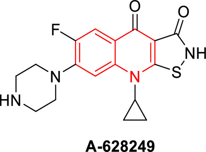
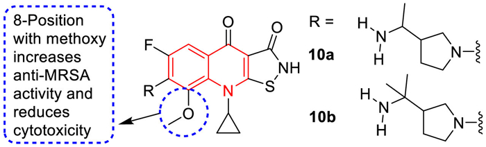
In 1987, isothiazoloquinolones (ITQs), originally reported by Chu and colleagues [47,48], were more potent than quinolones against S. aureus. The best represented of ITQs was the ciprofloxacin analogue A-628249 (Fig. 8). Wiles and Wang et al. [49-52] synthesized ITQ-related compounds containing functionalized aryl and heteroaryl groups connected by a C–C bond at 7-position and structurally modified at 6-, 8-, and 9-positions, such as compounds 8a, 8b, and 9 (Table 2). These compounds exhibited excellent in vitro antibacterial activity against MRSA as well as strong inhibitory activity against DNA rotamases and topoisomerase IV. In 2011, a series of 8-methoxy ITQs with a 7-aminocyclic ring was synthesized by Kim et al. [53]. The structures of 10a (R, S) and 10b (R) are shown in Fig. 9. The MICs of 10a and 10b against MRSA and their inhibitory activities against target enzymes from S. aureus, wild-type topoisomerase IV and DNA rotamase, and the corresponding enzymes from Staphylococcal mutants expressing fluoroquinolone resistance are outlined in Table 3. Compounds 10a (R, S) and 11b (R) exhibited potent anti-MRSA activity with an MIC value of 0.06 µg/mL. Notably, compared to ciprofloxacin, 10b (R) increased inhibition of wild-type DNA gyrase activity by 91-fold, while 10a (R, S) increased inhibition of wild-type topoisomerase IV activity by 75-fold. Similar improvements were observed for 10b (R) and 10a (R, S) inhibition of mutant DNA gyrase, with > 48-fold and > 62-fold increases, respectively. Compounds 10b (R) and 10a (R, S) increased the inhibition of mutant topoisomerase IV by 95-fold and 121-fold, respectively. The above data suggested that 8-position with methoxy could increase anti-MRSA activity and reduce cytotoxicity.
In 2010, O'Donnell et al. [54] performed an antimicrobial evaluation of haloquinoline derivatives, in which 4-hydroxy-3-iodoquinolin-2-one (11) showed strong to excellent antibacterial activity against eight clinical MRSA isolates (Table S1 in Supporting information). For clinical isolates EMRSA-16, CWMRSA, and HMRSA, the MIC value of 11 was equal to or lower than that of vancomycin, an antibiotic used in the clinical treatment of MRSA infection. While the antibacterial activity of 11 against other clinical MRSA isolates was better than vancomycin (MIC = 0.78–1.56 µg/mL).
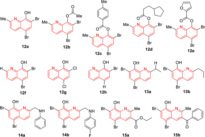
In 2014, Abouelhassan's group [55] synthesized a library of 21 quinoline analogues and evaluated their antimicrobial activities (Fig. 10). Among these 21 quinoline analogues, some quinoline analogues showed effective activity against MRSA-2. The quinoline derivative 12a–e (Fig. 10) exhibited very effective biofilm dispersion against MRSA-2 (EC50 = 2.80 µmol/L). In particular, quinoline derivative 12c (EC50 = 2.06 µmol/L) exhibited the most effective biofilm dispersion activity. The 2-position of the halogenated quinoline (HQ) scaffold played an important role in the corresponding antibacterial activity of the HQ analogues. Compared with quinoline derivative 12a, bromoxyquinoline 12f (Fig. 10) had a hydrogen atom at 2-position, and 12a had a methyl group at 2-position of the HQ scaffold (Fig. 10). This monomethyl difference resulted in a 16-fold increase in the antibacterial activity of 12a against Staphylococcal pathogens and a 128-fold elimination of antibacterial activity against the Gram-negative pathogens Acinetobacter baumannii. In addition, 12a can also eradicate MRSA biofilms. Based on the potent anti-MRSA activity, they subsequently synthesized a series of HQ analogues, which had different alkylation groups at the 2-position of the HQ scaffold [56]. In addition to 2-alkylated HQ analogues, they also designed an alternative pathway to synthesize reductive amination HQ analogues by reducing amination at 2-position of the HQ scaffold with various amines and anilines [56]. Some HQ analogues showed effective antibacterial activity, such as 13a and 13b (Fig. 10). It is worth mentioning that the MIC values of 14a (Fig. 10) and 12a (MIC ≤ 0.78 µmol/L) against MRSA-2 were identical. Among these HQ analogues, 14b (Fig. 10) exhibited the highest anti-MRSA activity with MIC of 0.39 µg/mL, and it did not show hemolysis toward red blood cells (RBC) at 200 µmol/L. In general, HQ analogues containing aniline halide groups displayed good to highly potent antibacterial activity, while HQ analogues containing alkyl and methoxylaniline groups usually showed low antibacterial activity. For example, 13b [minimum biofilm eliminating concentration (MBEC) = 93.8 µmol/L] and 14b (MBEC = 93.8 µmol/L) displayed improved biofilm eradication activity against MRSA-2 compared with 12a (MBEC = 188 µmol/L). These synthesized HQ analogues were 20 times more potent as MRSA-2 biofilm eradicators than current anti-MRSA therapeutic agents including vancomycin, daptomycin, and linezolid. Moreover, they displayed low hemolysis and low cytotoxicity to HeLa cells [56]. Eighty percent of bacterial infections are related to biofilm, and halogenated 8-hydroxyquinolines had the characteristics of biofilm degradation. Accordingly, they used Friedlander reactions to synthesize a series of halogenated quinolines, capable of eradicating bacterial biofilms while exhibiting minimal mammalian cytotoxicity and hemolytic activity [56]. In preliminary MIC assays, some HQ compounds exhibited higher antimicrobial activity compared to 12a. HQ compounds 15a and 15b (Fig. 10) showed improved activity against MRSA-2, with MIC of 0.39 and 0.59 µmol/L, respectively, and 15b proved to be one of the most effective biofilm eradicators ever reported against MRSA (MBEC = 3.9–23.5 µmol/L) [57-59]. Antimicrobial enhancement is defined as the ability of a non-growth inhibitory enhancer [the plant-derived chemical gallic acid (GA)] to reduce the MIC value of HQ by more than 4-fold. It is proven that combination therapy can improve antibacterial activity. Abouelhassan et al. [60] also evaluated enhancement assays in combination with GA against four clinical MRSA (MRSA-ATCC, MRSA-1, MRSA-2, and SA-156). Compounds 12f–h (Fig. 10) had a 2–1000-fold enhancement effect when combined with GA. When 12f was combined with GA, its activity was increased by 1000-fold potentiation against MRSA SA-156. They also studied the combination of GA with conventional antibacterial agents, such as ciprofloxacin and vancomycin. However, none of these antibacterial agents were enhanced by GA. These results indicated that the unique antibacterial mechanism of the HQ compounds was different from that of traditional antibiotics. HQ compounds operated through a metal (Ⅱ) dependent mechanism and unlike conventional antibiotics, exhibited good cytotoxicity when tested in HeLa cells in lactate dehydrogenase (LDH) release assay. In summary, Abouelhassan et al. [60] found that the selective combination of GA and HQ small molecules had strong antibacterial activity against a variety of pathogens, including multidrug-resistant clinical isolates. The combination therapy has also been effective in eradicating MRSA biofilms. The phytochemical-HQ combination provided a promising new platform for the development of clinically useful antibacterial combination therapy [60].
The indole part is an important basic subunit for a large number of natural products and drugs [61], and indole-containing molecules have been reported to possess some antibacterial activity [62,63]. Moreover, indoquinoline analogues also play an important role in antimicrobial activity.
Under 2,3-dichloro-5,6-dicyano-1,4-benzoquinone (DDQ)-mediated oxidation conditions, Challa et al. [64] prepared a library of indolo[2,3-b]quinolines, chromeno[2,3-b]indoles, and 3-enyloctadiols using readily available 3,3′-diindolylmethane (DIM) (the synthetic route of the representative compounds 16a and 16b was shown in Scheme S1 in Supporting information). The MIC and minimum bactericidal concentration (MBC) of indoloquinolines against four different methicillin-resistant S. aureus strains (three of which were clinical isolates) were determined. The results showed that 16a and 16b possessed excellent activity against methicillin-sensitive S. aureus and MRSA (Table 4). Methicillin was active against susceptible strains, and the MIC value was 1 µg/mL, but it had MIC values of 32 µg/mL to > 64 µg/mL for all MRSA strains. This result indicated that the MRSA strain was highly resistant to methicillin. In contrast, compounds 16a and 16b showed similar activity against both drug-sensitive and drug-resistant strains, while the MIC values of these two compounds against methicillin-sensitive strains were 1 µg/mL. For MRSA strains, the MIC values of 16a and 16b ranged from 1 µg/mL to 4 µg/mL, and the MBC values of 16a and 16b ranged from 2 µg/mL to 8 µg/mL, while methicillin had an MIC value of > 64 µg/mL in all tested MRSA strains (Table 4). The results manifested that compounds 16a and 16b were selective anti-MRSA agents, and their activities against MRSA were better than that against other bacteria. At the concentration of 16 µg/mL, both compounds 16a and 16b completely killed MRSA within 360 min.
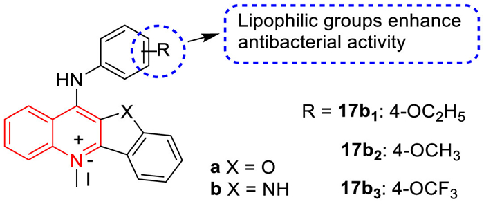
In 2017, the antibacterial activity and mode of action of some N-methylbenzofurano[3,2-b]quinolones and N-methylbenzo-indolo[3,2-b]quinoline derivatives containing quaternary pyridine centers were studied by Sun et al. [65]. The results showed that all compounds had good antibacterial activity compared to the 11 clinical antibiotics evaluated in this study. In general, the antibacterial activity of 11-aniline-substituted 5-N-methyl-10-H-indole[3,2-b] quinolinium derivatives (b series) was slightly higher than that of 11-aniline-substituted 5-n-methylbenzofuran[3,2-b]quinoline derivatives (a series) (Fig. 11). This result may be due to the interaction between the imine in the quaternary pyridine center and the amino acids in the filamentous temperature-sensitive protein Z (FtsZ). In addition, the addition of lipophilic groups at the 4-position of 11-aniline slightly enhanced the antibacterial activity of these N-methyl quaternary ammonium derivatives. Among these derivatives, 17b, and 17b (Fig. 11) were the most effective against MRSA, with MIC values of 2 µg/mL, and their antibacterial activities were about 100 times higher than that of berberine and methicillin. In the guanosine triphosphatase (GTPase) assay, 17b inhibited the GTPase activity of FtsZ in a dose-dependent manner. The binding of the quinoline derivatives to the C-terminal domain gap interfered with the GTPase activity of FtsZ, and then disrupted the formation of FtsZ polymer, leading to abnormal cell division and cell death.
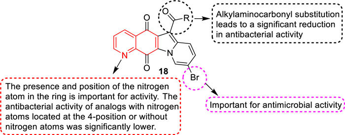
The ester analogues in 9-bromo-substituted indolylquinoline-5,12-dione derivatives showed strong antibacterial activity against Gram-positive strains, especially against MRSA [66]. The MIC values of compounds 18a, 18b, and 18c against MRSA were as high as 0.063 µg/mL, which was 16 times higher than that of vancomycin. Compound 18d exhibited the best activity with an MIC value of 0.031 µg/mL, which was 32 times higher than vancomycin (Table 5). Compound 18a had a strong inhibitory effect on the superhelix activity of DNA rotation of E. coli and the relaxation activity of topoisomerase IV of S. aureus, exhibiting a mechanism similar to ciprofloxacin [67,68]. Although compound 18a displayed good anti-MRSA activity in vitro, its bioavailability in mouse models was very low, probably because of its low water solubility. SARs (Fig. 12) studies have revealed that: (1) The substitution of 9-position with bromine can enhance the antibacterial activity, but the substitution of 9-position with chlorine shows a lower activity [69]. (2) Although 7-fluorinated analogues exhibited strong antibacterial activity with the same MIC value as compound 18a, it had high cytotoxicity, indicating that it may not be a good choice for further development of good antibacterial agents [70]. (3) The substitution of alkyl, amino, and carbonyl groups at the 6-position resulted in a significant decrease in antibacterial activity, which may be attributed to their reduced solubility. (4) The presence and position of nitrogen atoms in the ring were important for activity. The antibacterial activity of analogues with nitrogen atoms at 4-position or without nitrogen atoms decreased significantly. On the other hand, substitution of the 6-position by the alkoxycarbonyl group was important for enhancing the antibacterial activity. The modification of compound 18a was guided by SARs and 28 indolylquinoline-5,12-diketone derivatives were synthesized to obtain the derivatives with higher anti-MRSA activity and good water solubility. Compound 18a exhibited strong activity against clinical MRSA strains, with MIC50 and MIC90 values lower than 0.0078 µg/mL. Compound 18a was soluble in water with a solubility of 1.98 mg/mL and also displayed strong activity against clinical MRSA strains with an MIC50 value of 0.063 µg/mL and an MIC90 value of 0.125 µg/mL, which was 16 times higher than vancomycin (Table S2, Supporting information), suggesting that it could be further developed as an anti-MRSA lead compound.
In 1990, compound 19 was first synthesized by Yamato et al. [71] for the study of anti-tumor activity (Table 6). Zhao et al. [72-75] believed that compound 19, containing the new skeleton of indoquinoline and its analogues, might have antibacterial activity and then measured the anti-MRSA activity of compound 19 against MRSA strains OM481 and OM584. The results showed that compound 19 had no activity against MRSA. Subsequently, they determined the anti-MRSA activity of analogues of compound 19 and found that compound 20 had an anti-MRSA activity with MIC of 8 µg/mL (against MRSA OM481) and 16 µg/mL (against MRSA OM584). Afterwards, Zhao et al. [76] synthesized indolo[3,2-b]quinoline analogues, 4-(acridine-9-amino)phenol hydrochloride (21), benzofurano[3,2-b]quinoline, and indolo[1,2-b]quinoline based on the indolo[3,2-b]quinoline backbone of lead compound 20 and investigated their anti-MRSA activities against s-type OM481 and OM584 strains. The results showed that anti-MRSA activity of indoquinoline tetracyclic compounds against MRSA OM481 and OM584 strains was superior to that of tricyclic compound 4-(acridine 9-amino)phenol hydrochloride. The positions and types of substituents in indoquinoline analogues were important for the anti-MRSA activity against both strains. The indoquinoline analogues 20a, which introduced a hydroxyl group at the 4′-position showed obvious anti-MRSA activity against OM481 (MIC = 4 µg/mL) and OM584 (MIC = 2 µg/mL), revealing that 4′-hydroxyl group was important for the anti-MRSA activity towards both MRSA OM481 and OM584 (Table 6). The introduction of substituents at 7-position of indoquinoline analogues also exhibited good anti-MRSA activity, especially methoxy (20a) exhibited obvious anti-MRSA activity against both OM481 and OM584 with MIC of 2 µg/mL (Table 6). The anti-MRSA activity of indoquinoline (20a) and methylindoquinoline (20a) was weak, but benzofuran-quinoline (20a) displayed significant anti-MRSA activity against both strains, with an MIC of 2 µg/mL (Table 6). The SARs indicated that the indoquinoline ring, benzofuranquinoline ring, and 4-aminophenol group were the basic structures for the anti-MRSA activity.
In 2016, Shiri et al. [77] reported a series of diamide derivatives containing 2-chloroquinoline scaffolds by Ugi condensation reaction of 2-chloroquinoline-3-carboxaldehyde amines, carboxylic acids, and isonitriles. Among them, compound 22 exhibited the strongest antibacterial activity against MRSA with an MBC value of 0.62 mmol/L, which was stronger than ampicillin but lower than ciprofloxacin (Fig. 13).
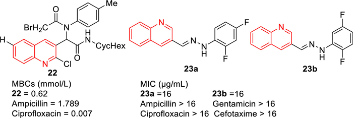
Compounds 23a and 23b are new compounds synthesized based on quinoline-3-formaldehyde hydrazone compounds (MIC: 16 µg/mL), which have a significant antibacterial effect against MRSA compared with ampicillin, gentamicin, ciprofloxacin, and cefotaxime (MIC: > 16 µg/mL) (Fig. 13). According to the results of MTT test, compounds 23a and 23b had no significant effect on McF-7 cells at 100 µmol/L concentration [78].
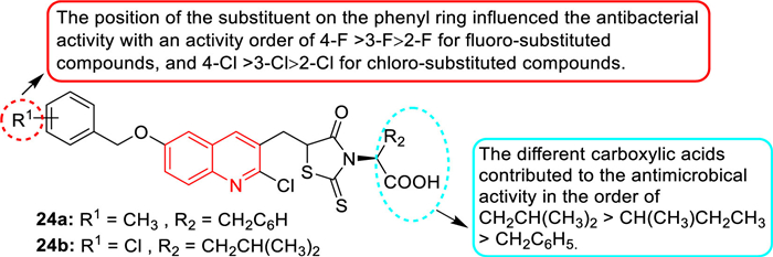
In 2013, Guo et al. prepared a series of rhodanine derivatives containing the quinoline fraction and evaluated their in vitro antibacterial activities [79]. The in vitro antibacterial assay showed that most of the rhodanine derivatives exhibited excellent antibacterial activity against different MDR Gram-positive bacteria (MIC value was 1–2 µg/mL). In particular, compounds 24a and 24b (Fig. 14) displayed the strongest level of inhibitory activity (MIC = 1 µg/mL), with a 4-8-fold increase in antibacterial activity compared with the standard drug norfloxacin (MIC = 4–8 µg/mL). A comparison of carboxylic acid derivatives at the N-position of the rhodanine ring indicated that the contribution order of different carboxylic acids to antibacterial activity was -CH2CH(CH3)2 > -CH(CH3)CH2CH3 > -CH2C6H5. In addition, the position of substituents on the benzene ring affected the antibacterial activity, and the sequence of antibacterial activity of fluorine-substituted compounds was 4-F > 3-F > 2-F, and the sequence of antibacterial activity of chlorine-substituted compounds was 4-Cl > 3-Cl > 2-Cl. Compound 24a did not affect the cell viability of human cervical (HeLa) cells when the concentration was equal to the MIC value but showed cytotoxicity at higher concentrations.
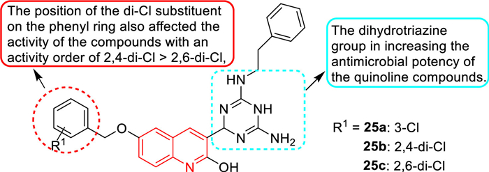
In 2019, Bai et al. [80] designed and synthesized some dihydrotriazine derivatives containing the quinoline fraction as novel antimicrobial agents. Dihydrotriazine derivatives 25a–c (Fig. 15) were the most active compounds against MRSA3506 with an MIC of 1 µg/mL. Based on the activity analysis of these dihydrotriazine derivatives, the following SARs were obtained. The dihydrotriazine group can improve the antibacterial activity of the quinoline compounds. The introduction of phenylethyl at the N-position of the dihydrotriazine ring resulted in significant differences in anti-MRSA activity, suggesting that the substituents of aromatic nuclei were critical for the activity of these dihydrotriazine compounds. In addition, the position of the 2-Cl substituent on the phenyl ring also affected the anti-MRSA activity. The 2,4-di-Cl substituted phenyl ring played a key role in the activity of these dihydrotriazine compounds, and its activity was greater than that of 2,6-Cl substituted compounds. These results were consistent with the previously reported results of a series of rhodanine and dihydrotriazine derivatives [81,11]. In vitro enzyme studies showed that compound 25a exhibited inhibition of dihydrofolate reductase (DHFR), and compound 25a did not show significant cytotoxic activity (HCT116 and LO2 cells IC50 > 100 µmol/L). These findings suggested that these dihydrotriazine derivatives containing the quinoline fraction could be potential lead compounds for rational development of novel quinoline-based antibacterial agents.
The antimicrobial screening of novel ring-substituted styrylquinolines and two oxorhenium complexes compounds was carried out by Cieslik et al. [82]. Compound 26 (Table 7) was the most effective anti-MRSA candidate, with MIC/IC90 values of 3.9 and 7.81 µmol/L at 24 and 48 h, respectively, which were more potent than the standard drugs bacitracin, penicillin V, and ciprofloxacin.
In 2018, Fang et al. [83] reported that 1-methylquinolinium iodide derivative (27) had good antibacterial activity against bacterial strains, and it had a synergistic effect in combination with β-lactam antibiotics against antibiotic-resistant strains of S. aureus. The antibacterial activity of 27 (Table 8, MIC = 1.5 µg/mL) against MRSA strains was 100-fold higher than that of methicillin and berberine. Compound 27 at 0.375 µg/mL significantly increased the antibacterial activity of methicillin against MRSA and decreased the MIC value from 1024 µg/mL to 32 µg/mL. The fractional inhibitory concentration index (FICI) of 27 with methicillin was 0.281. The combination of 27 with ampicillin or oxacillin also exhibited synergistic effects on MRSA with FICIs of 0.5 and 0.375, respectively. Compound 27 increased the antibacterial activity of ampicillin by 4 times (MIC decreased from 48 µg/mL to 12 µg/mL) and increased the antibacterial activity of oxacillin against MRSA by 8 times (MIC decreased from 256 µg/mL to 32 µg/mL) at a concentration of 0.375 µg/mL. In addition, when combined with compound 27, imipenem and ceftazidime exhibited enhanced antibacterial activity with an FICI of 0.75 (Table 8). Mechanistic studies revealed that compound 27 inhibited bacterial growth by inhibiting the GTPase activity of FtsZ.
 DownLoad:
CSV
DownLoad:
CSV

|
Teng et al. [84] evaluated the antibacterial activity of eight quinoline derivatives with daptomycin as a reference drug. As shown in Table 9, when both R1 and R2 groups were kept as p-methyl, the only weak activity of compound 28a was detected, and the MIC of 28a against MRSA was 12 µg/mL. When the R1 group was replaced by trifluoromethyl, the resulting compound 28b had better activity with an MIC of 3.0 µg/mL against MRSA. Subsequently, Teng's group [84] retained the R1 as the CF3 group and modified R2 as m-trifluoromethyl, p-trifluoromethyl, and 3-chloro-4-fluorine groups, respectively, yielding compounds 28c, 28d, and 28e. Compared with 28b, quinoline derivative 28c showed no change in antibacterial activity against MRSA, while compound 28d exhibited enhanced activity against MRSA with a MIC of 0.75 µg/mL, indicating the p-CF3 was more beneficial than the p-CH3 group in enhancing the anti-MRSA activity of these quinoline derivatives. Compared with compound 28b, the substitution of the 3-chloro-4-fluoro group on compound 28e also showed better activity against MRSA. Based on this result, the R2 group of compounds 28f, 28g and 28h were changed to 3-chloro-4-fluoro groups, but with various R1 groups. The quinoline compound 28f with a p-isopropyl phenyl ring also exhibited strong antibacterial activity against MRSA with an MIC value of 1.5 µg/mL. Additionally, compound 28g was synthesized by substituting the phenyl ring in the ortho position, and it also displayed strong efficacy against MRSA with an MIC value of 1.5 µg/mL. The introduction of an ethoxy spacer between the benzene ring and oxygen at 4-position resulted in a compound 28h, whose antibacterial activity was not completely destroyed, with an MIC value of 3.0 µg/mL.
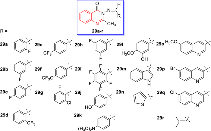
In 2021, compounds 29a–r (Fig. 16) were synthesized by Manhas et al. [85] using materials reported in the relevant literature to react with various aldehyde condensations. All synthesized quinazolin-4-one Schiff bases 29a–r were subjected to in vitro antibacterial screening, and the zone of inhibition was determined at a concentration of 1.0 mg/mL, and the zone of inhibition between 7 and 9 mm and was considered effective [86]. Monofluoroquinazoline-4-ketone was not effective against MRSA, but the p-trifluoromethyl derivative 29e, the 3-oxygenated trifluoromethyl derivative 29f, the 2-chloro-6-fluoro derivative 29g, and the 3,5-difluoro derivative 29h exhibited good antibacterial effects against MRSA. Notably, dimethylaminophenyl and 3-hydroxy-4-methoxyphenyl derivatives 29k and 29l were also active against MRSA. Therefore, these compounds could be used as lead compounds for modifying their structures and improving their anti-MRSA activities.
During 2000–2016, Dinakaran et al., Strigacova et al., and Wang et al. [87-89] synthesized three series of quinoline-4-carboxylic acid derivatives on the basis of quinoline-4-carboxylic acid and evaluated their antibacterial activities. Among them, compound 30a (Fig. 17) showed good antibacterial activity against MRSA with an MIC value of 512 µg/mL, which was comparable to the antibacterial activity of ampicillin (Fig. 17). Additionally, MTT assay showed that compound 30a had low cytotoxicity. SARs revealed that 2-phenyl ortho amide side chain length had a significant effect on the antibacterial activity, for the same end of the alkaline, with a long side chain (n = 2, three bonds between the basic N terminus and carbonyl group) compounds exhibited stronger antibacterial activity than those derivatives with shorter side chain and a carbonyl group (n = 1, two bonds between the basic N terminus and carbonyl group), respectively. The cyclic amino group at 2-phenyl group can increase the antibacterial activity of quinoline-4-carboxylic acid derivatives against MRSA.

In 2012, Sevgi et al. [90] synthesized several quinoline-based ethylenedione derivatives and N-(8-hydroxyquinolin-5-yl)-aminoethylenedione and evaluated their anti-MRSA activities. It was worth mentioning that compound 31 (Fig. 17) exhibited the most effective anti-MRSA activity with an inhibition zone of 12 mm and an MIC value of 32 µg/mL. Among the dihydropyrimidinone derivatives synthesized by Paul et al. [91], compound 32 (Fig. 17) displayed good antibacterial activity against MRSA with an MIC value of 3.5 µg/mL, superior to the positive control streptomycin (MIC = 3.9 µg/mL).
The compound 5-chloro-13-phenethyl-13,14-dihydro-2H-[1,3]oxazino[5,6-H]quinoline (33, Fig. 17) was first reported by Enquist's research group [92], which had a unique scaffold and was effective against Gram-negative bacteria, including E. coli and Pseudomonas aeruginosa. In 2019, Fu's group firstly elucidated the new mechanism of action of compound 33 against E. coli, mainly through blocking lipopolysaccharide (LPS) transport (Lpt) A-LptC interaction by targeting LptA [93]. Using compound 33 as a precursor, a series of oxazole-quinoline derivatives were designed and another series of quinoline derivatives with 5-chloro atoms were constructed, while the hybrid compound 33c was prepared by linking a quinolone fragment as a special substituent to the quinoline core (Fig. 17) [94]. The antibacterial experiments showed that compounds 33a, 33b, and 33c had potent effects on drug-resistant strain MRSA, with MIC values of 8 µg/mL. The results of bacterial molecular docking studies revealed that compound 33c had the advantage of dual-targeting mechanism of LptA and topoisomerase IV.
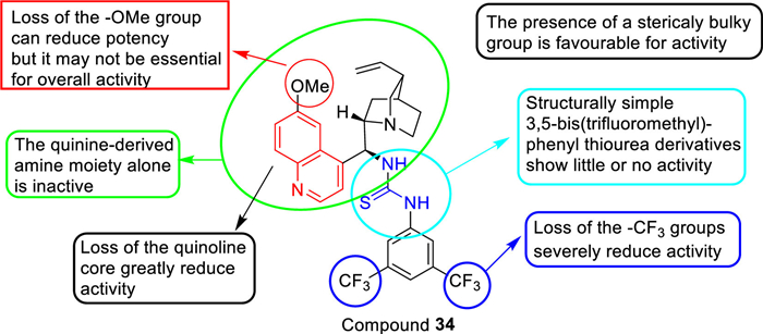
Among the thiourea-containing quinine derivatives (Fig. 18) synthesized by Dolan et al. [95], compound 34 was the most active quinine derivative with an MIC90 value of 17.74 µg/mL, which was higher than the reported MIC90 value of 1.35 µg/mL or 2 mg/L for vancomycin against MRSA. The SARs in Fig. 18 indicated that: (1) loss of the -OMe group can reduce antibacterial potency but it may not be essential for overall activity and loss of the quinoline and -CF3 severely reduce anti-MRSA activity; (2) The presence of a sterically bulky group was favorable for anti-MRSA activity and the quinine-derived amine moiety alone was inactive; (3) Structurally simple 3,5-bis(trifluoromethyl)-phenyl thiourea derivatives exhibit little or no anti-MRSA activity. Additionally, toxicity evaluation showed that 34 was not only non-toxic to Galleria mellonella larvae but also did not appear to impede larvae development. Furthermore, compound 34 was also non-toxic to G. mellonella larvae at concentrations up to 1000 µg/mL. Encouraged by these results, thiourea-containing quinine derivative 34 had great potential as a new antibacterial agent against MRSA.
Here, we briefly summarized the anti-MRSA mechanisms of some representative quinoline analogues based on the types of targets and the process of action between quinoline analogues and bacteria, mainly including the interaction of DNA topoisomerase, disruption of cell membranes and inhibition of the FtsZ associated with cell division.
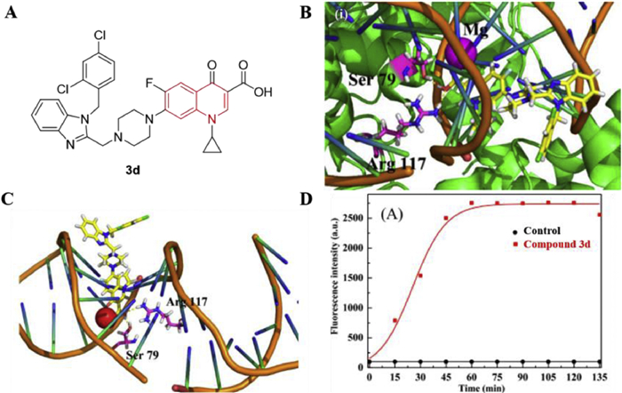
It is well known that DNA topoisomerases are the main targets of quinolone antibiotics. Herein, compound 3d was able to inhibit the relaxation activity of EcTopo IV at a concentration of 10 µmol/L [40]. Interactions of compound 3d (Fig. 19A) with DNA topoisomerase IV receptor were shown in Figs. 19B and C, the carboxyl group of this molecule was very close to residue Arg117 of the DNA topoisomerase IV complex. Compound 3d can also form hydrogen bonds with Ser79 of the DNA topoisomerase IV complex via the hydrogen atom of the carboxyl group. In addition, compound 3d can insert into the superhelical DNA of the enzyme-DNA complex. This synergistic binding may facilitate the formation of a stable quinolone-DNA enzyme complex, which may account for the strong inhibitory effect of 3d on MRSA. Moreover, Zhang et al. [40] used propidium iodide (PI) to study the ability of 3d to penetrate bacterial cell membranes and found that 3d can cross the damaged bacterial cell membranes and bind to DNA to emit stronger fluorescence (Fig. 19D).
The bacterial cell membrane is one of the important structures on which bacteria depend for survival and is rich in enzyme systems that perform many important metabolic functions. In 2010, Hu et al. reported that a quinoline derivative HT61 (1) can act on the plasma membrane of bacterial cells by disrupting the bacterial cell membrane potential of MSSA, resulting in the release of cellular contents [38]. The polarization effect of the quinoline derivative HT61 on MSSA bacterial membranes was detected by the fluorescent probe 3,3-dipropylthiadicarbocyanine iodide (DiSC3(5)). As shown in Fig. S1A (Supporting information), 1 min after the addition of HT61, the fluorescence intensity was enhanced in a concentration-dependent manner for the stationary phase MSSA. While the fluorescence intensity did not increase significantly after treatment for 1 min. This result suggested that the maximum depolarization of the cytoplasmic membrane of stationary phase MSSA occurred within 1 min to 4 min after being treated with HT61. However, for log phase MSSA (Fig. S1B, Supporting information), the release of fluorescence after HT61 treatment was slower than that of the stationary phase and achieved a peak after 45 min. It appears that the increased fluorescence intensity was concentration-dependent only at lower concentrations of HT61. When HT61 reached higher concentrations, the fluorescence release did not increase when the concentration of HT61 was higher than 20 mg/mL. Furthermore, transmission electron micrographs showed that after treatment with HT61 for 10 min, the cell membrane of S. aureus was disrupted and the cytoplasm exuded into the extracellular space (Fig. S2B, Supporting information). Additionally, after treatment with HT61 at higher concentrations, the cell wall cracked and the cell contents were expelled (Figs. S2C and D, Supporting information).
FtsZ is highly conserved in bacteria and is essential for cell division [96]. Recent studies have reported that quinoline derivatives induce cell death in antibacterial assays possibly due to their inhibitory effect on the GTPase activity of FtsZ, making FtsZ a novel target for the development of broad-spectrum antibiotics [97]. As shown in Fig. S3 (Supporting information), compound 17b strongly inhibited Sa-FtsZ polymerization in a dose-dependent manner, exhibiting a significant inhibition of FtsZ polymerization at a concentration of 16 µg/mL [65]. The effect of the N-methylbenzindolo[3,2-b]quinoline derivative 17b on FtsZ polymerization was also analyzed by transmission electron microscopy by Sun et al. [65]. As displayed in Fig. S4A (Supporting information), a dense network of FtsZ protofilaments with an average width of 104 ± 18 nm was observed in the absence of 17b. While a concentration of 8 µg/mL of 17b greatly reduced the size and thickness of FtsZ polymer and the bundling of FtsZ protofilaments (Fig. S4B, Supporting information). Since the GTPase activity of FtsZ correlates with the dynamic polymerization of FtsZ [98], the effect of compound 17b on the dynamic polymerization of FtsZ was investigated. In the GTPase assay, 17b showed about 20% inhibition at a concentration of 4 µg/mL (Fig. S4C, Supporting information). However, approximately 50% and 60% inhibition were reached at concentrations of 8 and 16 µg/mL, respectively. These results suggested that 17b significantly inhibited the GTPase activity of FtsZ in a dose-dependent manner. In 2018, Fang et al. [83] found that compound 27 inhibited bacterial growth by inhibiting the GTPase activity of FtsZ. They used transmission electron microscopy to find a significant increase in the size of the FtsZ polymer after incubation of FtsZ with 27 at a concentration of 1.5 mg/mL (Figs. S4D and E, Supporting information). Moreover, compound 27 at 0.75 mg/mL showed about 20% inhibitory activity, and 27 at concentrations of 1.5, 3, and 6 mg/mL can achieve 50%, 70%, and 80% inhibition effects (Fig. S4F, Supporting information). These results suggested that the anti-MRSA activity of compounds 17b and 27 may be due to their inhibitory effects on GTPase activity and FtsZ polymerization.
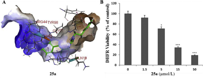
DHFR, is involved in the biosynthetic pathway of folate, a nitrogenous base and precursor of some amino acids. DHFR is one of the key components of this pathway and inhibition of DHFR can be lethal to bacteria. Thus, DHFR can be used as a target against pathogenic bacteria. Bai et al. [80] reported that compound 25a was well inserted into the active pocket of S. aureus DHFR (Fig. 20A) and it could reduce the activity of DHFR by 81% at a concentration of 50 µmol/L compared to the negative control (Fig. 20B). It was confirmed that compound 25a showed an inhibitory effect on DHFR, indicating that DHFR may be a potential antibacterial target [80].
In this review, we addressed what is known about the antimicrobial activity of quinoline analogues over the past two decades, particularly against MRSA, their SARs, as well as mechanisms. Quinoline analogues have a wide range of biological and pharmaceutical activities, and a number of quinoline analogues have been designed, developed, and screened against Gram-positive, Gram-negative, and MDR bacteria in recent two decades. Many quinoline analogues exhibited more potent antibacterial activity against MRSA than the reference antibiotics and were less toxic. The SARs suggested that the anti-MRSA activity of quinoline analogues is related to the position, species, and spatial relationships of the various substituents. Their abundant SARs may provide better insights for further rational development of quinoline-based antimicrobials to combat MRSA infections. Additionally, the elucidation of the anti-MRSA mechanism of some representative quinoline analogues will provide a reference for the design of novel quinoline-based antibacterial drugs in the future.
The authors declare that they have no known competing financial interests or personal relationships that could have appeared to influence the work reported in this paper.
We thank the National Natural Science Foundation of China (No. 32272575) and National College Student Innovation and Entrepreneurship Training Program (No. 202210459164) for financial support.
Supplementary material associated with this article can be found, in the online version, at doi:
A. Asadi, S. Razavi, M. Talebi, M. Gholami, Infection 47 (2019) 13–23. doi: 10.1007/s15010-018-1222-5
L.N. Silva, K.R. Zimmer, A.J. Macedo, D.S. Trentin, Chem. Rev. 116 (2016) 9162–9236. doi: 10.1021/acs.chemrev.6b00184
A.R. Brochado, A. Telzerow, J. Bobonis, et al., Nature 559 (2018) 259–263. doi: 10.1038/s41586-018-0278-9
E.D. Brown, G.D. Wright, Nature 529 (2016) 336–343. doi: 10.1038/nature17042
K. Bush, P. Courvalin, G. Dantas, et al., Nat. Rev. Microbiol. 9 (2011) 894–896. doi: 10.1038/nrmicro2693
W. Cheng, T. Xu, L. Cui, et al., J. Med. Chem. 66 (2023) 962–975. doi: 10.1021/acs.jmedchem.2c01793
N. Koyama, J. Inokoshi, H. Tomoda, Molecules 18 (2012) 204–224. doi: 10.3390/molecules18010204
H.F. Chambers, F.R. Deleo, Nat. Rev. Microbiol. 7 (2009) 629–641. doi: 10.1038/nrmicro2200
H. Grundmann, M. Aires-de-Sousa, J. Boyce, E. Tiemersma, Lancet 368 (2006) 874–885. doi: 10.1016/S0140-6736(06)68853-3
Z. Yi, X. Xu, X. Meng, et al., Chin. Chem. Lett. 34 (2023) 108238. doi: 10.1016/j.cclet.2023.108238
T.Y. Zhang, C. Li, Y.S. Tian, et al., Chin. Chem. Lett. 28 (2017) 1737–1742. doi: 10.1016/j.cclet.2017.05.022
R. Hope, D.M. Livermore, G. Brick, M. Lillie, R. Reynolds, J. Antimicrob. Chemother. 62 (2008) 65–74. doi: 10.1093/jac/dkn166
P.D. Stapleton, P.W. Taylor, Sci. Prog. 85 (2002) 57–72. doi: 10.3184/003685002783238870
Y. Guo, E. Hou, T. Wen, et al., J. Med. Chem. 64 (2021) 12903–12916. doi: 10.1021/acs.jmedchem.1c01073
C. Liu, H. Shen, S. Wang, et al., Chin. Chem. Lett. 29 (2018) 1824–1828. doi: 10.1016/j.cclet.2018.10.025
R. Yang, E. Hou, W. Cheng, et al., J. Med. Chem. 65 (2022) 16879–16892. doi: 10.1021/acs.jmedchem.2c01674
Y. Bouzian, Y. Sert, K. Khalid, et al., J. Mol. Struct. 1246 (2021) 131217. doi: 10.1016/j.molstruc.2021.131217
S.I. Eissa, A.M. Farrag, S.Y. Abbas, et al., Bioorg. Chem. 110 (2021) 104803. doi: 10.1016/j.bioorg.2021.104803
L. Xu, X. Mu, M. Liu, et al., Chin. Chem. Lett. 34 (2023) 108063. doi: 10.1016/j.cclet.2022.108063
B. Sureshkumar, Y.S. Mary, C.Y. Panicker, et al., Arab. J. Chem. 13 (2020) 632–648. doi: 10.1016/j.arabjc.2017.07.006
H. Kumar, V. Devaraji, R. Joshi, et al., RSC Adv. 5 (2015) 65496–65513. doi: 10.1039/C5RA08778C
A.M. Ghanim, A.S. Girgis, B.M. Kariuki, et al., Bioorg. Chem. 119 (2022) 105557. doi: 10.1016/j.bioorg.2021.105557
K. Douadi, S. Chafaa, T. Douadi, M. Al-Noaimi, I. Kaabi, J. Mol. Struct. 1217 (2020) 128305. doi: 10.1016/j.molstruc.2020.128305
K.B. Patel, P. Kumari, J. Mol. Struct. 1268 (2022) 133634. doi: 10.1016/j.molstruc.2022.133634
R.D. Overacker, S. Banerjee, G.F. Neuhaus, et al., Bioorg. Med. Chem. 27 (2019) 3595–3604. doi: 10.1016/j.bmc.2019.06.044
T.G. Shruthi, S. Eswaran, P. Shivarudraiah, et al., Bioorg. Med. Chem. Lett. 29 (2019) 97–102. doi: 10.1016/j.bmcl.2018.11.002
J. Mo, H. Yang, T. Chen, et al., Bioorg. Chem. 93 (2019) 103310. doi: 10.1016/j.bioorg.2019.103310
V.G. Ugale, H.M. Patel, S.J. Surana, Arab. J. Chem. 10 (2017) S1980–S2003. doi: 10.1016/j.arabjc.2013.07.026
A.A. Boteva, O.P. Krasnykh, Chem. Heterocycl. Compd. 45 (2009) 757–785. doi: 10.1007/s10593-009-0360-1
S.B.S. Kumar, H. Gupta, Med. Chem. 9 (2009) 1648–1654.
T.L. Lauderdale, Y.R. Shiau, J.F. Lai, H.C. Chen, C.H. King, Antimicrob. Agents Chemother. 54 (2010) 1338–1342. doi: 10.1128/AAC.01197-09
C.R. Li, Y. Li, G.Q. Li, et al., J. Antimicrob. Chemother. 65 (2010) 2411–2415. doi: 10.1093/jac/dkq341
J.M. Remy, C.A. Tow-Keogh, T.S. McConnell, J.M. Dalton, J.A. Devito, J. Antimicrob. Chemother. 67 (2012) 2814–2820. doi: 10.1093/jac/dks307
B. Li, J. Yao, J. Wei, et al., J. Global. Antimicrob. Resist. 12 (2018) 77–78. doi: 10.1016/j.jgar.2017.12.004
Y.Q. Hu, S. Zhang, Z. Xu, et al., Eur. J. Med. Chem. 141 (2017) 335–345. doi: 10.1016/j.ejmech.2017.09.050
G.F. Zhang, S. Zhang, B. Pan, X. Liu, L.S. Feng, Eur. J. Med. Chem. 143 (2018) 710–723. doi: 10.1016/j.ejmech.2017.11.082
Y. Hu, A.R. Coates, J. Antimicrob. Chemother. 68 (2013) 374–384. doi: 10.1093/jac/dks384
Y. Hu, A. Shamaei-Tousi, Y. Liu, A. Coates, PLoS One 5 (2010) e11818. doi: 10.1371/journal.pone.0011818
L.Z. Gao, Y.S. Xie, T. Li, W.L. Huang, G.Q. Hu, Chin. Chem. Lett. 26 (2015) 149–151. doi: 10.1016/j.cclet.2014.09.017
L. Zhang, D. Addla, J. Ponmani, et al., Eur. J. Med. Chem. 111 (2016) 160–182. doi: 10.1016/j.ejmech.2016.01.052
X. Huang, Y. Bao, S. Zhu, et al., Bioorg. Med. Chem. Lett. 25 (2015) 3928–3932. doi: 10.1016/j.bmcl.2015.07.044
T. Zhang, W. Shen, M. Liu, et al., Eur. J. Med. Chem. 104 (2015) 73–85. doi: 10.1016/j.ejmech.2015.09.030
H.J. Yun, Y.H. Min, J.A. Lim, et al., Antimicrob. Agents. Chemother. 46 (2002) 3071–3074. doi: 10.1128/AAC.46.9.3071-3074.2002
S.F. Cui, Y. Ren, S.L. Zhang, et al., Bioorg. Med. Chem. Lett. 23 (2013) 3267–3272. doi: 10.1016/j.bmcl.2013.03.118
S.F. Cui, D. Addla, C.H. Zhou, J. Med. Chem. 59 (2016) 4488–4510. doi: 10.1021/acs.jmedchem.5b01678
M. Norouzbahari, S. Salarinejad, M. Güran, et al., Daru, J. Pharm. Sci. 28 (2020) 661–672. doi: 10.1007/s40199-020-00373-6
D.T. Chu, Patent, US4687770, 1987.
D.T. Chu, Patent, US4689325, 1987.
J.A. Wiles, Q. Wang, E. Lucien, et al., Bioorg. Med. Chem. Lett. 16 (2006) 1272–1276. doi: 10.1016/j.bmcl.2005.11.065
J.A. Wiles, Y. Song, Q. Wang, et al., Bioorg. Med. Chem. Lett. 16 (2006) 1277–1281. doi: 10.1016/j.bmcl.2005.11.064
J.A. Wiles, A. Hashimoto, J.A. Thanassi, et al., J. Med. Chem. 49 (2006) 39–42. doi: 10.1021/jm051066d
Q.L. Wang, E. Lucien, A. Hashimoto, et al., J. Med. Chem. 50 (2007) 199–210. doi: 10.1021/jm060844e
H.Y. Kim, J.A. Wiles, Q. Wang, et al., J. Med. Chem. 54 (2011) 3268–3282. doi: 10.1021/jm101604v
F. O'donnell, T.J. Smyth, V.N. Ramachandran, W.F. Smyth, Int. J. Antimicrob. Agents. 35 (2010) 30–38. doi: 10.1016/j.ijantimicag.2009.06.031
Y. Abouelhassan, A.T. Garrison, G.M. Burch, et al., Bioorg. Med. Chem. Lett. 24 (2014) 5076–5080. doi: 10.1016/j.bmcl.2014.09.009
A. Basak, Y. Abouelhassan, V.M. Norwood, et al., Chemistry 22 (2016) 9181–9189. doi: 10.1002/chem.201600926
D.J. Musk Jr, P.J. Hergenrother, Curr. Med. Chem. 13 (2006) 2163–2177. doi: 10.2174/092986706777935212
U. Römling, C. Balsalobre, J. Intern. Med. 272 (2012) 541–561. doi: 10.1111/joim.12004
A.T. Garrison, Y. Abouelhassan, H. Yang, et al., MedChemComm 8 (2017) 720–724. doi: 10.1039/C6MD00381H
Y. Abouelhassan, A.T. Garrison, F. Bai, et al., ChemMedChem 10 (2015) 1157–1162. doi: 10.1002/cmdc.201500179
J.P.H. Fan, M.T. Hamann, J.F. Hu, Chem. Rev. 108 (2008) 264–287. doi: 10.1021/cr078199m
T.K. Wood, S.H. Hong, Q. Ma, Trends. Biotechnol. 29 (2011) 87–94. doi: 10.1016/j.tibtech.2010.11.001
J.H. Lee, J. Lee, FEMS Microbiol. Rev. 34 (2010) 426–444. doi: 10.1111/j.1574-6976.2009.00204.x
C. Challa, J. Ravindran, M.M. Konai, et al., ACS Omega 2 (2017) 5187–5195. doi: 10.1021/acsomega.7b00840
N. Sun, R.L. Du, Y.Y. Zheng, et al., Eur. J. Med. Chem. 135 (2017) 1–11. doi: 10.1016/j.ejmech.2017.04.018
H. Yang, H.W. Wang, T.W. Zhu, et al., Eur. J. Med. Chem. 127 (2017) 166–173. doi: 10.1016/j.ejmech.2016.12.054
L.M. Oppegard, B.L. Hamann, K.R. Streck, et al., Antimicrob. Agents. Chemother. 53 (2009) 2110–2119. doi: 10.1128/AAC.01440-08
X.W. Wu, Z.P. Wu, L.X. Wang, et al., Eur. J. Med. Chem. 46 (2011) 4625–4633. doi: 10.1016/j.ejmech.2011.07.042
L.K. An, Z.C. Li, X.W. Wu, X.Y. Zou, L.Q. Gu, Afr. J. Pharm. Pharmacol. 7 (2013) 1020–1025. doi: 10.5897/AJPP12.544
Y. Cheng, L.K. An, N. Wu, et al., Bioorg. Med. Chem. 16 (2008) 4617–4625. doi: 10.1016/j.bmc.2008.02.036
M. Yanato, Y. Takeuchi, M.R. Chang, K. Hashigaki, et al., Chem. Pharm. Bull. 38 (1990) 3048–3052. doi: 10.1248/cpb.38.3048
A. Paulo, A. Duarte, E.T. Gomes, J. Ethnopharmacol. 44 (1994) 127–130. doi: 10.1016/0378-8741(94)90079-5
L.G. Mardenborough, X.Y. Zhu, P. Fan, et al., Bioorg. Med. Chem. 13 (2005) 3955–3963. doi: 10.1016/j.bmc.2005.04.008
X.Y. Zhu, L.G. Mardenborough, S. Li, et al., Bioorg. Med. Chem. 15 (2007) 686–695. doi: 10.1016/j.bmc.2006.10.062
S. Shiota, M. Shimizu, T. Mizushima, H. Ito, et al., Biol. Pharm. Bull. 22 (1999) 1388–1390. doi: 10.1248/bpb.22.1388
M. Zhao, T. Kamada, A. Takeuchi, et al., Bioorg. Med. Chem. Lett. 25 (2015) 5551–5554. doi: 10.1016/j.bmcl.2015.10.058
M. Shiria, A. Nejatinejhad-Arani, Z. Faghihi, S.A. Shintre, N.A. Koorbanally, Org. Chem. Res. 2 (2016) 113–119.
M.O. Puskullu, I. Celik, M. Erol, et al., Bioorg. Chem. 101 (2020) 104014. doi: 10.1016/j.bioorg.2020.104014
M. Guo, C.J. Zheng, M.X. Song, et al., Bioorg. Med. Chem. Lett. 23 (2013) 4358–4361. doi: 10.1016/j.bmcl.2013.05.082
X. Bai, Y. Chen, Z. Liu, et al., Chem. Biodivers. 16 (2019) e1900056. doi: 10.1002/cbdv.201900056
H.R. Piao, Med. Chem. 4 (2014) 441–448.
W. Cieslik, R. Musiol, J.E. Nycz, et al., Bioorg. Med. Chem. 20 (2012) 6960–6968. doi: 10.1016/j.bmc.2012.10.027
Z. Fang, L. Ban, Y. Li, et al., J. Pharmacol. Sci. 137 (2018) 283–289. doi: 10.1016/j.jphs.2018.07.005
P. Teng, C. Li, Z. Peng, et al., Bioorg. Med. Chem. 26 (2018) 3573–3579. doi: 10.1016/j.bmc.2018.05.031
N. Manhas, P. Singh, N.A. Koorbanally, Polycycl. Aromat. Compd. 42 (2022) 5183–5195. doi: 10.1080/10406638.2021.1926293
D.B. Farag, N.A. Farag, A. Esmat, et al., MedChemComm 6 (2015) 283–299. doi: 10.1039/C4MD00392F
M. Dinakaran, S. Palaniappan, Y. Perumal, et al., Med. Chem. 4 (2008) 482–491. doi: 10.2174/157340608785700225
J. Strigacova, D. Hudecová, L'. Varečka, A. Lásiková, D. Végh, Folia Microbiol. 45 (2000) 305–309. doi: 10.1007/BF02817551
X. Wang, X. Xie, Y. Cai, et al., Molecules 21 (2016) 340. doi: 10.3390/molecules21030340
F. Sevgi, A.D. Bedük, World. Appl. Sci. J. 19 (2012) 192–197.
D. Paul, R.G. Reddy, S.P. Rajendran, J. Chil. Chem. Soc. 63 (2018) 3974–3982. doi: 10.4067/s0717-97072018000203974
P.A. Enquist, A. Gylfe, U. Hägglund, et al., Bioorg. Med. Chem. Lett. 22 (2012) 3550–3553. doi: 10.1016/j.bmcl.2012.03.096
X. Zhang, Y. Li, W. Wang, et al., Int. J. Antimicrob. Agents 53 (2019) 442–448. doi: 10.1016/j.ijantimicag.2018.11.016
H.G. Fu, Z.W. Li, X.X. Hu, et al., Molecules 24 (2019) 548. doi: 10.3390/molecules24030548
N. Dolan, D.P. Gavin, A. Eshwika, et al., Bioorg. Med. Chem. Lett. 26 (2016) 630–635. doi: 10.1016/j.bmcl.2015.11.058
J. Lutkenhaus, S. Pichoff, S. Du, Cytoskeleton 69 (2012) 778–790. doi: 10.1002/cm.21054
G.M. Nepomuceno, K.M. Chan, V. Huynh, et al., ACS Med Chem. Lett. 6 (2015) 308–312. doi: 10.1021/ml500497s
X Yang, Z. Lyu, A. Miguel, et al., Science 355 (2017) 744–747. doi: 10.1126/science.aak9995

Figure 18 SARs of quinine-based thiourea 34. Reproduced with permission [95], Copyright 2016, Elsevier publications.

Figure 19 (A) Structure of 3d; (B, C) Three-dimensional conformation of 3d docked in topoisomerase IV–DNA complex; (D) Mechanism of antibacterial action of 3d at concentrations of 12× MIC. Reproduced with permission [40], Copyright 2016, Elsevier publications.

Figure 20 Inhibition of DHFR activities of compound 25a. Reproduced with permission [80], Copyright 2019, John Wiley publications. *P < 0.05, ***P < 0.001, significant with respect to the control (0 µmol/L) group.

 扫一扫看文章

扫一扫关注我们


 下载:
下载:


