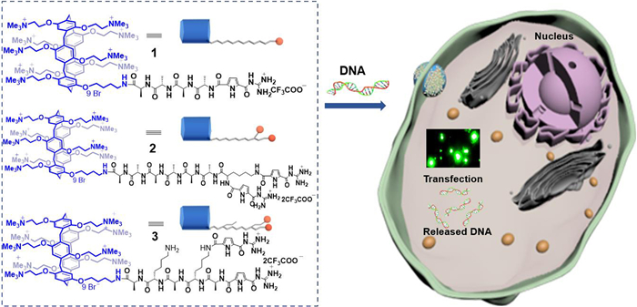
Scheme 1.
Chemical structures and cartoon representations of peptides 1–3. Schematic illustration of uptake and release process mediated by peptide plasmid DNA (pDNA) nanoparticles.
Pillar[5]arene–modified peptide-guanidiniocarbonylpyrrol amphiphiles with gene transfection properties
Kaiya Wang , Minzan Zuo , Tao Zhang , Huilan Yue , Xiao-Yu Hu

The concept of gene therapy by transferring exogenous DNA into cells was initially proposed by visionary scientists nearly half a century ago [1]. Efficient and controlled delivery of genetic information into living cells is crucial for both fundamental research and biomedical applications like genome analysis and gene therapy [2]. Due to the existence of barriers and poor stability in biological media, naked nucleic acids cannot directly penetrate lipid membranes and thus exhibit limited capacity to transfect cells [3]. Therefore, specially designed artificial gene delivery vectors are highly demanding and of great interest. Recombinant viruses such as replication-defective retro- and adeno-associated viruses have been proved to be efficient gene delivery vehicles. Nevertheless, their widespread applications and scale-up are limited by severe drawbacks such as toxicity, intrinsic immunogenicity, and high production costs [4]. Alternatively, the use of nonviral vectors can in principle circumvent these issues, and can compact and transport more genetic information [5]. Two main traditional classes of nonviral vectors are designed based on cationic lipids or polymers [6], among which cationic amphiphilic lipids have become the most promising vectors for gene transfection [7]. Supramolecular macrocyclic amphiphiles, featuring persistent shapes and highly ordered assembly, have developed several strategies for this application [8-11]. In particular, pillararenes, among countless supramolecular hosts [12-18], are a family of well-studied macrocycles with symmetric and rigid cavities that can bind with a variety of guests [19-23]. The ability of cationic pillararenes acting as gene carrier to bind, condense, and deliver DNA across cell membranes has been previously investigated [24-29]. Nevertheless, their gene transfection ability is only sporadically reported. In addition, it is also well-known that weakly basic guanidinocarbonylpyrrole (GCP) can efficiently bind to cell membrane and penetrate cells, delivering a large variety of genetic cargos and exhibiting excellent transfection efficiency [30-34]. We have previously reported that pillar[5]arene-modified amphiphilic peptide 1 (Scheme 1) bearing one GCP moiety was able to condense plasmid DNA and enable cell transfection in a manner that was dependent on the self-assembled morphology and conformation [35].
We thus hypothesize that better performing vectors will be engineered by incorporating more GCP moieties into the skeleton of pillar[5]arene to improve the gene transfection efficiency. In this work, peptides 2 and 3 bearing two GCP units were synthesized and expected to demonstrate superior transfection efficiency (Scheme 1). However, to our surprise, peptide 2 showed only little transfection activity, whereas peptide 3 was proved to perform cell transfection quite efficiently. The rationale for this divergence is that the two GCP moieties in peptide 2 are attached to the rims with four methylene length difference, which inhibited them from interacting with cytomembrane simultaneously. In contrast, the two GCP moieties in peptide 3 can adopt parallel conformation to bind with cytomembrane cooperatively, thus making it an efficient gene vector. The subtle structural difference in peptides 2 and 3 could result in distinct transfection efficacy, making it possible to gain an in-depth understanding of their structure-activity relationship. This research presents a good example of rational structural design in achieving effective gene transfection vectors.
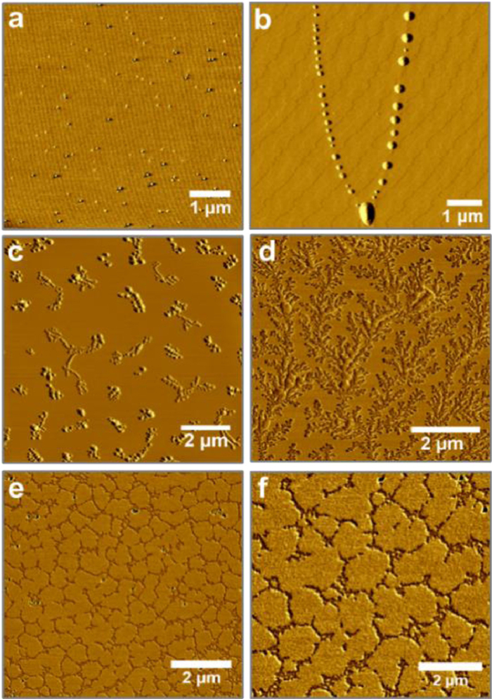
Peptide 1 was synthesized according to the previously reported methods and used as a comparison in this study [35]. Peptides 2 and 3 were achieved by coupling mono-alkylamine modified nona-cationic pillar[5]arene with GCP-conjugated peptides, which were prepared by Fmoc solid peptide synthesis. The resulting peptide amphiphiles were characterized by NMR spectroscopy and HPLC (Figs. S1–S15 in Supporting information). Their self-assembled morphologies were then characterized by atomic force microscopy (AFM). As shown in Fig. 1, at a concentration of 0.2 mmol/L, peptide 1 formed tiny dispersed nanoparticles and pearl-necklace-like assembly could be observed after one day incubation. Freshly prepared samples of peptides 2 and 3 formed aggregated large-sized nanoparticles and cross-linked networks, respectively. Flower-like nanofibers were observed for peptide 2 and there was not much change in morphology for peptide 3 after aging for one day. These morphology differences were ascribed from the fractional difference of the GCP moieties in the structures, resulting in distinct DNA delivery and gene transfection properties.
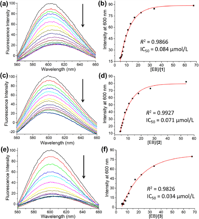
To explore the potential application of peptides 1–3 as gene transfection vectors, their binding properties with calf thymus DNA (ctDNA) were then assessed by ethidium bromide (EB) displacement assay and isothermal titration calorimetry (ITC) measurements. The IC50 values (concentration of peptide required to replace 50% of the initially bound EB, showing fluorescence at 600 nm) of 1 and 2 were roughly the same (0.084 and 0.071 µmol/L), whereas IC50 of peptide 3 was 0.034 µm (Fig. 2). This indicated that the binding affinity of 3 towards DNA was higher than that of 1 and 2, owing to the presence of two parallel GCP moieties. Furthermore, the binding constants for peptides 1–3 with DNA were determined by ITC experiments (Figs. S16–S18 in Supporting information). Both data of peptides 2 and 3 could be fitted by sequential binding site model. The first binding constants (K1) are both in the range of 104 L/mol, whereas the second binding constants (K2) are larger than K1, indicating the positive cooperativity of the two GCP moieties [36]. The cooperative effect is more profound in the case of peptide 3. Moreover, based on our previous studies [30], the morphology of the peptides would change to compact nanoparticles after their binding with DNA. These above results disclosed that peptides 1–3 could bind tightly with DNA and may potentially be applied in gene delivery and transfection.
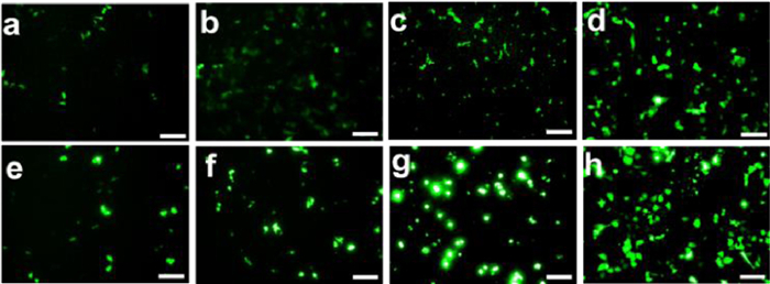
Subsequently, transfection experiments were performed with HeLa cells by fluorescence microscopy (Fig. 3). The commercially available branched polycationic polyethyleneimine (PEI) was used as a control. The cells were treated with the DNA-loaded aggregates formed by peptides 1, 2, and 3, respectively at 37 ℃. Successful delivery of plasmid DNA into cells was monitored by the green fluorescence of green fluorescent protein (GFP) after 24 h and 48 h incubation, respectively. We are pleased to see that peptide 3 is a very efficient transfectant, which is comparable to PEI owing to the cooperative interaction of paired GCPs. It is also noteworthy that no helper lipid was needed in our system, which was commonly applied in other transfection vectors. In contrast, little transfection activity was observed for peptides 1 and 2 due to the single valid GCP moiety in their structures, which confirmed our assumption that GCP moiety is crucial in the transfection process, and more paired GCP moieties will lead to higher transfection efficiency. The transfection results were also confirmed by using a normal cell line HEK 293T (Fig. S19 in Supporting information). The transfection efficacy of peptides 2 and 3 was comparable to PEI after 48 h incubation, but the brightfield images indicated great amount of cell death in the PEI group (Fig. S20 in Supporting information). Cytotoxicity of peptides 1–3 was then assessed on HeLa cells; it was found that peptides 1–3 had negligible cytotoxicity, whereas cell viability of the PEI group was reduced more than 70% (Fig. 4).

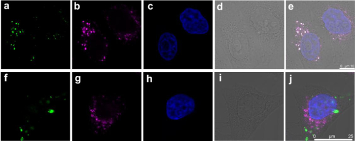
Finally, as shown in Fig. 5, the cellular uptake of the vectors was investigated by confocal laser scanning microscope (CLSM). LAMP1-AF633 antibody and Hoechst were used to label endo/lysosomes (purple fluorescence, Figs. 5b and g) and nuclei (Figs. 5c and h) of HeLa cells, respectively. HeLa cells were treated with the DNA-loaded aggregates formed by peptide 3 at 37 ℃ for 2 h and 6 h, respectively. After incubating for 2 h, green fluorescence could be observed (Fig. 5a), indicating the successful uptake of DNA by HeLa cells. And the green fluorescence overlapped very well with purple fluorescence (Fig. 5e), indicating the endocytosis process was first localized in endo/lysosomes. After 6 h, some green fluorescent signals were observed in cytosol (Figs. 5f and j), which disclosed successful escape of DNA from endo/lysosomes. These findings are ubiquitous among the tested cell line (Fig. S21 in Supporting information), which suggest peptide 3 is able to successfully transfer the labelled DNA into the cell and perform the subsequent gene transfection processes in the cytoplasm.
In conclusion, we have discovered that attaching GCP groups in the rim of water-soluble pillar[5]arenes demonstrates the possibility to enhance the cell transfection ability. Peptides 1–3 were synthesized by coupling water-soluble pillar[5]arenes with short peptide bearing one or two GCP moieties. These amphiphilic peptides can bind to DNA and form condensed complexes, owing to the high positive charges and excellent recognition ability of GCP group. In the case of peptide 2, the two GCP moieties cannot interact with cytomembrane synergistically, making it less efficient and have comparable transfection efficiency with mono-GCP modified peptide 1. Nevertheless, aggregates formed by peptide 3 are proved to be efficient gene transfection vector, owing to cooperative interaction of the two GCP moieties with suitable length. As for the functionalization of the pillararene cavity, positively charged guest molecules may be encapsulated and lead to better performance in gene transfection efficacy. The research is currently underway in our laboratory. The results obtained in this work warrant further investigation of macrocycle incorporated gene delivery platforms and elucidate the structure-activity relationship in this regard.
The authors declare no competing financial interests.
This work was supported by the Natural Science Foundation of Jiangsu Province (Nos. BK20200432, BK20211179), National Natural Science Foundation of China (Nos. 22271154, M-0411), and the Fundamental Research Funds for the Central Universities (Nos. NG2022003, NS2021040).
Supplementary material associated with this article can be found, in the online version, at doi:
T. Friedmann, R. Roblin, Science 175 (1972) 949–955. doi: 10.1126/science.175.4025.949
C.E. Dunbar, K.A. High, J.K. Joung, et al., Science 359 (2018) eaan4672. doi: 10.1126/science.aan4672
K.A. Whitehead, R. Langer, D.G. Anderson, Nat. Rev. Drug Discov. 8 (2009) 129–138. doi: 10.1038/nrd2742
C.E. Thomas, A. Ehrhardt, M.A. Kay, Nat. Rev. Genet. 4 (2003) 346–358. doi: 10.1038/nrg1066
J.P. Behr, Acc. Chem. Res. 45 (2012) 980–984. doi: 10.1021/ar200213g
S. Bhattacharya, A. Bajaj, Chem. Commun. 45 (2009) 4632–4656. doi: 10.1039/b900666b
P.L. Felgner, T.R. Gadek, M. Holm, et al., Proc. Natl. Acad. Sci. U. S. A. 84 (1987) 7413–7417. doi: 10.1073/pnas.84.21.7413
Y.C. Pan, X.Y. Hu, D.S. Guo, Angew. Chem. Int. Ed. 60 (2021) 2768–2794. doi: 10.1002/anie.201916380
R.V. Rodik, A.S. Klymchenko, Y. Mely, V.I. Kalchenko, J. Incl. Phenom. Macrocycl. Chem. 80 (2014) 189–200. doi: 10.1007/s10847-014-0412-8
W.C. Geng, Q. Huang, Z. Xu, R. Wang, D.S. Guo, Theranostics 9 (2019) 3094–3106. doi: 10.7150/thno.31914
X. Chen, H. Yang, X. Song, et al., Chin. Chem. Lett. 34 (2023) 107753. doi: 10.1016/j.cclet.2022.107753
Y. Ding, J. Jiao, B. Sun, et al., Chin. Chem. Lett. 32 (2021) 3539–3543. doi: 10.1016/j.cclet.2021.05.045
K. Wang, J.H. Jordan, X.Y. Hu, L. Wang, Angew. Chem. Int. Ed. 59 (2020) 13712–13721. doi: 10.1002/anie.202000045
J.R. Wu, Y.W. Yang, J. Am. Chem. Soc. 141 (2019) 12280–12287. doi: 10.1021/jacs.9b03559
K. Wang, X. Cai, W. Yao, et al., J. Am. Chem. Soc. 141 (2019) 6740–6747. doi: 10.1021/jacs.9b02287
K. Wang, J.H. Jordan, B.C. Gibb, Chem. Commun. 55 (2019) 11695–11698. doi: 10.1039/c9cc06501f
X. Wu, L. Gao, J. Sun, X.Y. Hu, L. Wang, Chin. Chem. Lett. 27 (2016) 1655–1660. doi: 10.1016/j.cclet.2016.05.004
K. Wang, X. Huang, M. Mohan, et al., Chem. Commun. 58 (2022) 6196–6199. doi: 10.1039/d2cc01491b
T. Ogoshi, S. Kanai, S. Fujinami, T.A. Yamagishi, Y. Nakamoto, J. Am. Chem. Soc. 130 (2008) 5022–5023. doi: 10.1021/ja711260m
K. Wang, J.H. Jordan, K. Velmurugan, et al., Angew. Chem. Int. Ed. 60 (2021) 9205–9214. doi: 10.1002/anie.202010150
K. Wang, X. Tian, J.H. Jordan, et al., Chin. Chem. Lett. 33 (2022) 89–96. doi: 10.1016/j.cclet.2021.06.026
M. Zuo, W. Qian, M. Hao, et al., Chin. Chem. Lett. 32 (2021) 1381–1384. doi: 10.1016/j.cclet.2020.09.033
F. Gao, X. Yu, L. Liu, et al., Chin. Chem. Lett. 34 (2023) 107558. doi: 10.1016/j.cclet.2022.05.072
A.L. Barran-Berdon, M. Martinez-Negro, L. Garcia-Rio, et al., J. Mater. Chem. B 5 (2017) 3122–3131. doi: 10.1039/C6TB02939F
Y. Chang, K. Yang, P. Wei, et al., Angew. Chem. Int. Ed. 53 (2014) 13126–13130. doi: 10.1002/anie.201407272
I. Nierengarten, M. Nothisen, D. Sigwalt, et al., Chem. Eur. J. 19 (2013) 17552–17558. doi: 10.1002/chem.201303029
W. Feng, M. Jin, K. Yang, Y. Pei, Z. Pei, Chem. Commun. 54 (2018) 13626–13640. doi: 10.1039/c8cc08252a
S. Guo, Q. Huang, Y. Chen, et al., Angew. Chem. Int. Ed. 60 (2021) 618–623. doi: 10.1002/anie.202013975
S. Guo, Q. Huang, J. Wei, et al., Nano Today 43 (2022) 101396. doi: 10.1016/j.nantod.2022.101396
H.Y. Kuchelmeister, S. Karczewski, A. Gutschmidt, S. Knauer, C. Schmuck, Angew. Chem. Int. Ed. 52 (2013) 14016–14020. doi: 10.1002/anie.201306929
M. Li, S. Schlesiger, S.K. Knauer, C. Schmuck, Angew. Chem. Int. Ed. 54 (2015) 2941–2944. doi: 10.1002/anie.201410429
M. Li, M. Ehlers, S. Schlesiger, et al., Angew. Chem. Int. Ed. 55 (2016) 598–601. doi: 10.1002/anie.201508714
H. Jiang, X.Y. Hu, S. Mosel, et al., ChemBioChem 20 (2019) 1410–1416. doi: 10.1002/cbic.201800728
X. Liu, K. Wang, M. Externbrink, et al., Chin. Chem. Lett. 31 (2020) 1239–1242. doi: 10.1016/j.cclet.2019.10.036
X.Y. Hu, M. Ehlers, T. Wang, et al., Chem. Eur. J. 24 (2018) 9754–9759. doi: 10.1002/chem.201801315
C.A. Hunter, H.L. Anderson, Angew. Chem. Int. Ed. 48 (2009) 7488–7499. doi: 10.1002/anie.200902490

Scheme 1 Chemical structures and cartoon representations of peptides 1–3. Schematic illustration of uptake and release process mediated by peptide plasmid DNA (pDNA) nanoparticles.

Figure 1 AFM images of (a, c, e) freshly prepared samples of peptides 1–3, and (b, d, f) the samples after incubating for one day.

Figure 2 (a, c, e) Ethidium bromide (EB) displacement titration with 10 µmol/L stock solution of peptides 1, 2 and 3, and (b, d, f) the data is fitted with exponential decay first order function with the IC50 values of 0.084, 0.071 and 0.034 µmol/L, respectively.

Figure 3 Transfection experiments performed with 2 µg pF143-GFP plasmid DNA and peptide 1 (a, e), 2 (b, f), 3 (c, g), PEI (d, h) to HeLa cells for different periods (a–d: 24 h, e–h: 48 h). Scale bar: 50 µm.

Figure 4 Concentration-dependent cytotoxicities of peptides 1, 2, 3, and PEI against HeLa cells after 48 h incubation.

Figure 5 Confocal microscopy images of HeLa cells incubated with peptide 3 (100 µm) for 2 h (a–e) and 6 h (f–j), respectively. (a, f) Green fluorescence (GFP-labelled); (b, g) purple fluorescence (LAMP1-AF633 labelled); (c, h) blue fluorescence (Hoechst labelled); (d, i) bright field and (e, j) all merger together.

 扫一扫看文章

扫一扫关注我们
 DownLoad:
 下载:
下载: