Figure 1.
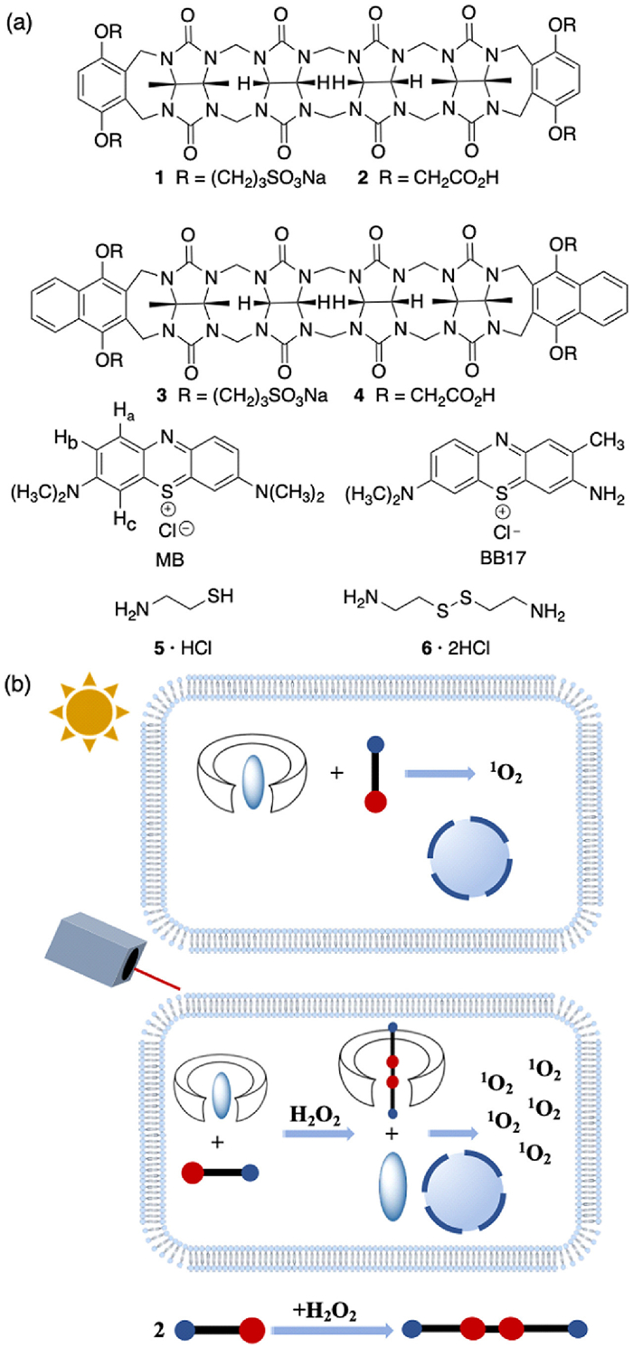
(a) Chemical structures of hosts 1-4, MB, BB17, pro-guest 5 and guest 6. (b) Cartoon depicting reactive oxygen specie activates photodynamic therapy.
Reactive oxygen specie-induced photodynamic therapy activation by supramolecular strategy
Shuyi Wang , Zizhen Zhao , Jiayang Yao , Siyang Jiang , Zhan-Ting Li , Da Ma

Photodynamic therapy (PDT) is an effective treatment method for multiple types of cancer and infectious diseases [1-4]. Several PDT practices have been approved for clinical uses [5-9]. Although known for its negligible systematic toxicity and high selectivity, PDT agents are known to accumulate in skin tissues, which may cause severe skin cytotoxicity [10-13]. To avoid this side effect, patients have to wear protective gears or stay in the dark room for an extended period of time. Therefore, it is highly desired to develop PDT treatment, which only activates PDT effect in the pathological tissues.
Strategies based on nanoscale drug delivery systems (DDSs) and photo quenchers have been reported to reduce side effects of skin cytotoxicity [14-20]. Supramolecular encapsulation is known to have a major impact towards pharmaceutical efficacy [21-29]. Very recently, supramolecular strategy is reported to suppress skin cytotoxicity by removing PDT agents with the assistance of supramolecular organic frameworks [30-33]. We decide to explore a simple and reliable supramolecular strategy to activate PDT in pathological microenvironment by using pro-guest. Pro-guest is designed not to bind with host molecules, which may convert to be competitive guest under suitable conditions to displace encapsulated drugs or dyes inside the host cavity [34-36]. By choosing the appropriate host and pro-guest molecules, pathological microenvironment may trigger the conversion from pro-guest to competitive guest, which leads to the activation of PDT effect.
Here, as a proof of concept study, we report a pro-guest-based supramolecular strategy for PDT selectively activated by reactive oxygen specie (ROS). ROS is often overexpressed in tumor and inflammatory tissues [37-44]. We discover that supramolecular encapsulation is capable of "turning off" the photo cytotoxicity. ROS is found to trigger the release of encapsulated PDT agents, which activates PDT. The cytotoxicity of PDT is evaluated in human cancer and normal cells before or after ROS activation.
As shown in Fig. 1a, two PDT agents methylene blue (MB) and basic blue 17 (BB17) are used as model drugs [45,46]. Acyclic cucurbit[n]uril (CB[n]) hosts 1-4 are used to encapsulate PDT drugs. These host molecules have phenylene or naphthylene walls, and sulfonate or carboxylate side chains. The different backbones and side chains render host molecules varied influences toward encapsulated PDT drugs. Carboxylated acyclic CB[n]s 2 and 4 have a modest solubility in water (0.9 and 0.7 mmol/L, respectively). 2-Thiol ethylamine is used as pro-guest 5. We expect pro-guest 5 will convert to be guest 6 under ROS condition, which may displace encapsulated PDT drugs. As depicted in Fig. 1b, supramolecular encapsulation will shut down the generation of singlet oxygen (1O2) and turn off photo cytotoxicity in normal tissues. By contrast, in ROS overexpressed pathological tissues, PDT drugs are displaced to acitivate the generation of 1O2 and turn on the PDT effect.
First, we investigated the host-guest chemistry. The value of Ka for the complexes formed between host 1 and PDT drugs/pro-guest/guest was determined by direct titration or indicator displacement assay. As listed in Table 1, the value of Ka for the complexes of 1·MB and 1·BB17 was determined to be (6.2 ± 1.0) × 106 L/mol and (9.2 ± 0.5) × 106 L/mol, respectively. The high binding affinity ensured a stable complexation in cellular or even tissue environment. The binding affinity for the complex of host and guest 6 was significantly higher compared to that for the complex of host and pro-guest 5. Hosts 2 and 3 demonstrated similar Ka values as that of host 1. By comparison, host 4 showed a relatively smaller Ka value with MB and BB17, while its binding with compounds 5 and 6 was tighter. Therefore, the conversion of pro-guest 5 to guest 6 may result in a displacement of encapsulated PDT drug.
 DownLoad:
CSV
DownLoad:
CSV

|
Next, the rate of 1O2 generation was determined. The PDT efficacy is often related to the generation of ROS formation rate, especially the formation of 1O2 [47,48]. The rate of 1O2 generation was determined in water with 9,10-anthracenediyl-bis(methylene)dimalonic acid (ABDA) as the indicator.
As shown in Figs. 2a and b, the encapsulation by host molecules showed different impacts towards the 1O2 generation rate of MB and BB17. Hosts 1-3 significantly reduced the 1O2 generation rate. By contrast, host 4 barely changed the 1O2 generation. These observations indicated that different side walls and side chains of the host molecules might have a major impact towards the influence of 1O2 generation.
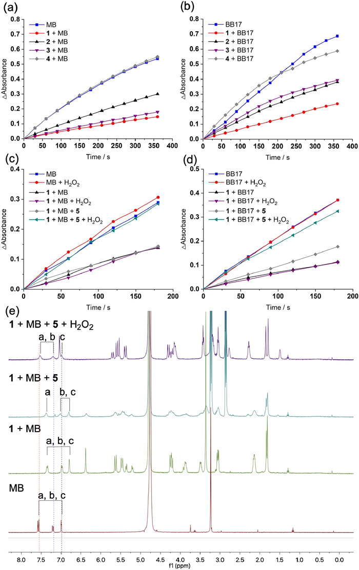
The H2O2-induced recovery of 1O2 generation efficiency was investigated. As shown in Figs. 2c and d, after host 1, PDT drug and pro-guest 5 were incubated in a solution of H2O2 for 4 h, the 1O2 generation rate recovered to a level similar to that of PDT drug alone. These observations showed that a ROS-induced PDT drug displacement had taken place. By contrast, in the absence of H2O2, the 1O2 formation remained on a low level, indicating pro-guest alone was unable to displace the encapsulated drug.
The pro-guest to guest conversion and drug displacement were monitored by fluorescence and 1H NMR spectroscopy. As shown in Fig. S12 (Supporting information), the fluorescence intensity of MB and BB17 was significantly reduced when encapsulated by host molecules. Therefore, the fluorescence intensity could serve as an indicator for the drug release. When incubated in an aqueous solution of H2O2 (100 µmol/L), the fluorescence intensity rapidly increased. 1H NMR spectra also indicated a H2O2-triggered displacement of drugs. As shown in Fig. 2e, when encapsulated by host molecules, proton resonances of Ha-Hc shifted upfield. When incubated in H2O2, the upfield shifted proton resonances of MB shifted downfield, demonstrating the displacement occurred.
Cell study was used to investigate the efficacy of supramolecular strategy based on pro-guest. Human cancer B16 cells were used to mimicking the PDT efficacy in pathological tissues. Briefly, cancer cells were incubated in drug with varied concentration, drug+host 1, drug+host 1+pro-guest 5, or drug+host 1+pro-guest 5+H2O2 for 4 h. Cells were washed and irradiated with laser at 655 nm (0.12 W/cm2) for 5 min each well, and subsequently incubated for another 20 h. Cells were then washed and the viability was evaluated by CCK-8 assay. As shown in Fig. 3a, the addition of host 1 significantly reduced the photo cytotoxicity of MB. The addition of pro-guest 5 in the absence of H2O2 did not significantly reverse the photo cytotoxicity inhibition. By sharp contrast, when incubated with H2O2, the photo cytotoxicity recovered to a level slightly lower than that of MB alone. These observations that the mimicked ROS overexpressed tumor microenvironment is able to activate the photo cytotoxicity of PDT agent MB. Similar observations were shown for BB17 in Fig. 3b.
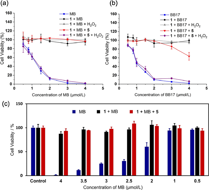
Normal human L02 cells were used to mimic the skin photocytotoxicity. A solar simulator was used to mimic the sunlight and solar photo cytotoxicity. As shown in Fig. 3c, the encapsulation of MB by host 1 was able to significantly suppress the photo cytotoxicity. Since normal tissues lack overexpressed ROS, a solution of MB, host 1 and pro-guest 5 was used to mimick the PDT drugs distributed to normal tissues. As expected, negligible cytoxocity was observed, which confirmed that pro- guest 5 was unable to displace MB and normal tissues could avoid sunlight-caused photo toxicity.
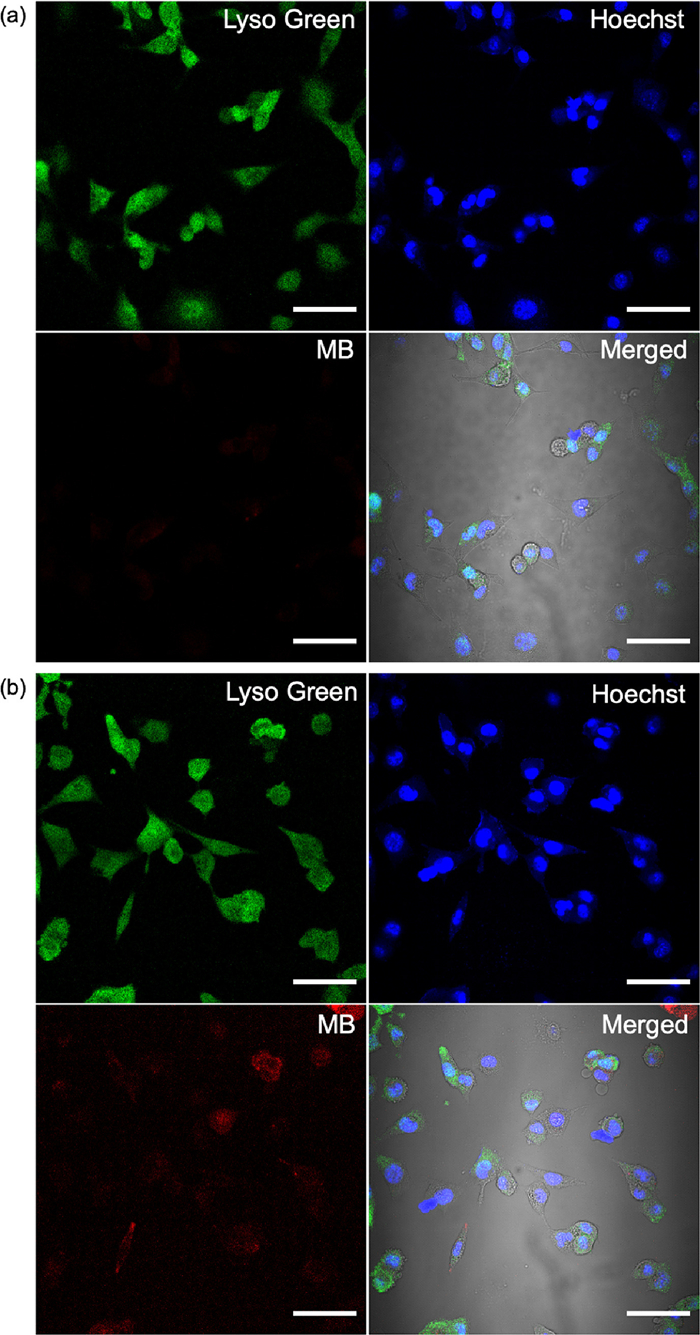
Lastly, confocal laser scanning microscopy (CLSM) was used to image the uptake of MB, which may help explain the mechanism of photo cytotoxicity suppression and recovery. Briefly, B16 cells were incubated with MB, host 1 and pro-guest 5 in the absence or presence of H2O2 for 2 h. Subsequently, cells were stained with Hoechst and Lysotracker Green, and imaged by CLSM. As shown in Fig. 4, in the absence of H2O2, minimal MB uptake was observed. By sharp contrast, when in presence of H2O2, the uptaken efficiency of MB was greatly enhanced. As shown in Fig. S19 (Supporting information), free MB is more capable of being internalized by cells compared to host-encapsulated MB. Therefore, the H2O2-induced displacement of MB may improve cell uptake efficiency and photo cytotoxicity.
In summary, we report a new supramolecular strategy to inhibit photo cytotoxicity of PDT drugs, and reactivate the PDT efficacy in the presence of H2O2. Cell study confirms the rapid recovery of photo cytotoxicity when H2O2 was added. Mechanistic investigation shows that PDT drug displacement and cell uptake enhancement are the reasons for the recovery of cytotoxicity. This strategy may be explored to develop supramolecular PDT drugs with reduced photo toxicity to skin tissues.
The authors declare that they have no known competing financial interests or personal relationships that could have appeared to influence the work reported in this paper.
The authors are grateful to National Natural Science Foundation of China (No. 21921003) for financial support.
Supplementary material associated with this article can be found, in the online version, at doi:
Z. Huang, Cancer Res. Treat. 4 (2005) 283–293.
D.E.J.G.J. Dolmans, D. Fukumura, R.K. Jain, Nat. Rev. Cancer 3 (2003) 380–387. doi: 10.1038/nrc1071
M. Triesscheijn, P. Baas, J.H.M. Schellens, et al., Oncologist 11 (2006) 1034–1044. doi: 10.1634/theoncologist.11-9-1034
P. Agostinis, K. Berg, K.A. Cengel, et al., CA Cancer J. Clin. 61 (2011) 250–281. doi: 10.3322/caac.20114
A.M.R. Fisher, A.L. Murphree, C.J. Gomer, Lasers Surg. Med. 17 (1995) 2–31. doi: 10.1002/lsm.1900170103
H. Abrahamse, M.R. Hamblin, Biochem. J. 473 (2016) 347–364. doi: 10.1042/BJ20150942
S. Kwiatkowski, B. Knap, D. Przystupski, et al., Biomed. Pharmacother. 106 (2018) 1098–1107. doi: 10.1016/j.biopha.2018.07.049
B.C. Wilson, Can. J. Gastroenterol. 16 (2002) 393–396. doi: 10.1155/2002/743109
H. Chen, Y. Wan, X. Cui, S. Li, C. Lee, Adv. Healthc. Mater. 10 (2021) 2101607. doi: 10.1002/adhm.202101607
M. Alexiades-Armenakas, Clin. Dermatol. 24 (2006) 16–25. doi: 10.1016/j.clindermatol.2005.10.027
F.H. Sakamoto, L. Torezan, R.R. Anderson, J. Am. Acad. Dermatol. 63 (2010) 195–211. doi: 10.1016/j.jaad.2009.09.057
P. Lehmann, Hautarzt 58 (2007) 597–603. doi: 10.1007/s00105-007-1363-4
G. Bozzini, P. Colin, N. Betrouni, et al., Photodiagn. Photodyn. Ther. 9 (2012) 261–273. doi: 10.1016/j.pdpdt.2012.01.005
Y. Wang, Y. Lin, H. Zhang, et al., J. Cancer Res. Clin. Oncol. 142 (2016) 813–821. doi: 10.1007/s00432-015-2066-3
X. Li, B.Y. Zheng, M.R. Ke, et al., Theranostics 7 (2017) 2746–2756. doi: 10.7150/thno.18861
G. Lavie, T. Barliya, M. Mandel, et al., Photochem. Photobiol. 83 (2007) 1270–1277. doi: 10.1111/j.1751-1097.2007.00171.x
J. Dillon, J.C. Kennedy, R.H. Pottier, et al., Photochem. Photobiol. 48 (1988) 235–238. doi: 10.1111/j.1751-1097.1988.tb02815.x
B. Huang, P. Wang, Y. Ouyang, et al., ACS Appl. Mater. Interfaces 12 (2020) 41038–41046. doi: 10.1021/acsami.0c10372
B. Bae, K. Na, Biomaterials 31 (2010) 6325–6335. doi: 10.1016/j.biomaterials.2010.04.030
Y. Zhou, H. Li, Y. Yang, Chin. Chem. Lett. 26 (2015) 825–828. doi: 10.1016/j.cclet.2015.01.038
K. Yang, J. Wen, S. Chao, et al., Chem. Commun. 54 (2018) 5911–5914. doi: 10.1039/C8CC02739K
K.X. Teng, L.Y. Niu, Q.Z. Yang, Chem. Sci. 13 (2022) 5951–5956. doi: 10.1039/D2SC01469F
J. Gao, J. Li, W.C. Geng, et al., J. Am. Chem. Soc. 140 (2018) 4945–4953. doi: 10.1021/jacs.8b02331
Q. Duan, Y. Cao, Y. Li, et al., J. Am. Chem. Soc. 135 (2013) 10542–10549. doi: 10.1021/ja405014r
J. Chen, S. Li, Z. Wang, et al., Chem. Sci. 12 (2021) 7727–7734. doi: 10.1039/D1SC01139A
M. He, L. Chen, B. Jiang, et al., Chin. Chem. Lett. 30 (2019) 131–134. doi: 10.1016/j.cclet.2018.10.035
J. Yi, W. Liang, X. Wei, et al., Chin. Chem. Lett. 29 (2018) 87–90. doi: 10.1016/j.cclet.2017.05.004
Y. Yang, Y. Yu, H. Chen, et al., ACS Nano 14 (2020) 13536–13547. doi: 10.1021/acsnano.0c05541
Y. Yang, X. Liu, W. Ma, et al., Biomaterials 265 (2021) 120456. doi: 10.1016/j.biomaterials.2020.120456
Y. Liu, C.Z. Liu, Z.K. Wang, et al., Biomaterials (2022) 121467.
T. Xiao, L. Zhou, X.Q. Sun, et al., Chin. Chem. Lett. 31 (2020) 1–9. doi: 10.1016/j.cclet.2019.05.011
L. Yu, Z. Wang, Z. Mo, et al., Acta Pharm. Sin. B 11 (2021) 2004–2015. doi: 10.1016/j.apsb.2021.02.001
X. Liu, Y. Yang, M. Ling, et al., Adv. Funct. Mater. 31 (2021) 2101709. doi: 10.1002/adfm.202101709
S. Jiang, S. Lan, D. Mao, et al., Chem. Commun. 54 (2018) 9486–9489. doi: 10.1039/C8CC05552A
Y. Jiao, S. Lan, D. Ma, Chin. Chem. Lett. 32 (2021) 1025–1028. doi: 10.1016/j.cclet.2020.08.001
Y.F. Xiao, W.C. Chen, J.X. Chen, et al., ACS Appl. Mater. Interfaces 14 (2022) 5112–5121. doi: 10.1021/acsami.1c23797
Q. Yuan, J. Huang, C. Xian, et al., ACS Appl. Mater. Interfaces 13 (2021) 2165–2178. doi: 10.1021/acsami.0c15133
L. Shi, Y. Wang, C. Zhang, et al., Angew. Chem. Int. Ed. 60 (2021) 9562–9572. doi: 10.1002/anie.202014415
L. Wang, B. Zhu, Y. Deng, et al., Adv. Funct. Mater. 31 (2021) 2101804. doi: 10.1002/adfm.202101804
T. Jin, D. Cheng, G. Jiang, et al., Bioact. Mater. 14 (2022) 42–51. doi: 10.1016/j.bioactmat.2021.12.009
L. Ming, K. Cheng, Y. Chen, et al., Cancer Med. 10 (2021) 257–268. doi: 10.1002/cam4.3592
K.B. Kennel, F.R. Greten, Redox Biol. 42 (2021) 101891. doi: 10.1016/j.redox.2021.101891
N.S. Aboelella, C. Brandle, T. Kim, et al., Cancers 13 (2021) 986. doi: 10.3390/cancers13050986
M. Shu, J. Tang, L. Chen, et al., Biomaterials 268 (2021) 120574. doi: 10.1016/j.biomaterials.2020.120574
J.P. Tardivo, A. Del Giglio, C.S. de Oliveira, et al., Photodiagn. Photodyn. Ther. 2 (2005) 175–191. doi: 10.1016/S1572-1000(05)00097-9
R. Wiench, D. Skaba, J. Matys, et al., Antibiotics 10 (2021) 349. doi: 10.3390/antibiotics10040349
Z. Liu, H. Zou, Z. Zhao, et al., ACS Nano 13 (2019) 11283–11293. doi: 10.1021/acsnano.9b04430
X. Li, S. Lee, J. Yoon, Chem. Soc. Rev. 47 (2018) 1174–1188. doi: 10.1039/C7CS00594F
S. Jiang, J. Yang, L. Ling, et al., Anal. Chem. 94 (2022) 5634–5641. doi: 10.1021/acs.analchem.1c05647

Figure 1 (a) Chemical structures of hosts 1-4, MB, BB17, pro-guest 5 and guest 6. (b) Cartoon depicting reactive oxygen specie activates photodynamic therapy.

Figure 2 1O2 generation of (a) MB (4 µmol/L) and (b) BB17 (4 µmol/L) with or without hosts 1-4 (20 µmol/L) in H2O after different time intervals of 655 nm laser irradiation using ABDA as an indicator; 1O2 generation of (c) MB, host 1+MB, host 1+MB+5, host 1+MB+H2O2 and host 1+MB+5+H2O2; (d) BB17, host 1+BB17, host 1+BB17+5, host 1+BB17+H2O2 and host 1+BB17+5+H2O2, [PDT drug] = 4 µmol/L, [PDT drug]: [host 1]: [pro-guest 5]: [H2O2] = 1:2:25:25; (e) 1H NMR spectra (400 MHz, D2O, 298 K) of MB (1.0 mmol/L), host 1: MB = 1:1, host 1: MB: pro-guest 5 = 1:1:10, host 1: MB: pro-guest 5: H2O2 = 1:1:10:10.

Figure 3 Photocytotoxicity of different concentration of (a) MB and (b) BB17 with [1] = 4 µmol/L, [5] = 100 µmol/L and [H2O2] = 100 µmol/L to B16 cells (655 nm, 0.12 W/cm2, 5 min per well). (c) Photocytotoxicity of different concentration of MB, with [1] = 8 µmol/L and [5] = 100 µmol/L to L02 cells (solar simulator, 0.2 W/cm2, 10 min/well). All cell viability is displayed as mean ±SD (n = 4).

Figure 4 CLSM Images of B16 cells treated with MB (20 µmol/L), host 1 (40 µmol/L) and pro-guest 5 (400 µmol/L) (a) without H2O2 and (b) with H2O2 (200 µmol/L) for 2 h, and the cells were stained with Hoechst and Lysotracker green. Scale bar: 50 µm.
Table 1. Binding constants (Ka, L/mol) for the interaction of hosts 1-4 with MB, BB17, 5 and 6.
|
|
 下载: 导出CSV

 扫一扫看文章

扫一扫关注我们