Citation:
Chao Yang, Dan Li, Shaohong Zang, Yingtang Zhou, Lei Zhang, Zhangfeng Zhong. Nanocoating of CsgA protein for enhanced cell adhesion and proliferation[J]. Chinese Chemical Letters,
2023, 34(4): 107733.
doi:
10.1016/j.cclet.2022.08.013
Nanocoating of CsgA protein for enhanced cell adhesion and proliferation
English
Nanocoating of CsgA protein for enhanced cell adhesion and proliferation
National Engineering Research Center for Marine Aquaculture, Institute of Innovation & Application, Zhejiang Ocean University, Zhoushan 316022, China
b.
State Key Laboratory of Southwestern Chinese Medicine Resources, School of Pharmacy, Chengdu University of Traditional Chinese Medicine, Chengdu 611137, China
c.
Department of Chemical Engineering, Waterloo Institute for Nanotechnology, University of Waterloo, Waterloo, ON N2L3G1, Canada
d.
Macau Centre for Research and Development in Chinese Medicine, Institute of Chinese Medical Sciences, University of Macau, Macao 999078, China
Received Date:
08 June 2022 Accepted Date:
08 August 2022 Revised Date:
31 July 2022 Available Online:
15 April 2023
Abstract:
Immune rejection, poor biocompatibility and cytotoxicity have seriously stalled the widespread application of biometallic materials. To overcome these problems, biometallic materials with fast and sufficient osseointegration, antibacterial properties and long-term stability have attracted the attention of researchers worldwide. Surface modification is currently used as a general strategy to develop material coatings that will overcome these challenging requirements and achieve the successful performance of implants. In this study, we proposed a substrate surface-modification strategy based on biofilm CsgA proteins that promote rapid cell attachment, proliferation, and stabilization of the cytoskeleton. CsgA-based nano-coating is easy to fabricate and has superior performance, which is expected to expand the application of medical implants.
Surface modification of scaffolds and substrates plays an important role in tissue engineering and biomedicine [1,2]. The inert surface of polymer materials leads to their poor biocompatibility, which seriously affects the adhesion, proliferation, and migration of cells, such as polystyrene, polyurethane, and polytetrafluoroethylene [3-5]. Subsequently, many surface-modification strategies have been proposed to enhance the biological activity of materials, mainly including chemical modification and physical adsorption [6-9]. For example, the surface of the material is first treated with physical techniques (i.e. using plasma) so that it can be grafted with active molecules (i.e. active peptides) [10-12]. This method is not only relatively cumbersome and complicated but also affects the original properties of the substrate. Alternatively, physical adsorption (i.e. adsorption of the extracellular matrix or active peptides) is also a common but relatively inefficient method [13-15]. Therefore, developing simple and efficient methods for substrate surface modification is imperative.
Biofilm proteins help bacteria adhere firmly to surfaces, such as functional amyloid Curli fibers, which are important in biofilm structure and adhesion to various surfaces [16,17]. As a subunit of Curli, the CsgA protein not only has an adhesion function but also self-assembles to form an amyloid fiber structure with good mechanical properties, environmental tolerance, and thermal stability [18-20]. By soaking, CsgA can form conformable coatings on nearly any substrate and surface. Therefore, as a very potential surface-modification material, CsgA has been applied in the fields of energy, electronic devices, material synthesis, and so on [21-23]. Since CsgA interacts with various human proteins that contribute to bacterial virulence, we speculate that CsgA has good biocompatibility and pro-cell adhesion activity [24].
In this work, we proposed a surface-modification strategy for cell culture substrates based on biofilm CsgA protein. CsgA-based nano-coating significantly promoted cell proliferation and angiogenesis, induced cells to secrete epidermal growth factors, and alleviated fractal-like morphological features. We believe that CsgA-based nano-coatings can be used as useful medical materials in the future for the treatment and prevention of diseases, such as diabetic wounds.
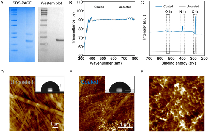
We first expressed CsgA proteins as inclusion bodies in E. coli BL21 (DE3) cells and obtained these proteins from the cell lysate through Ni-NTA affinity chromatography. The sodium dodecyl sulfate–polyacrylamide gel electrophoresis (SDS–PAGE) image showed a clear band between 10 kDa and 15 kDa, which corresponds with its theoretical molecular weight (Fig. 1A). The Western blot image also confirmed the successful purification of CsgA proteins (Fig. 1A). Then, we deposited fresh-made CsgA monomer protein solution in the 3-cm Petri dish, allowing coating formation at the bottom through a spontaneous assembling process. After these preparation procedures, we visually observed almost no significant changes at the bottom of the Petri dishes. The UV–vis spectra further confirmed that our coating is transparent (Fig. 1B). To verify the coating in the Petri dish, several material analyses were conducted. Initially, we used XPS to investigate the differences in the surface chemistry of the dish before and after coating formation. The spectrum of the coated dish exhibited distinctive O 1s and N 1s peaks, which were attributed to the amino acid, highly indicating that the CsgA proteins were coated at the bottom of the dish (Fig. 1C).
Figure 1
Figure 1.
Characterization of the CsgA protein and the CsgA nanofibre-coated Petri dish. (A) SDS−PAGE and Western blot images of the expressed CsgA protein. (B) UV−vis spectra of the uncoated and CsgA nanofibre-coated Petri dishes. (C) XPS uncoated and CsgA nanofibre-coated Petri dish. (D) AFM height image and contact angle image (insert) of the uncoated Petri dish. (E) AFM height image and contact angle image (insert) of the CsgA nanofibre-coated Petri dish. (F) Zoomed in AFM height image of the coated Petri dish.
Significantly, we conducted AFM morphology measurements. Height images indicated the absence of nanofibre features on the surface of the dish with uncoated CsgA protein, only PA/PE composite membranes on the surface of the dish were detected (Fig. 1D). As reported previously, peptides could be assembled to construct nanostructures on a particular surface, which highly depends on the chemistry properties of the surface [25]. By contrast, coated dishes with CsgA protein exhibited typically intertwined nanofibres, which indicated the surfaces-assistant assembly of CsgA proteins on the Petri dish (Fig. 1E). As shown in Fig. 1F, the assembled pattern was uniform, which might contribute to the promotion of cell adhesion and growth.
Moreover, surface-assistant assembly of peptide/protein could switch the hydrophilic properties of the surface [25]. Accordingly, when the CsgA coating was on the surfaces, we speculated that the polar residues of amino acids, such as the carboxyl and hydroxy groups, were covered on the Petri dish surface, leading to the decrease in the water contact angle. In our results (Figs. 1D and E), the contact angle is confirmed to become more hydrophilic after coating and assembled formation (97.1° ± 0.5° vs. 78.3° ± 0.9°). Collectively, we solidly established the feasibility of our CsgA protein coatings on the bottom surface of the Petri dish by a surface-assistant assembly process and form a uniform pattern.
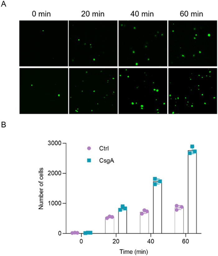
To examine the biological activity of CsgA-based nano-coatings, we first performed a rapid adherence experiment. GFP-HUVECs, at a concentration of 5000 cells/mL, were added to the dish (without cell and tissue culture-treated) with or without CsgA-based nano-coating, and cell morphology and adherence were observed with a confocal microscope at the indicated times (Fig. 2A). As the incubation time increased, the number of cells in both dishes increased significantly. Among them, the number of cells in the CsgA nano-coated culture dish reached a peak after 60 min of culture, which was three times the number of cells in the control dish (Fig. 2B). The results suggested that CsgA-based nano-coating could significantly promote cell adhesion.
Figure 2
Figure 2.
CsgA-based nano-coating promotes rapid cell attachment. (A) Confocal imaging of the fast adherence of GFP-expressing human umbilical vein endothelial cells (GFP-HUVECs) in control (upper) and CsgA nano-coated dishes (lower). (B) Number of fast adherent GFP-HUVECs in control and CsgA nano-coated dishes.
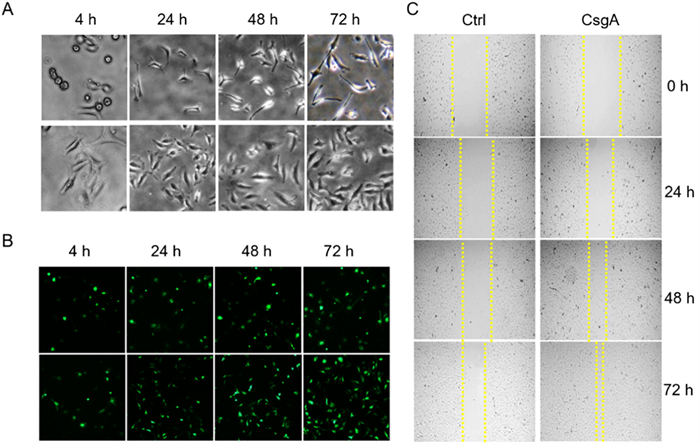
Subsequently, we examined the effects of CsgA-based nano-coating on HUVEC proliferation. HUVECs were seeded in CsgA nano-coated and control dishes and observed and photographed under a microscope after culturing for a specified period. After 4 h of culture, the cells in the CsgA nano-coated dishes were all adherent, whereas those in the control dish were round and appeared to have just adhered.
The morphology of HUVECs was perfect after 3 days of culture in CsgA dishes, and the cell density was significantly increased (Fig. 3A). By contrast, the cells in the culture dishes of the control group were still in a diseased state, and the number of cells did not change. Moreover, the confocal imaging results of GFP-HUVECs confirmed this effect (Fig. 3B and Fig. S1 in Supporting information). The results indicate the potential use of CsgA-based nano-coating as an effective agent for promoting cell adhesion and proliferation. To further investigate the effect of CsgA-based nano-coating on cell proliferation, we conducted wound-healing experiments [26]. As shown in Fig. 3C, the cells in the CsgA nano-coated dish were almost confluent after 72 h of culture, whereas the confluence rate of the cells in the control dish was only approximately 42%. The above results again indicate that the CsgA-based nano-coating has good cell adhesion and cell proliferation activities.
Figure 3
Figure 3.
CsgA-based nano-coating promotes cell proliferation and migration. (A) Morphology of HUVECs in control dishes (upper) and CsgA nano-coated dishes (lower). (B) Confocal images of GFP-HUVECs in control dishes (upper) and CsgA nano-coated dishes (lower). (C) CsgA-based nano-coating promotes HUVEC migration.
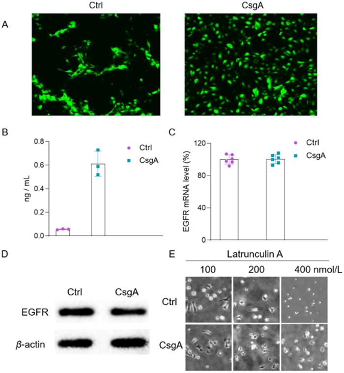
Cells exhibit fractal-like morphologies in unsuitable environments [27]. Accordingly, we validated the effect of CsgA-based nano-coating on fractal-like morphologies using GFP-HUVECs. GFP-HUVECs at a density of 50,000 cells/mL were observed and recorded under a confocal microscope after culture in both dishes for 3 days. The cells in the control dish presented fractal-like morphologies, whereas the cells in the CsgA nano-coated dish did not show a similar situation, indicating that CsgA-based nano-coating provided a good growth environment for HUVECs (Fig. 4A). Given that EGF is important in this process, we investigated the effect of CsgA-based nano-coatings on cellular EGF [26]. After HUVECs were grown in two culture dishes for 24 h, the cells were collected and treated with lyase, and the supernatant was collected after centrifugation. EGF content was detected using an EGF kit. Expectedly, the concentration of EGF in CsgA-based nano-coated dishes was twice that in control dishes, indicating that CsgA-based nano-coating could promote cell growth and proliferation by inducing EGF secretion (Fig. 4B). Moreover, we examined the effects of nano-coating on epidermal growth factor receptor (EGFR) expression. The results of the quantitative polymerase chain reaction and Western blot showed that the nano-coating material barely affected the expression of EGFR in HUVECs (Figs. 4C and D).
Figure 4
Figure 4.
CsgA-based nano-coating promotes HUVEC growth. (A) CsgA-based nano-coating alleviates fractal-like morphologies induced by EGF deficiency. (B) CsgA-based nano-coating promotes EGF secretion from HUVECs. (C) EGFR mRNA levels in HUVECs with or without CsgA-based nano-coating. (D) EGFR protein levels in HUVECs with or without CsgA-based nano-coating. (E) CsgA-based nano-coating alleviates the inhibitory effect of the cell adhesion inhibitor Latrunculin A.
Given the good growth-promoting and proliferative properties of CsgA-based nano-coating, we speculated that the coating might affect the growth of the cytoskeleton. Latrunculin A is a reversible inhibitor of actin assembly that inhibits cell growth [28]. Therefore, we examined the effects of latrunculin A on HUVEC growth. As shown in Fig. 4E, latrunculin A repressed cell attachment and unfolded in a dose-dependent manner. CsgA-based nano-coating significantly alleviated the inhibitory effect of the latrunculin A on cell adhesion. The results indicate that CsgA can stabilize the cytoskeleton, thereby promoting cell adhesion.
In this study, we reported a convenient modification strategy based on the self-assembly of CsgA proteins. CsgA proteins formed a typical interwoven nanofibre coating on the bottom of the Petri dish through self-assembly. CsgA-based nano-coating promoted rapid cell adhesion, proliferation and migration and alleviated the maladaptive state of cells by inducing EGF secretion instead of affecting EGFR expression. In addition, it significantly alleviated the inhibitory effects of cell adhesion inhibitors on cells. We believe that this efficient and convenient surface-modification strategy can help to improve the clinical application of biomaterials, especially biometallic materials.
Declaration of competing interest
The authors declare that they have no known competing financial interests or personal relationships that could have appeared to influence the work reported in this paper.
Acknowledgments
The work was supported by the National Natural Science Foundation of China (Nos. 82104477, U19A2010, and 81891012), special support from China Postdoctoral Science Foundation (Nos. 2019M663456 and 2019TQ0044), Xinglin Scholar Research Promotion Project of Chengdu University of TCM (No. BSH2019008), National Interdisciplinary Innovation Team of Traditional Chinese Medicine (No. ZYYCXTD-D-202209), the Macao Science and Technology Development Fund (No. FDCT 007/2020/ALC), the Shenzhen-Hong Kong-Macau S&T Program (Category C) (No. SGDX2020110309420200), and the Research Fund of University of Macau (No. CPG2022-00005-ICMS). We thank Dr. Yingfeng Li of Nanjing Normal University for providing assistance.
Supplementary materials
Supplementary material associated with this article can be found, in the online version, at doi:10.1016/j.cclet.2022.08.013.
H.M. Swasthi, K. Bhasne, S. Mahapatra, S. Mukhopadhyay, Biochemistry 57 (2018) 6270–6273. doi: 10.1021/acs.biochem.8b00841
[25]
L. Zhang, Y. Sheng, A.Zehtab Yazdi, et al., Nanoscale 11 (2019) 2999–3012. doi: 10.1039/c8nr08397e
[26]
R. Li, K. Liu, X. Huang, et al., Adv. Sci. 9 (2022) e2105152.
[27]
S.E. Leggett, Z.J. Neronha, D. Bhaskar, et al., Proc. Natl. Acad. Sci. USA 116 (2019) 17298–17306. doi: 10.1073/pnas.1905958116
[28]
J. Würtemberger, D. Tchessalova, C. Regina, et al., PLoS One 15 (2020) e0238572. doi: 10.1371/journal.pone.0238572
Figure 1
Characterization of the CsgA protein and the CsgA nanofibre-coated Petri dish. (A) SDS−PAGE and Western blot images of the expressed CsgA protein. (B) UV−vis spectra of the uncoated and CsgA nanofibre-coated Petri dishes. (C) XPS uncoated and CsgA nanofibre-coated Petri dish. (D) AFM height image and contact angle image (insert) of the uncoated Petri dish. (E) AFM height image and contact angle image (insert) of the CsgA nanofibre-coated Petri dish. (F) Zoomed in AFM height image of the coated Petri dish.
Figure 2
CsgA-based nano-coating promotes rapid cell attachment. (A) Confocal imaging of the fast adherence of GFP-expressing human umbilical vein endothelial cells (GFP-HUVECs) in control (upper) and CsgA nano-coated dishes (lower). (B) Number of fast adherent GFP-HUVECs in control and CsgA nano-coated dishes.
Figure 3
CsgA-based nano-coating promotes cell proliferation and migration. (A) Morphology of HUVECs in control dishes (upper) and CsgA nano-coated dishes (lower). (B) Confocal images of GFP-HUVECs in control dishes (upper) and CsgA nano-coated dishes (lower). (C) CsgA-based nano-coating promotes HUVEC migration.
Figure 4
CsgA-based nano-coating promotes HUVEC growth. (A) CsgA-based nano-coating alleviates fractal-like morphologies induced by EGF deficiency. (B) CsgA-based nano-coating promotes EGF secretion from HUVECs. (C) EGFR mRNA levels in HUVECs with or without CsgA-based nano-coating. (D) EGFR protein levels in HUVECs with or without CsgA-based nano-coating. (E) CsgA-based nano-coating alleviates the inhibitory effect of the cell adhesion inhibitor Latrunculin A.

 DownLoad:

 下载:
下载:


