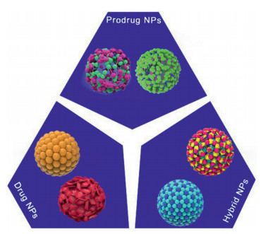
Figure 1.
Schematic diagram of self-assembled carrier-free nanomedicines.
Advances of nanoparticles as drug delivery systems for disease diagnosis and treatment
Rui Liu , Cong Luo , Zhiqing Pang , Jinming Zhang , Shaobo Ruan , Meiying Wu , Lei Wang , Tao Sun , Nan Li , Liang Han , Jinjin Shi , Yuanyu Huang , Weisheng Guo , Shaojun Peng , Wenhu Zhou , Huile Gao

It has been over half century since the first illustration of liposome in 1964 [1], and over 50 nanoparticles-based medicines have been approved by FDA [2]. It is undoubted that nanoparticles open a new category of drug delivery and bring it into a new height, especially after decades when doxorubicin liposomes and paclitaxel-albumin nanoparticles emerged on the market [3]. Thereafter, much attention was drawn to the nanoparticles-based medicine and efforts were put to push them into research and development. These nanomedicines were generally simple carriers, for instance, liposomes served as nano-capsules to help drug disperse well and elevate tolerance. Besides, antibiotics and antitumor agents were the major drugs delivered by nanomedicines [4]. Thanks to the natural affinity and specific binding of designed ligands, nanomedicines showed potential target capability to enhance efficacy and reduce adverse effect. Numerous ligands with superior active targeting property have been utilized, such as endogenous folate acid and hyaluronic acid [5-7], but the efficiency is not satisfactory, which might be attributed to competitive inhibition, inactivation of engineering ligands and corona coating [8-10]. Increasing efforts are made on these aspects to optimize the active targeting effect of nanomedicines [11]. For solid tumor, nanomedicines are born with talent of targeting delivery, based on a so-called enhanced permeability and retention (EPR) effect, which is a consequence of specific leakage of abnormal neovascularization in tumor [12]. Since the blood vessels in normal tissue are intact, the EPR effect makes nanomedicines accumulate more in tumor region, which is considered as positive targeting delivery without ligands. However, there were theories querying the EPR effect, and arguing the trans-vascular effect of nanomedicines is a dose- and receptor-dependent active transport [13, 14]. Investigations remain needed to clarify the truth.
Furthermore, drug delivery nanoparticles received dramatic advances in last ten years, emerging kinds of well-designed and intelligent drug delivery systems to overcome complicated barriers in the treatment of stubborn diseases. These nanoparticles are custom, complicated in compositions and various in functions. Taking solid tumor into consideration, as tumor grows rapidly and loses control, the tumor tissue is commonly dense, and hypoxia occurs in the inner region, leading to apoptosis, acidic circumstance and abnormal expression of stress proteins, which are typical characteristics of tumor microenvironment [15-17]. Novel nanoparticles have been designed to fit and utilize the tumor microenvironment, for example, using linkage with acidic response for drug payload realizes specific drug release in tumor [18], degradation by overexpressed enzyme shrinks nanoparticles size for deep tumor penetration [19] and increasing size enhances tumor retention [20]. These smart drug delivery systems are not only carriers solving drugs' concern, but significantly enhance their potential in therapy, and enable drugs with capabilities that are impossible to achieve by drug optimization.
Although the classification and delivery strategy of nanoparticles are not freshly made, numerous excellent works have been reported in these areas in recent years, hence a systemic and comprehensive review is required to summarize these works to demonstrate the frontiers and trends in drug delivery nanoparticles. In this review, we first classify the nanoparticles by their own characteristics, finely illustrate their different compositions and correlated properties and functions; besides, some smart strategies that promote the drug delivery efficiency are introduced, including active targeting, stimuli responsive, size and shape tunable, self-propelled and hijacking strategy; furthermore, since the carriers-based vaccines have attracted broad attention and exhibited potentials of drug delivery nanoparticles, we make critical illustrations about nanoparticles-based delivery of these specific therapeutic agents, including protein, nucleic acid, gas, and metals-involved artificial nanoenzyme. Nanoparticles as carriers to delivery these specific therapeutic agents are potential to resolve many unexpected concerns, such as toxicity, immunogenicity and instability. Oral administration is a newly developed strategy for nanoparticles delivery, which showed elevated compliance and reduced toxicity, and related works and advances are also concluded. Last, an overview of outlook and future expectation are made to show the trend of drug delivery nanoparticles.
Conventional nanomedicines are ordinarily composed of carrier materials and theranostic agents, such as liposomes, micelles, organic or inorganic nanoparticles (NPs) [21]. The versatile features and functions of carrier materials endow nano-vehicles with the ability to achieve the efficient diagnosis and treatment of diseases [22]. Despite many distinct advantages, the clinical translation of carrier material-based nanomedicines could be significantly restricted by time-consuming and costly development of carrier materials into pharmaceutical excipients. In addition to conventional nanocarriers, carrier-free nanoassemblies formed by small-molecule prodrugs and/or pure drugs have emerged as a unique branch of nanomedicines for biomedical applications [21, 23, 24]. These inimitably engineered NPs are characterized by carrier-free assembly, facile fabrication technique and high drug loading capacity [21, 23]. In this section, the latest updates in self-assembled carrier-free nanomedicines will be overviewed, including prodrug-nanoassemblies, pure drug-nanoassemblies and co-assembled hybrid nanoassemblies (Fig. 1).
Rational design of prodrugs has been extensively appreciated as an efficient and promising strategy [23]. Undeniably, the multitude of common challenges encountered in drug delivery process can be effectively addressed by prodrug strategy, such as low water-solubility, poor stability, inferior pharmacokinetics, as well as insufficient transmembrane transport [23]. Notably, suitable chemical modification has been found to bestow drug molecules with self-assembly capacity [23]. To date, a great number of small-molecule prodrugs has been acknowledged to have self-assembly ability, including amphiphilic monomeric prodrugs, hydrophobic monomeric prodrugs and dimeric prodrugs [23].
Similar with amphiphilic polymers, hydrophobic force drives the nanoassembly of amphiphilic small-molecule prodrugs [23]. By contrast, the underlying nanoassembly mechanism of hydrophobic prodrugs is unique even quite difficult to comprehend, such as the small-molecule oleate prodrugs of paclitaxel (PTX) [23, 25]. In recent years, a universally accepted mechanism is that chemical modification on drug molecules prevents drug crystallization during the nano-precipitation process [23]. Moreover, multiple intermolecular forces driving the assembly of hydrophobic prodrugs into stable NPs have been figured out with the assistance of molecular docking software, mainly including hydrophobic force, hydrogen bond, π-π stacking interaction and π-cation interaction [23]. In addition to monomeric prodrugs, nanoassemblies formed by heterodimeric and homodimeric prodrugs have also attracted intense attention [26, 27]. Among them, heterodimeric prodrug-nanoassemblies are regarded as a versatile nanoplatform for combination therapy [26]. For instance, a ROS-responsive nanoassembly of heterotypic PPa-PTX dimer was exploited for synergistic chemo-photodynamic therapy [26]. Of note, homodimeric prodrugs with strictly symmetrical dumbbell structures frequently suffer from unfavorable self-assembly capability, while co-assembly with photosensitizers could contribute to improve their assembly ability and stability [23, 27]. More importantly, precise activation of prodrugs to release parent drugs at the target sites is imperative for the molecular design of prodrugs [23, 25-27]. To achieve this goal, various chemical linkages have been inserted in prodrugs for stimuli-responsive drug release, such as thioether and selenide bonds [23, 25-27].
Despite the extensive investigation on prodrugs and prodrug-nanoassemblies, prodrug strategy still has some limitations: (ⅰ) Only a fraction of drugs are suitable for prodrug design, owing to the prerequisite of available chemical structures for chemical modification; (ⅱ) the potential security concerns of side chains and/or metabolic intermediates should also be taken into consideration; and (ⅲ) the complexity and costs of chemical synthesis also pose obstacles to the broad application of prodrug strategy. In recent years, nanosystems self-assembled by pure drugs have attracted on-going attention, owing to the simple preparation process and high drug delivery efficiency [21]. For instance, PPa was found to self-assemble into stable NPs without the assistance of any carrier materials [28]. Moreover, core-matched modification on the nanoassemblies with a PPa-PEG polymer has been proved to be a promising strategy for imaging-guided photodynamic cancer therapy [28].
Intriguingly, some drug molecules without self-assembly ability are found to be able to co-assemble with another kind of drug molecules and then form stable NPs, which provides an alternative nanoplatform for combination therapy [21]. More importantly, it is convenient to adjust the dose ratios of two or more drugs in the nanoassemblies to achieve optimal synergistic effects [21]. Based on pure drug nanoassembling nanotechnology, a series of self-assembled or co-assembled nanosystems have been constructed for the diagnosis and treatment of cancer and thrombus [29-32]. For instance, a dual-drug nanoassembly of photothermal photosensitizer and antiplatelet drug was fabricated for site-specific deep thrombus penetration and thrombolysis [32]. Briefly, ticagrelor (TGL) and DiR could readily co-assemble into stable NPs, and the surface of the nanoassembly was further modified with a PEG polymer and fibrin-targeting peptide for long circulation in the blood and site-specific accumulation in the clots [32]. Significantly, the drug loading rates of both DiR and TGL in the PEGylated nanoassemblies were up to 37.5 wt% [32]. The dual-drug nanoassembly demonstrated potent thrombolysis efficacy in a FeCl3-induced rat carotid arterial thrombosis model, owing to its favorable stability, antiplatelet activity, pharmacokinetic behaviors, thrombus-targeting ability, as well as photothermal-potentiated thrombus penetration [32]. Additionally, biomimetic hybrid dual-drug nanoassemblies could be constructed by cell membrane-camouflaging technique [33].
To date, a multitude of organic/inorganic nanoparticles have been developed for drug delivery to solve the physicochemical problems associated with drugs, such as low solubility, low stability, off-target deposition, and weak penetration across biological barriers. Although great success in nanomedicines has been achieved in preclinical research and clinical application, the clinical use of nanomedicines is still away from the optimal effect. As "foreign objects" to the body, most synthetic nanoparticles tend to easily cleared by the immune system and have low accumulation in the target site. Although PEGylation on nanoparticles could extend the nanoparticle circulation in vivo, the accelerated blood clearance phenomenon reduces the passive targeting of PEGylated nanoparticles and limits their application in clinics. In light of these issues, biomimicry seems to be a rational approach towards effective nanoparticle designs.
In 2011, Zhang's group firstly reported the cell membrane coating technology which opened the era of cell membrane-based biomimetic nanoparticles [34]. They coated red blood cell (RBC) membranes onto the surface of PLGA nanoparticles and revealed that the resultant nanoparticles had a superior circulation half-life of approximately 12–24 h, outperforming a PEGylated nanoparticle control. More importantly, RBC membrane-coated nanoparticles did not induce accelerated blood clearance after repeated injection. Following these reports, cell membrane coating on nanoparticles has since been a generality strategy of biomimetic nanoparticles. Cell membranes derived from various cell types including RBCs, stem cells, cancer cells, macrophages, neutrophils, natural kill cells, T cells, and platelets could be utilized to coat different nanoparticles. Moreover, besides cell membranes, intracellular organelle plasma membranes, extracellular vesicle membranes, cell membrane proteins, and even exogenous substances derived from viruses and bacteria are expanded to prepare biomimetic nanoparticles. The cell membrane is mainly composed of a mixture of lipids, proteins, and carbohydrates. It forms a bilayer structure with a thickness of 7–8 nm and is responsible for the interactions between cells and with surrounding environments, such as antigen recognition, cellular signaling, nutrient absorption, metabolic waste excretion, and protein transportation. More importantly, each type of cells has distinctive bioactivity and characteristics. For instance, platelets function in hemostasis, stem cells home to injury site, leukocytes infiltrate to inflammation site, and some malignant tumor cells penetrate the blood-brain barrier. Thus, cell membrane coating on nanoparticles could inherit the structural and functional complexity from original cells to nanoparticles, not only endowing them with common functions such as long circulation, low immunogenicity, and biocompatibility, but also granting them with special functions such as inflammation targeting, penetration through biological barriers, homing to injury site, and homogenous targeting. For instance, recently we have developed a macrophage-derived microvesicle (MMV)-coated nanoparticle (MNP) with bioactivity similar to rheumatoid arthritis (RA)-targeting macrophages for targeting drug delivery to RA [35]. It has been shown biocompatible MNP bears long circulation property and has a significantly enhanced targeting effect in vivo in a collagen-induced arthritis (CIA) mouse model compared with bare nanoparticles and RBC membrane coated-nanoparticles. It is found that Mac-1 and CD44 contribute to the outstanding targeting effect of the MNP and tacrolimus-loaded MNP could significantly suppress the progression of RA in mice. This study demonstrates that MNP mimicking macrophages is an efficient biomimetic vehicle for RA targeting and treatment.
On account of the limited functionalities of single-cell type, hybrid cell membranes with multi-functions of several cell types are explored to develop biomimetic nanoparticles. For instance, erythrocyte-cancer cell hybrid membrane-coated nanoparticles can simultaneously achieve long blood circulation and superior targeting to homologous tumors [36]. Leukocyte-platelet hybrid membrane-coated nanoparticles could specifically bind with circulating tumor cells and home to tumors. To leverage both advantages of cell membranes and artificial lipid membranes, cell-lipid hybrid membranes are also involved in the design of biomimetic nanoparticles [37-39]. For example, inspired by the increase in circulating platelet-monocyte aggregates in patients′ post-myocardial ischemia-reperfusion (MI/R) injury, we designed a platelet-lipid hybrid membrane-coated nanoparticle for targeting delivery of miR-21 to the ischemic heart and improved myocardial remodeling through reprogramming macrophages post MI/R injury [40]. Generally, cell membranes are coated on nanoparticles by the sonication, extrusion and microfluidics method. The cell secretion method by which cells internalize nanoparticles into cells, wrap them in exosomes, and exocytose nanoparticle-loaded exosomes into extracellular space have also attracted increasing interest as a biomimetic strategy [41].
In addition to cell membrane-based biomimetic nanoparticles, other biomimetic nanoparticles such as lipoprotein-inspired nanoparticles which retain extended circulation time and active targeting to lipoprotein receptors by mimicking the shape and structure of endogenous lipoproteins are also ideal nanoplatforms for drug delivery [42]. In general, biomimetic nanoparticles especially cell membrane-coated nanoparticles represent an emerging class of nanoparticles through mimicking source cells and display great potential in drug delivery, detoxification and targeting imaging. However, the quality control cell membranes, the coating integrity, and the scale-up manufacturing of these biomimetic nanoparticles still present some unique challenges concerning the safety, effectiveness and controllability and need to be fully investigated.
Despite the remarkable drug delivery accomplishments achieved by these commonly-used synthetic polymers, the high immunogenicity or toxic degradation products still greatly impede their application. Additionally, it is difficult to control the stereochemistry, structure, and molecular weight of synthetic polymers, which would impact the drug's biodistribution and pharmacokinetics. Due to the complicated synthetic process of polymeric carriers, the polymer production including synthesis and purification can be difficult and expensive to scale up. In view of these challenges, naturally occurring biodegradable polymers, obtained from plants, animals, and micro-organisms, arouse the increasing interest as an attractive alternative to these synthetic polymers. A variety of natural polymers for drug delivery include polysaccharides [43] such as chitosan, agarose, dextran, hyaluronic acid, alginate, carrageenan and cyclodextrin, and proteins [44] such as silk, albumin, keratin, collagen, gelatin, elastin and resilin. These polymers possess a number of unique advantages including biocompatibility, bioavailability, ease of synthesis and purification, plasticity and scalability, efficient drug loading efficiency, prolonged retention time, and non-immunogenicity. Furthermore, unlike none of physiological activity of synthetic polymers, some proteins or polysaccharides as drug delivery vehicles also exhibit the pharmacological activities and tissue targeting capacity. For example, Lactoferrin possesses a wide array of functions including anticancer, anti-inflammatory, immunomodulatory, cognitive function improvement and wound healing effects [45]. Hyaluronic acid, involved in a variety of cellular processes like angiogenesis and regulation of inflammatory pathways, has been widely used in drug delivery micro-nanosystems. What is more, some polysaccharides could specifically bind with these overexpressed receptors like asialoglycoprotein receptor, galectins, selectins, mannose receptors and CD44 receptors on specific cells, which are involved in cancer, enterocytes, and blood brain barrier.
Based on the unique structural and physico-chemical properties of natural polymers, manifold drug delivery vehicles have been developed with exceptional drug loading and release profiles, pharmacokinetics, targeting capacity, and biosafety. According to the drug administration routes, the drug delivery carriers produced by natural polymers could be employed for intravenous, oral, transdermal, and in situ application. The most representative drug delivery vehicles composed by natural polysaccharides or proteins for parenteral administration are micro-/nano-particles to improve the systemic circulation and enhance the tissue targeting of payloads. Nowadays, some novel carriers derived from natural polymers were developed for non-intravenous injection with the higher drug delivery efficiency. The first drug delivery option is the oral colon-targeting systems [46], based on the gastrointestinal (GI) protection as well as colon mucoadhesion of natural agents. Taking the complex physical environment into consideration, either protein or polysaccharide alone is not suitable enough to protect drugs from GI degradation and to keep drugs entrapped in the carrier until it reaches to colon. Some polysaccharides with the opposite charges such as chitosan/alginate, chitosan/pectin, and anionic carboxymethyl starch/cationic quaternary ammonium starch, were used to generate layer-by-layer assembled polymeric film by the sequential adsorption of polyelectrolytes. The combination of protein and polysaccharide as a nanohybrid complexes also could avoid to release drugs prematurely in the stomach and result in higher colon-targeting efficacy. Cheng et al. developed an enzyme-triggered fuse-like microcapsule, by means of layer by layer self-assembly of alginate and protamine via the electrostatic absorption, to help the colon-targeted of probiotics [47]. To avoid the premature early drug release, Zhang et al. prepared the NPsinMPs system for ulcerative colitis treatment, by embedding zein NPs coated with chondroitin sulfate into hydrogel microspheres via an electrospraying technology [48]. Liu et al. innovatively designed a colon-targeted adhesive core-shell hydrogel microsphere in combination of the anti-acid and colon-targeted property of an alginate calcium hydrogel shell and the mucoadhesive ability of the thiolated-hyaluronic acid hydrogel core [49]. Additionally, biodegradable microneedles (MNs) fabricated from natural polymers have become the center of attention because of the good patient compliance of MNs and the recognized biodegradability, biocompatibility, and sustainable character of natural materials. Various polysaccharides like alginates, chitosan, chondroitin sulfate, xanthan gum, pullulan, and proteins such as zein, collagen, gelatin, fish scale and silk fibroin, based biomaterials have been employed to fabricate biodegradable MNs [50]. Recently, dissolvable MN arrays composed of carboxymethyl cellulose were used to fabricate the recombinant coronavirus vaccine incorporating the protein MERS-S1f, MERS-S1fRS09, MERS-S1ffliC, SARS-CoV-2-S1 or SARS-CoV-2-S1fRS09, eliciting the potent antigen-specific antibody responses as early as week 2 after immunization [51]. Interestingly, Bletilla striata polysaccharide (BSP) is a natural hydrosoluble glucomannan, with wound healing, procoagulant, anti-inflammatory, and antioxidant activities. Researchers designed the BSP-fabricated dissolvable MNs for hypertrophic scar repair, which shown higher mechanical strength and better physical stability than MNs made of hyaluronic acid [28]. Furthermore, due to the unique three-dimensional network structure of natural polysaccharides and proteins, they are apt to generate in situ hydrogel in response to temperature, ionic strength, pH value, and multiple sensitive characters in target site [52]. These injectable scaffold hydrogels have been widely used in ocular drug delivery, wound healing, subcutaneous implantation, and in-situ tumor treatment [53, 54]. To sum up, despite these progress made, the application of drug delivery formulations using natural polysaccharide-/protein-based materials still confront some bottlenecks, such as the inhomogenous molecular weight, undefinable spatial structure, and the lack of solubility in most organic solvents, restricting the chemical modification and drug loading approaches.
Over the past decades, polymeric materials have been extensively explored as the drug delivery system for biomedical application, which has gained tremendous attention. Depend on the types of drugs and their requirements for a particular administration route, polymeric drug delivery platforms can be customized, and a variety of options are now available, including polymeric NPs, dendrimer, polyplex and polymersome. As one of the most studied delivery platforms, polymeric NPs including polymeric micelles, polymeric nanospheres and polymeric nanocapsules are ideal platform for delivering various bioactive drugs, such as hydrophobic drugs, hydrophilic drugs, nucleic acid, peptide and protein [55]. Given to the unique structure and composition, polymeric NPs drug delivery system possesses several advantages compared to free drug. For example, the structure of polymeric micelle is generally characterized by continuous hydrophobic core and hydrophilic shell. On the one hand, the hydrophobic core can non-covalently encapsulate hydrophobic drugs during the fabrication process, leading to improved drug solubility and stability. On the other hand, the hydrophilic shell can help to both avoid unexpected drug degradation from serum components and prevent opsonization by the complement system and opsonization-induced rapid clearance of drugs by mononuclear phagocytosis system (MPS), leading to prolonged circulation time [56]. Paclitaxel, a hydrophobic chemotherapeutic, has been widely proven to non-covalently encapsulated in polymeric micelles to improve its pharmacokinetic profile in systemic circulation while alleviate drug-induced side effects in both preclinical studies and clinical trials. Currently, four polymeric micelle formulations encapsulating paclitaxel (Genexol-PM, NK105, Paccal Vet and Paxceed) are under clinical evaluation for different indicators, of which Genexol-PM has been already approved by FDA [57]. In addition, by pre-conjugation, water-insoluble drugs or peptides can also be covalently encapsulated into hydrophobic core, representing a more stable encapsulation. Furthermore, owing to the feasibility for design, modification and fabrication, polymeric NPs delivery system can be rationally tailored to possess unique physiochemical properties, such as size, charge, morphology, or modified with targeting ligand to meet specific delivery requirements or obtain desirable delivery performance, such as reduced clearance rate, targeting delivery, enhanced cellular uptake and enhanced penetration or retention at diseased site.
Recently, the increased number of approved nucleic acid drugs demonstrated their therapeutic potential against disease by targeting genetic bases in vivo, while in vivo delivery of nucleic acids remains challenging [58]. Polymeric NPs with specific compositions and physiochemical properties are also ideal platform for therapeutic nucleic acid delivery. One common design feature for polymer-based nucleic acid delivery system is the incorporation of cationic groups that can bind negatively charged nucleic acid to form polyplexes through electronic interaction [59]. Moreover, cationic polymers can also promote the escape of nucleic acids from endosome/lysosome by exploiting proton sponge's effect if they are internalized through endocytic pathway. These cationic polymers generally contain many proton-accepting groups including primary, secondary, and tertiary amines that can induce protonation in acidic endosome/lysosome, leading to an influx of chloride and water into endosome/lysosome. This influx further leads to subsequent bursting of the endosome/lysosome from osmotic pressure and membrane destabilization, referring as proton sponge effect [60]. For example, as a well-known proton sponge polymer, polyethylenimine (PEI) and PEI-derivatives have been commercialized for nucleic acids delivery, such as pDNA, siRNA and miRNA [61]. In addition to the increased osmotic pressure induced by proton sponge effect, the direct permeabilization and thermomechanical disruption induced by swelling of PEI may also involve in the endosomal/lysosomal escape of nucleic acid [62]. However, the high cation density has been reported to cause non-specific cellular uptake, cell membrane damage and systemic toxicity in vivo. To troubleshoot this issue, strategies such as increasing branch density or surface shielding with poly(ethylene glycol) (PEG) or heparin have been demonstrated. Besides, other synthetic cationic polymers with the ability of pH-triggering disassembly are also suitable for nucleic acid delivery, such as poly(2-(diisopropylamino)ethyl methacrylate) (PDPA), poly(2-dimethylaminoethyl acrylate) (PDMAEA) and oligo-ethyleneimines (OEIs). They can stay stable during circulation while undergo pH-triggering disassembly, further leading to disruption of polymeric micelle and cargo release in the tumor microenvironment [63].
With the advances of polymer science, more functional polymeric delivery system or other polymeric platforms can be pursed, such as poly-lipid hybrid NPs, poly-drug/peptide/protein conjugates. Although polymeric drug delivery systems are promising, there remain several concerns for their potentials to be clinically applied and translated, including but not limited to biocompatibility and safety, premature drug release, colloid stability and manufacturing. To ensure high safety, the use of nontoxic and biodegradable naturally derived polymers such as polysaccharides (chitosan and hyaluronic), alginate, dextran, collagen, or FDA-approved synthetic polymers such as poly(lactide-co-glycolic acid) (PLGA), poly(Ɛ-caprolactone) (PCL), poly(lactide) (PLA) and poly(glycolic acid) (PGA) can be good options. To enable spatiotemporal drug release, one promising strategy is to develop stimulus-responsive drug delivery system by either using stimulus-responsive polymer or conjugating drug onto polymer through a stimulus-responsive linker [64]. However, the more sophisticated design of polymeric delivery system, the less possibility for clinical application. Therefore, when designing nanomedicines based on polymeric materials, these concerns should be taken into consideration.
Lipid nanoparticles have gained increasing attention as prospective nanocarriers for the delivery of various therapeutics including small molecules, peptides and nucleic acids. Especially, lipid nanoparticles are currently in the spotlight of mRNA vaccine research and two COVID-19 mRNA vaccines, mRNA-1273 and BNT162b based on lipid nanoparticles were approved by FDA in 2020 to prevent pandemic COVID-19 [65, 66]. Many other lipid nanoparticle-based nanomedicines are under research and development for cancer chemotherapy, mRNA vaccines against virus infection, and gene-editing therapies of genetic diseases.
As the earliest generation of lipid nanoparticles and the earliest nanomedicine platform applied clinically, liposomes with a vesicular structure were found in the 1960s and were proved to be a multifunctional nanocarrier to deliver a variety of drugs. Liposomes are generally composed of phospholipids along with stabilizers such as cholesterol. PEGylated lipids are also often integrated in liposomes to endow them with the "stealth" function in vivo. Liposomes can be classified into multilamellar vesicles (MLVs) and unilamellar vesicles (ULVs), which can be further classified into large unilamellar vesicles (LUVs) and small unilamellar vesicles (SUVs). To enhance the effectiveness of targeting drug delivery to the specific site, targeting ligand modification and stimuli-responsive drug release strategies can be incorporated into liposomes. Great success has been made in liposomal drug delivery and many liposomal formulations have been approved (e.g., Doxil, DepoCyt, DepoDur, Mepact, Exparel, Marqibo, and Onivyde) [66, 67] or are under clinical trials. While liposomes are versatile nanovehicles, the complex production methods, a relatively low drug-loading capacity, and difficulty in large-scale manufacture limit the biomedical applications of liposomes to some extent.
Subsequent generations of lipid nanoparticles including solid lipid nanoparticles, nanostructured lipid carriers, and cationic lipid nanoparticles were developed to address some shortcomings of liposomes. Solid lipid nanoparticles are solid lipid nanospheres consisting of solid lipids such as lecithin, triacylglycerol and waxes while nanostructured lipid carriers are constituted of solid lipids and liquid lipid or oil. Compared with liposomes, solid lipid nanoparticles and nanostructured lipid carriers have higher drug-loading capacity and are manufactured easily and massively. However, solid lipid nanoparticles bear some disadvantages such as low loading capacity of hydrophilic drugs and drug leakage with long term storage because of the crystallization of solid lipids to expel the encapsulated cargoes out of solid lipid nanoparticles. For nanostructured lipid carriers, solid lipids immobilize interior cargoes and prevents nanoparticle coalesce, while liquid lipids or oil increase the drug loading-capacity and restrain drug expulsion from nanocarriers though reducing the lipid crystallinity.
With the development of genetics, scientists have discovered numerous nucleic acids potential as gene therapy agents and RNA therapeutics. However, as hydrophilic and negatively charged large molecules, nucleic acids cannot penetrate plasma membranes and are easily degraded by endogenous nucleases. Thus, gene vectors that can protect nucleic acids from degradation and deliver them into the cytoplasm of target cells are vital for the delivery of nucleic acids. Besides viral vectors, cationic lipid nanoparticles are the most widely used nonviral vector for nucleic acid delivery because they are easy to manufacture, are low immunogenic, can carry large payloads, and can be designed for multiple dosages. Cationic lipid nanoparticles are formed by complexing between synthetic cationic lipids and anionic nucleic acids based on electrostatic interaction. They can protect nucleic acids from nuclease degradation during circulation, facilitate them to enter target cells through lipid nanoparticle endocytosis and help to release them from endosomes into the cytoplasm by the electrostatic interactions with negatively charged plasma membranes. To date, amounts of cationic lipids (or ionizable lipids) have been explored for cationic lipid nanoparticle preparation and nucleic acid delivery. Lipid properties, such as physiochemical diversity, molecular architecture, and biodegradability could contribute for the improvement of nucleic acid delivery. For instance, Lipofectamine, a commercialized transfection agent containing a key cationic lipid, 2,3-dioleyloxy-N-[2-(sperminecarboxamido) ethyl]-N,N-dimethyl-1-propanaminium trifluoroacetate (DOSPA), could deliver mRNA in diverse cell types. MC3, (6Z, 9Z, 28Z, 31Z)-heptatriaconta-6,9,28,31-tetraen-19-yl 4-(dimethylamino) butanoate, a typical ionizable lipid protonated and positively charged at low pH but keeping neutral at physiological pH, was well-known as the crucial ingredient of Onpattro, the first authorized siRNA drug. Increasing the biodegradability of lipids could result in better delivery efficacy and faster elimination from the liver and plasma in vivo [68]. For instance, heptadecan-9-yl 8-((2-hydroxyethyl)(6-oxo-6-(undecyloxy)hexyl)amino) octanoate (SM-102), a key ionizable lipid in mRNA-1273, and ((4-hydroxybutyl)azanediyl)bis(hexane-6,1-diyl) bis(2-hexyldecanoate) (ALC-0315), a vital ionizable lipid in BNT162b, have better in vivo delivery efficacy and pharmacokinetics than MC3 [65]. Endosomal escape is a fundamental barrier impeding cytoplasm delivery of nucleic acids. To address this problem, Daniel J. Siegwart's group developed zwitterionic ionizable lipids which could assemble into a cone in endosomal acidic environments, enabling membrane hexagonal transformation, and releasing cargoes from endosomes to the cytoplasm [69]. It was shown these zwitterionic ionizable lipid-based nanoparticles can enable efficient organ-selective mRNA delivery and genome editing in vivo after intravenous administration. Besides cationic lipids, other lipids such as phospholipids, cholesterol and PEGylated lipids are incorporated in cationic lipid nanoparticles to improve nanoparticle properties including particle stability in vitro and in vivo, circulation profiles, biodistribution, safety, and delivery efficiency to target tissues or cells.
With the ability to enhance drug solubility, control drugs release, and improve the pharmacokinetics and distribution, lipid nanoparticles have been explored and optimized to deliver a variety of drugs. Due to the superior lipid properties such as physiochemical diversity, multifunctionality (e.g. adjuvants to boost vaccine efficacy [70]) and biodegradability, lipid nanoparticles will achieve impressive progress in modern drug therapy against many diseases.
Inorganic nanosystems are of great interest in the biomedical applications due to their structural and functional diversity, such as tunable size, shape, surface and composition properties. The unique optical properties of inorganic nanosystems endow them excellent performance in disease diagnosis. The distinctive magnetic characteristics of inorganic nanomaterials make them huge potential for contrast imaging, magnetic targeting or magnetic hyperthermia. More importantly, inorganic nanosystems exhibit plentiful nanostructures beneficial to therapeutic drug delivery, including zero-dimensional nanoparticles, two-dimensional nanosheets, and three-dimensional implants, etc.
Optical imaging has become a widely used imaging technique in clinical practice owing to its fast and easy-to-use advantages. In recent years, benefiting from the high stability and versatility in chemical design of inorganic nanosystems, they have emerged as fascinating optical imaging agents in the biomedical fields [71]. Compared to traditional organic fluorophores, inorganic quantum dots (QDs) reveal narrow emission bands and high photostability, thus exhibiting unique superiorities in optical imaging. For example, nitrogen-doped carbon dots (N-CDs) have been demonstrated to be used as specific fluorescent probes for detecting the occurrence and development of tumors, which could be realized based on the abnormal glycolysis metabolism in tumor tissues and high sensitivity to nicotinamide adenine dinucleotide (NAD+, oxidized) levels of N-CDs [72]. The N-CDs probes could notably distinguish tumor cells from normal cells and be used to assess their proliferation activity with high specificities of 96.15% in 13 types of tumor cells and 90.90% in orthotopic xenograft models.
The intriguing physicochemical characteristics and structural advantages of inorganic nanosystems make them promising carrier platforms for therapeutic cargos, including small molecule drugs, nucleic acids, and proteins. In particular, two-dimensional inorganic nanomaterials, a newly emerging class of nanomaterials, have attracted tremendous attention from the scientific community in recent years due to their extraordinary properties distinct from their nanoparticle and bulk counterparts, such as the most widely studied black phosphorus (BP) nanosheets [73]. The inherent biodegradability and single phosphorus composition of BP nanosheets make them promising inorganic nanosystems for clinical translation. Fluoxetine, a clinical medication for antidepressant, was successfully loaded on the surface of BP nanosheets by electrostatic interaction, which notably shortened the therapy time of depression under NIR laser irradiation and reduce side effects of free drugs [74].
In addition to using inorganic nanosystems as carriers for chemotherapeutic drug delivery, they have also been exploited to be efficient nanocatalysts for disease-specific nanocatalytic therapeutic models, which are reviewed in Section 8.
With the advent of the era of intelligent medical treatment, inorganic nanosystems have been applied in increasingly complex biomedical fields through constructing nanomotors with autonomous movement capabilities. The nanomotors can execute designated tasks by converting external energy into their own mechanical energy, thereby overcoming the challenges faced by traditional drug delivery systems. For example, platelet membrane-coated Pt-modified mesoporous/macroporous silica nanomotors (MMNM) achieved excellent thrombolytic performance by sequential delivery of thrombolytic and anticoagulant drugs [75]. The active components on MMNM were Pt nanoparticles, like driving engines, endowing MMNM with powerful motion ability and increased penetration depth under the irradiation of NIR light, thus achieving improved thrombolysis effect in multiple thrombus models.
Despite great achievements have been made in the field of biomedical applications of inorganic nanosystems, their translation from bench to bedside still remains great challenge. The first concern is the nano-bio interactions, which cover the interactions between nanoparticles and proteins or other components in the blood-stream. These interactions affect the physicochemical and biological properties of the nanomaterials, including their distribution, pharmacokinetics, and metabolism, etc. Another major hurdle for inorganic nanosystems is the potential long-term safety concern, especially those inorganic nanomaterials without biodegradability. More systematic and quantitative evaluations are needed to be conducted to screen biocompatible inorganic nanomaterials for potential clinical use in the future.
Usually, the function of single nanosystem is limited in biomedical field. The study on nano-bio interface has promoted the development of hybrid nanoparticles. Hybrid nanoparticles refer to an integration of two or more distinct components into one nanosystem, e.g., liposomes/polymers, polymer/inorganic silica, silica/magnetic nanocrystal. These hybrid nanosystems not only exhibit the characteristics of individual components, but also may bring out novel functionalities or enhancement of theranostic efficiency. Researches show that the hybridization endows nanosystem with multi-functions such as safety, targeting effect, on-demand drug release.
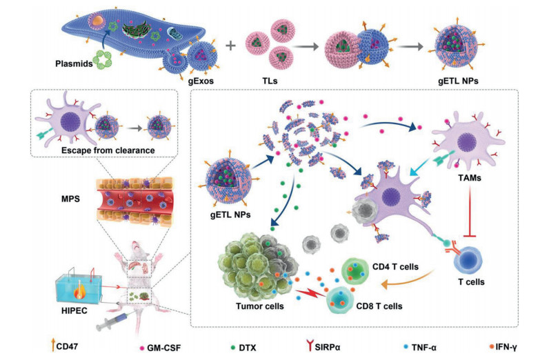
Hybrid nanoparticles can be synthesized by many methods. Physical encapsulation, surface chemical modification, doping and fusion method are usually used in the construction of nanoparticles. Lv et al. developed engineered exosomes-thermosensitive liposomes hybrid NPs via fusion method (Fig. 2) [21]. The hybrid NPs possessed both the immune escape effect due to CD47 on exosome and thermosensitive drug release profile due to liposome. This strategy promoted the accumulation of NPs in tumor, enhanced chemoimmunotherapy of metastatic peritoneal cancer. Qin et al. reported a dual-enzyme-loaded hybrid nanogel [76]. Firstly, Fe3O4 NPs were encapsulated into indocyanine green (ICG) loaded polystyrene-block-poly(acrylic acid) (PS-b-PAA) micelles, and then introduced into supramolecular hydrogel. Lactate oxidase (LOx) and catalase (CAT) were immobilized into hydrogel through charge adsorption effects. The hybrid nanogel exhibited cascade catalytic ability to produce ROS. LOx catalyzed endogenous lactate to generate H2O2, which promoted Fe3O4 NPs with oxidase-like activity to convert H2O2 into •OH. Meawhile, CAT catalyzed H2O2 into O2, which improved 1O2 production due to ICG mediated photodynamic therapy. The study claimed high reactive oxygen species (ROS) level in tumor, inhibited tumor growth. From a functional point of view, the hybrid nanoparticles can be rational designed by integration of diagnostic and therapeutic components to achieve such dual functions. Shen et al. constructed traceable nano-biohybrid complexes for theranostics in neurodegenerative diseases [77]. Biopolycations (polylysine) with dopamine modification were firstly synthesized for loading CRISPR-chem drugs. Then these polymers were anchored onto iron oxide nanoparticles (IONP) through the chelation of dopamine and iron. The high relaxation rate of IONP guaranteed magnetic resonance imaging (MRI) signals, which contributed to trace the distribution of nanoplatform in vivo. The resulted nano-biohybrid complexes provided MRI guidance for Alzheimer's disease, showing great potential in theranostics.
In recent years, hybrid nanoparticles with bioactive materials as structural components become a hot research topic [78]. Compared with traditional nanoparticles, bioactive materials derived from natural cell or bacteria have attractive properties, because they inherit characteristics of parent cells/bacteria, such as inflammation/tumor targeting effect, immunogenicity, barrier penetration. Besides, bioactive materials are rich in functional groups and are easy to synthesize hybrid nanoparticles for drug delivery. Various biohydrid nanoparticles derived from viruses, spores and probiotics have emerged and been applied in nanomedicine. Guo et al. developed a hydrid microneedle patch containing virus like particles (VLPs) with immunogenicity [72]. Tumor antigen peptide OVA sequence was first inserted into the hepatitis B core (HBc) antigen. The plasmid expressing these designed amino acid sequence was introduced into E. coli BL21. The produced OVA-HBc was extracted from E. coli and self-assembled into hydrid VLPs. At last, vaccine adjuvant CpG-DNA loaded mesoporous silica nanoparticles and immunogenic OVA-HBc VLPs were co-capsulated into microneedles together. The microneedles acted as tumor-specific vaccination and stimulated the maturation of dendritic cells, improved antitumor effect and immune memory effect. Song et al. reported an in situ assembled hybrid nanoparticles by spore capsid protein and deoxycholic acid (DA) [79]. Interestingly, Bacillus coagulans spore was modified with DA and adsorbed doxorubicin (DOX). The spore colonized and germinated to probiotics in intestine, accompanying with spore capsid protein abscission. Then self-assembly of hydrophobic spore capsid protein and hydrophilic DA resulted in hybrid nanoparticles generation. The spore capsid protein endowed NPs with enhanced intestinal mucus penetration ability, due to that sulfhydryl groups of tyrosine in spore capsid protein could cleave disulfide bond between mucin glycoproteins. In this sense, hybrid nanoparticles with bioactive materials are promising in nanomedicine, however, still remain challenging. The complexity of bioactive materials implies the difficulty to define their mechanism in vivo. Additionally, the safety and biocompatibility are also noteworthy aspects. Therefore, there is still much to be explored about biohybrid nanoparticles before clinical application.
Theranostic is composed from therapy and diagnosis, where in most cases the two procedures are sequentially programmable and tempo-spatially discrete. Theranostic systems are regarded as a systematical combination, which can realize an efficient therapeutic result and meanwhile provide diagnostic information. Particularly, a theranostic system for drug delivery represents an emerging nanoplatform that can primarily lead a therapeutic efficacy, meanwhile giving reliable real-time tempo-spatial information on the drug distribution, drug-release occurrence and/or in-vivo drug-release kinetics in a non-invasive manner. From the design principle, the theranostic nanoparticles as drug delivery systems can be categorized into 1) drug vehicles from inorganic carriers suitable for medical imaging; 2) labeling the drug carriers with an "always-on" type probe; and 3) prodrug strategy linking with a quenched luminophore that can be activated once being triggered via an "off-to-on" procedure.
Inorganic carriers specially designed for theranostic nanoparticles should normally possess inherent characteristics in medical imaging (such as CT, MRI, fluorescence, photoacoustic tomographyz), and meanwhile can be endowed with drug-carrying capability (porous structure, surficial covalent modification or adsorption). The reported theranostic inorganic carriers can be derived from several bio-compatible elements with various micromorphology, including: gold (nanorod, nanoparticle, nanocage, triangular/hexagonal nanoplate and hollow nanostructure) [80], iron (normally as Fe3O4 or hybrid nanoparticle) [81], carbon (quantum nanodots, fullerene, nanotube, graphene and nanodiamond), manganese (MnO2, MnSiO3 or hybrid nanoparticle), copper (CuOx nanoparticles), silver (hybrid nanoparticle based on Ag2Se, Ag cluster or AgBiS2 hollow nanospheres),
Another type of inorganic carriers are based on upconversion nanoparticles, which can eliminate the auto-fluorescence from living tissues under NIR version for better and deeper bioimaging [82] through a unique nonlinear process of sequential energy absorbance and transfer with more than two photons, with the potential applications in synchronous bioimaging, photodynamic therapy (PDT), and photothermal therapy (PTT). Upconversion nanoparticles could be prepared from lanthanide-based metals (Er3+, Ho3+, Tm3+ and Nd3+).
Drug vehicles from inorganic carriers usually can give long-term or multi-modal imaging ascribed from their fine stability, while the drug loading efficiency can be modulated by artificially changing the shape and mesoporous rate. For instance, metals ions can bind organic ligands to form metal-organic framework with adjustable specific surface area and anchoring capability [83]. However, the inorganic carriers are still far enough from being denoted as ideal drug delivery systems, due to the non-biodegradability and possible toxcity in vivo.
Directly labeling the drug carrier with photophore to form a theranostic nanoparticle is a traditional strategy in endowing the drug carriers with visibility, which has already been widely applied in understanding the in vivo distribution, destination and cell-drug interactions. During the decades, covalently or non-covalently linking the drug vehicles with new versatile photophore to yield diagnose characteristic represents a new direction. Coumarin-, fluoroprene (BODYPY)-, rhodamine- and cyanine (including semicyanine)-type small molecules are common photophores with high fluorescent quantum yields, fine light stability and accepted biocompatibility. Differenced by the anchored photophore, the probe labeled nanoparticles could be observed in a real-time mode under fluorescence microscope, in vivo fluorescence imaging system, photoacoustic imaging system or fluorescence assisted laparoscope [72].
It should be noted that the drug carriers labeling with a photophore is an "always-on" mode, meaning that the constructed theranostic nanoparticles possess a perennially luminescence property irresponsive to the surrounding microenvironment. Besides, fluorophore labeled onto the metal-based nanoparticles or nitro group-contained matrix could be quenched somehow. A phenomenon of aggregation-induced quenching could also affect the fluorescence efficiency.
In order to sense the microenvironment and reflect the variation, especially when in vivo, a smart "off-to-on" prodrug strategy has been recently developed, where the reporter is in an "off" state, and turned-on upon the drug-release [84]. Normally, the drug and reporter were covalently-linked onto the same nanoplatform (prodrug plus prodye) or encapsulated into the same drug vehicle, where the occurrence of the drug-release and dye-activation are precisely simultaneous upon being triggered. Thus, recognizing the drug-release kinetics from the theranostic prodrug-based nanoparticles by collecting the "off-to-on" signal could be achieved to give rich information on "when, where and how much" of the drug-delivery and distribution.
Notably, the drug-dye conjugated theranostic system could be modularly design and constructed synthesized from multiple synthetic steps. We used to report a symmetrical self-immolative drug-dye conjugated prodrug using a disulfide bond as the trigger to respond the tumor microenvironment [85]. The prodrug can be initiated by the disulfide cleavage to release the drug and dye simultaneously in a strict one-to-one mode. The activated probe can emit near-infrared fluorescence to report the prodrugs' activation and biodistribution in vivo in a non-invasive way.
Targeting nanoparticle carriers to sites of disease is critical for their successful use as drug delivery systems. In recent years, the active targeting strategy has been widely investigated for disease diagnosis and treatment, which refers to the modification of the surface of nanoparticles with targeted ligands (such as proteins, peptides, nucleic acid, small molecules, cell membranes) [86]. Compared to passive targeting, active targeting strategy mainly relies on biological interactions between the cell targets and ligands on the surface of nanoparticles, which may improve therapeutic efficacy by promoting binding and cellular uptake for precision diseases therapy, as well as reducing the damage to normal cells [87]. In this part, the main targeting strategies will be summarized, including receptor-mediated, antibody-mediated and cell membrane-based targeting strategies.
The receptor-mediated targeting strategy involves the integration of the corresponding ligands onto the surface of the nanoparticles, thereby targeting cell surface or overexpressed receptors, delivering drugs to cells through receptor and ligand-specific response [87]. For example, Yan et al. established an epidermal growth factor receptor (EGFR) targeted nanophotosensitizer to investigate its active ability and therapeutic efficacy. After sequential regulation of tumor microenvironment (TME) by sequential thalidomide (THD) and pre-PDT treatments, the synergistic enhancement of the tumor accumulation and targeting ability of nanophotosensitizers was achieved, further highlighting the superiority of the active targeting strategy [33]. In 2021, Wang et al. engineered hemoglobin-poly(ε-caprolactone) (Hb-PCL) conjugate self-assembled tumor-associated macrophage (TAM)-targeted nano red blood cell (RBC) biomimetic system to reprogram tumor immunosuppressive microenvironment (TIME) for enhanced chemo-immunotherapy. The Hb could bind to plasma haptoglobin (Hp) and then be recognized by CD163-expressing M2-type macrophages, thereby targeting TAMs specifically [48].
Antibody-mediated targeting strategy refers to the modification of corresponding antibodies on the surface of nanoparticles, thereby using the specific recognition mechanism between antibody and antigen on the cell surface to actively target specific cells [87]. Recently, Merino et al. designed PD-L1 targeted DOX-loaded immunoliposomes to promote the enhanced efficacy of the antitumor immune response. PD-L1, commonly over-expressed in many solid tumors and cell exhaustion, represents an attractive target for immunoliposomes. Consequently, the immunoliposomes induced the reversion of the immunosuppressive tumor microenvironment by blocking the PD-1/PD-L1 interaction and contributing to increasing cell internalization of DOX [88]. In another example, Ji et al. developed a mesenchymal stem cell (MSC)-targeting siRNA-encapsulated nanocarrier system capable of specifically delivering siRNA to the lung-resident mesenchymal stem cells (LR-MSCs). LR-MSC targeting was achieved by functionalizing the micelle surface with an anti-stemcell antigen-1 antibody fragment. Therefore, therapeutic benefits are obtained by successful suppression of myofibroblast differentiation of LR-MSCs in bleomycin-induced pulmonary fibrosis model mice [89].
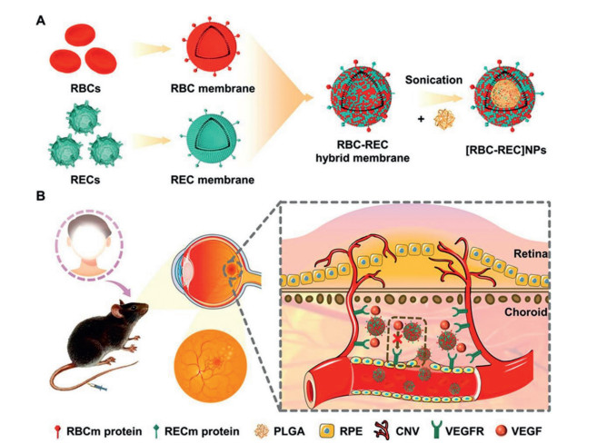
Inspired by materials of natural origin, researchers recently have designed various cell membrane-based nanoparticles (CMBNPs), which can combine chemical properties of membrane materials with advantages of proteins on the surface of cell membranes, offering various merits such as good biocompatibility and low immunogenicity, as well as prolonging the circulation time. More importantly, cell membranes of different origins have homologous active targeting ability for different disease sites, which greatly enhances the enrichment of nanoparticles in the foci, thus improving the therapeutic efficiency and reducing the toxic side effects. Until now, the cell membrane-based targeting strategy has been used for the treatment of various diseases, including tumor, inflammation, cardiovascular or other diseases [90]. For example, Xu et al. reported platelet membrane-cloaked polymeric nanoparticles conjugated with recombinant tissue plasminogen activator (rt-PA) to achieve clot targeting thrombolytic therapy. Compared to conventional thrombolytic strategies, the hybrid biomimetic nanocarrier could prolong the half-life of the therapeutic drug and improve the targeting ability, eliciting a significantly enhanced thrombolysis activity [91]. In 2021, Ouyang et al. developed a macrophage membrane-functionalized nanosystem featuring inflammatory site target ability, biomarker activatability, fluorescence and optoacoustic imaging, as well as therapy for inflammatory diseases. In this nanosystem, the macrophage membrane ensured effective targeting to the site of inflammation. Besides, under the ROS stimulation, the chromophores of this nanoparticle could be activated for fluorescence and photoacoustic imaging, releasing drugs for inflammation-targeted therapy [92]. Recently, Li et al. designed a biomimetic antiangiogenic agent based on hybrid cell-membrane-cloaked nanoparticles for noninvasively targeted treatment of choroidal neovascularization (CNV) (Fig. 3). Due to the predominantly expression of vascular endothelial growth factor (VEGF) receptor 2 on endothelial cells, authors constructed retinal endotheliocyte (REC) and RBC membranes-derived anti-VEGF nanoagents ([RBC-REC]NPs). The self-recognition capability of membrane-cloaked nanoparticles enabled them to target retinal endotheliocytes, thus contributing to their enhanced accumulation in the CNV region [93]. Similar with the cell membrane-mediated target delivery, there are researches using living cell to home diseases or overcome biophysical barriers [94, 95], which rely on the membrane binding to target cells.
In summary, the active targeting strategy is developing and a focus of research today. It has become one of the crucial strategies for constructing ideal drug delivery systems. With the continuous research of active targeting strategy and the development of related disciplines, the use of targeted drug delivery systems for various diseases will become mainstream in the future.
Although most developed nanomedicines showed improved targeting and delivery efficiency compared to conventional drugs, they still face the issues that the loaded drug can be prematurely released during delivery process or drug cannot be released at desirable time window. Recently, the development of stimulus-responsive drug delivery system to improve drug release profiles and delivery efficiency has gained increasing attention. The stimulus-responsive drug delivery system can stay stable during delivery process while respond to specific stimulus at site-of-interest, leading to changes of physiochemical properties, disassembly or cleavage of specific linker [96]. These stimuli used for activation are generally based on differences between the environment of the diseased tissues/cells and normal tissues/cells. For example, the tumor microenvironment is often characterized by various heterogenicities, such as acidic pH, upregulated enzyme expression, high redox potential, high ROS level and high ATP, compared to normal tissues/cells, which can be used as endogenous stimuli. These endogenous stimuli are also found existing in intracellular space of diseased cells, such as acidic pH condition in lysosome, high glutathione and ROS level in cytosol compared to extracellular space [97]. In comparison, the exogenous stimuli including light, ultrasound, electronic and magnetic field have also been widely explored and are more easily to be accessible because they can be simply applied at diseased site [98]. To enable stimulus-responsive drug release in a spatiotemporally manner, drugs are usually required to be either appropriately conjugated onto NPs delivery system via stimulus-responsive linker (referring as pre-drug) or be encapsulated into stimulus-responsive NPs. However, the preparation of stimulus-responsive pre-drug should not affect the pharmacological properties of drug, such as therapeutic mechanism and biological activity [96].
The stimulus-responsive drug delivery system also offers many other superiorities, such as prolonging circulation time, overcoming biological barrier, enhancing diseased site penetration and retention, intracellular assembly, thus leading to better delivery efficiency [99, 100]. It is now well accepted that the physiochemical properties (e.g., size, charge, morphology and surface modification) of nanoparticles play a critical role in determining not only their delivery behavior in the circulating system but also distribution profile at diseased site [101]. In the context of tumor, small size nanoparticles are often characterized by better penetration through tumor interstitium but poor retention. In contrast, lager size nanoparticles are more likely to be retained while have poor penetration efficiency. To integrate the advantages of both large size and small size, stimulus-responsive drug delivery system with either size-shrinkable [102, 103] or size-increasing ability [104] have been developed. These size-changeable drug delivery system showed enhanced penetration and retention profile within tumor site, thus leading to enhanced drug accumulation. Moreover, the biological barriers, such as blood-brain barrier, pulmonary mucus layer and cell membrane, pose a challenge for nanomedicine entering diseased tissue/cell. To overcome these biological barriers, strategies using stimulus-cleavable ligand [105] and stimulus-triggering charge-reversable have been proposed and demonstrated much improved drug delivery efficiency. In addition to be used for enhancing therapeutic outcome, stimulus-responsive delivery system has also been exploited as imaging agents for disease diagnosis. For example, inorganic NPs modified with functional groups can undergo in-situ self-assembly in the presence of specific stimulus, which showed distinct change of optical properties [106]. To date, various endogenous and exogenous stimulus-responsive drug delivery system based on liposome, polymeric nanoparticle, micelles, inorganic nanoparticle have been developed as theranostic platform.
As aforementioned, the diseased site or cell generally has a variety of different heterogenicities. To further improve the drug delivery spatiotemporally, researchers are now focusing on developing dual or multiple stimuli-responsive drug delivery system. The combination of two or more stimuli can be rationally chosen based on either the pathological condition or exogenous, such as pH/redox, pH/ROS, or pH/ROS/redox. Meanwhile, the combination of endogenous and exogenous stimuli is also presented to be flexible [107]. By step-by-step stimulus-responsiveness, the dual/multiple stimuli-responsive drug delivery system can not only improve the delivery efficiency specificity but also enable controlled drug release simultaneously, leading to much improved therapeutic effect and reduced dose-related side effect. For example, Gao's group developed an enzyme/pH dual-responsive drug delivery system (AuNPs-D&H-R&C) for the treatment of breast cancer. After delivering to breast tumor site via enhanced permeability and retention (EPR) effect, AuNPs-D&H-R&C could form in situ aggregate in response to the overexpressed furin within tumor interstitial space or intracellularly. The aggregates in turn restrict their back-flow to bloodstream and exocytosis by tumor cell, resulting in higher accumulation. Meanwhile, DOX and hydroxychloroquine (HCQ) could be released in response to acidic pH condition in endo/lysosome because they were conjugated onto gold nanoparticles through imine bond (a pH-liable linker). Taking together, this dual-responsive drug delivery system can significantly improve drug accumulation at breast tumor site to enable enhanced combination therapeutic effect [108]. With the advance of material science, biology and chemistry, novel stimulus and more efficient stimulus-responsive drug delivery system can be pursed. However, more sophisticated design may increase the difficulty of clinical translation, which should be taken into consideration.
Size and shape are the most important physiochemical properties of nanoparticles, as they commonly decide nanoparticles' in vivo behaviors, including distribution, activation, function and excretion. However, when nanoparticles are achieving drug delivery for diagnosis or treatment, the successive and complicated in vivo processes make the nanoparticles highly demanding. For instance, since tumor tissue is highly pressured with dense matrix and interstitial fluid [109], nanoparticles in small size show strong penetration in tumor tissue but are easily clearable, while large size makes them retain in tumor region but hard to penetrate [110, 111]. Generally, spherical nanoparticles are good in circulation but poor in retention, while linear ones depend on their aspect ratio (AR) [112-114]. Therefore, instead of engineering an invariable and defined size or shape for nanoparticles to suit complicated demands, utilizing size or shape tunable strategy is more intelligent and efficient [93, 115].
Size tunable strategy commonly include two aspects, aggregation and shrinkage. The aggregation increases size to make nanoparticles retain in tissue, prolonging the effective time. Xie et al. aggregated gold nanoparticles (AuNPs-D&H-R&C) by click reaction, which needed the pre-activation of tumor-overexpressed furin [116]. The AuNPs-D&H-R&C were uniform spherical around 40 nm to distribute and penetrate in tumor, but formed 260 nm aggregation under the triggering by furin, avoiding back-flow to blood stream and increasing cellular internalization. Consequently, by release cargos (DOX and HCQ), aggregated AuNPs-D&H-R&C showed strong and longtime function of modulating tumor-associated macrophage and killing tumor cells. Besides, for many metal nanoparticles, aggregation strengthen their photothermal effect, such as furin-mediated aggregation of Fe3O4 nanoparticles, which showed stronger photothermal therapy and MRI T2 imaging compared to monomeric nanoparticles [117]. Size shrinkage is another critical strategy, it successively meets the needs of circulation, distribution, retention and penetration, suitable for the drug delivery to solid tissue with stress. Zhang et al. constructed RLQLKL peptide-composed nanoparticles (LANPs) [102], the cleavage by neutrophil elastase (NE) reduced the PCL core, shrinking size from 212.1 nm to 72.7 nm, which allowed nanoparticles penetrating in brain tumor deeply and releasing drugs for therapy. Hyaluronic acid (HA) is an endogenous materials with negative charge, it composes the intercellular matrix and can be degraded by hyaluronidase (HAase). These properties make HA convenient as the key materials of size-shrinkable nanoparticles. Gao's group designed a kind of classical nanoparticles system by cationic small nanoparticles (gold nanoclusters, AuNCs or dendrigraft poly-l-lysine, DGL) and HA, which fabricated into large nanoparticles [118-121]. The coverage of HA on large nanoparticles ensured good circulation and tumor target effect in vivo, and allowed contained cationic small nanoparticles releasing after HA was degraded by tumor-expressed HAase. Consequently, the nanoparticles shrank size from over 200 nm to about 20 nm, deeply penetrated in tumor tissue for homogenous antitumor effect. Similar works based on HA degradation or dissociation were also found [122, 123]. Besides, there was report that HAase composed with dextran by pH sensitive maleimide linker, forming large nanoparticles (over 110 nm) but releasing monomeric HAase (~10 nm) to digest tumor matrix and deep penetrate [124]. Furthermore, the size shrinkage can be directly used in diagnosis, Stevens and co-workers designed a MMP-9 cleavable AuNCs-neutravidin conjugates (~11 nm), which could not enter urine through renal excretion. When the tumor associated MMP-9 was presence in blood, the conjugates was cleaved and released AuNCs (~1.5 nm) into urine, direct colorimetric readout of MMP-9 level was practicable by utilizing the nanozyme property of AuNCs [125].
There are also effective works made by shape tunable strategy. Shape change from sphere to line with suitable AR is reasonable, since it meets the successive demands of in vivo circulation, distribution, tumor retention and penetration. For example, Zhang et al. constructed a spherical micelle by bis-pyrene (BP), FFVLK peptide and human epidermal growth factor receptor 2 (HER2) ligand (NPs 1), and the NPs 1 could bind HER2 in tumor tissue and rearrange the structure into nanofiber (NFs 1) by ligand-receptor affinity, thus achieving the shape change. Dominant NFs 1 cross-linked into fiber net wrapping tumor region and strongly inhibited tumor growth [126]. The transformed nanofiber can also achieve enhanced tumor retention for longtime drug delivery. Gao's group designed a spherical nanostructure by Ce6-CD (Ce6-β-cyclodextrin) and ferrocene (Fc)-FFVLG3C-PEG [127]. Upon laser irradiation, Ce6 generated ROS to oxidize hydrophobic Fc into hydrophilic Fc+, resulting in nanofiber formation by Fc+-FFVLG3C-PEG and size shrinkage of Ce6-CD to enhance both tumor retention and penetration. Consequently, the continuous and homogenous ROS generation and Fenton reaction by Fc strongly promoted the antitumor effect. Similar shape change based on FFVL peptide to enhance tumor retention was also reported [53, 128-130]. Besides, shape change from line to sphere was also of significance. Yang et al. deformed spherical nanoparticles to short nanofibers by cross-linkage, with precise control of the AR (from 1.1 to 9.2), in which the AR7.4 showed the strongest blood vessel leakage and tumor penetration, and recovered into spherical nanomicelles in deep tumor for volume-dependent retention [131].
Self-propelled drug delivery system (DDS) boosts drug delivery efficiency by themselves and can be divided into two types according to different self-promotion mechanisms. Type 1 is self-propelled micro- and nanomotors (MNMs) that convert the surrounding chemical or external energy into mechanical forces to promote transport via the produced autonomous motion [132]. Type 2 is autocatalytic DDS that simultaneously regulates transport barriers and delivers drugs through the engineered versatility. Both types possess potential in mediating membrane diffusion and deep tissue penetration for active and targeted drug delivery.
The autonomous motion of MNMs can facilitate its efficiency of overcoming transport barriers (e.g., the blood-brain barrier (BBB) and dense extracellular matrix) to enhance drug delivery. For example, Joseph et al. constructed glucose oxidase and catalase co-powered asymmetric MNMs for BBB overcoming via glucose gradient-mediated navigation in the blood [133]. The propulsion of MNMs was mainly attributed to the decomposition of glucose by glucose oxidase into endogenous d-glucono-δ-lactone and water without forming harmful wastes. Through this glucose-chemotactic feature and large glucose consumption by the brain, MNMs autonomously move along the glucose concentration gradient from the center to the blood vessel wall and enter the brain more efficiently than passive nanoparticles.
The autonomous motion can also accelerate cellular internalization and lysosome escape of MNMs [134], which endows MNMs with the potential of delivering gene therapy. Tumor hypoxia often leads to high metastasis and inertness to chemotherapy while insufficient oxygen delivery and the confronting "Warburg effect" compromise the therapeutic efficacy of hypoxia alleviation. Recently, Yu et al. construct glucose oxidase and catalase co-powered nanomotor to simultaneously deliver sufficient oxygen to deep tumor and inhibit the aerobic glycolysis via the co-loaded hexokinase-2 siRNA to potentiate anti-metastasis in chemotherapy [135]. The production of oxygen bubbles propels the nanomotor to move along H2O2 gradient towards deep tumor and alleviates hypoxia in the meantime. The autonomous movement also mediates efficient hexokinase-2 silencing to inhibit glycolysis. The cascade enzyme powered nanomotor provides a potential for reversing tumor hypoxia and abnormal metabolic pathway for reinforced anti-metastasis of chemotherapy.
Autocatalytic DDS is developed with multi-functionality to simultaneously regulate transport barriers and delivers drugs. Among various tumor microenvironment factors (e.g., acidic pH, hypoxia, intercellular pressure, extracellular matrix and resistant protein), extracellular matrix significantly affects drug delivery and therapy. Huang et al. modified DOX-loaded nanoparticles with clusterin and collagenase Ⅳ to simultaneously reduce the nonspecific protein adsorption during the circulation and degrade type Ⅳ collagen of tumor extracellular matrix for mediating drug penetrating dense tumor tissues [136].
Receptor-mediated transcellular vesicle transport is often used to overcome the BBB and mediate intracranial drug accumulation owing to the closing of BBB paracellular diffusion by tight junctions and the limitation of BBB transcellular diffusion by efflux transporters. However, the efficiency of receptor-mediated transcellular vesicle transport is limited by the low density of BBB receptors and Mfsd2a-mediated low transcytosis rate. Inspired by the fact that statins can suppress cholesterol synthesis and then compensatorily induce expression of low-density lipoprotein receptor-related protein 1 (LRP1), Guo et al. designed LRP1-targeting simvastatin and DOX co-loaded nanoparticles to upregulate the BBB LRP1 expression and boost intracranial accumulation of the engineered nanoparticles [137]. LRP1 on the abluminal side of the BBB can remove intracranial nanoparticles. To escape abluminal LRP1-mediated BBB clearance, Khan et al. developed responsive nanoparticles to detach LRP1-targeting ligand after entering the brain and expose tumor-targeting ligand for drug delivery to intracranial lesions [138]. Recently, Ju et al. constructed tunicamycin and DOX co-loaded BBB-targeting nanoparticles to enhance intracranial nanoparticle accumulation via directly inhibiting the upstream regulator Mfsd2a of BBB low transcytosis [139, 140].
Specific features of diseased BBB can be utilized to mediate specific drug transport to intracranial lesions and avoid side effects to normal brain [141]. For example, prostate-specific membrane antigen (PSMA) is specially expressed in vascular endothelium around breast cancer brain metastases to promote tumor growth and angiogenesis. Based on the specific PSMA expression, Ni et al. engineered PSMA-targeted NPs to mediate specific drug delivery to breast cancer brain metastases [142]. ATP-sensitive potassium channel (KATP) is specially expressed in the blood-brain tumor barrier (BTB) and its activation can enhance BTB permeability via up-regulating caveolin-1 and down-regulating tight junctions. Miao et al. designed KATP activator minoxidil and therapeutic DOX co-loaded hyaluronic acid-modified nanoparticles to increase specific nanoparticle accumulation in brain metastases [143].
Despite enormous potentials for future clinical translation, there still exist many challenges for self-propelled DDS. MNMs are challenged by a series of issues including difficult and sophisticated synthesis technology, toxic exhaust gas, waste and extra fuel, short lifetime, quick blood elimination, and inaccurate cell targeting [132]. The development of zero-waste symmetric MNMs with exact mechanism of autonomous motion, in vivo long circulation and accurate cell targeting may be able to remarkably promote the clinical translation and biomedical application. For autocatalytic DDS, the modulated body components (e.g., tumor extracellular matrix, BBB LRP1 and Mfsd2a and BTB KATP), also have important physiological roles. The modulation of these body components may significantly affect normal physiological activities. Future biomedical application and clinical translation should be focused on autocatalytic DDS with more transient and reversible modulation effects.
Systematic administration of small molecule drugs is hindered by low bioavailability, untargeting, and biological barriers, which impeded drug efficiency and local delivery to affected tissue. Nanoparticle-based drug delivery systems are powerful tools for targeted delivery of small molecule drugs to diseased tissues. However, satisfactory local delivery is still challenging. The main obstacles include the removal of nanoparticles by phagocytes and biological barriers, such as endothelial cells that hinder the infiltration and accumulation of diseased tissues [144-146]. Inflammation is associated with many diseases, such as cancer, stroke and atherosclerosis. The inflammatory predisposition of immune cells can bring cellular hitchhikers directly to the disease tissue in a highly targeted manner [147]. Employing immune cells for active transport of drugs and drug-loaded nano-carriers to a target site is a promising recent approach. The advantages of these strategies are due to the natural transport capabilities of living cells, such as systemic circulation, active crossing of biological barriers, and chemotaxis to the disease site [148].
Neutrophils are one of the most abundant circulating leukocytes, and also one of the first leukocytes to reach inflammatory tissue [144, 149]. Upon activation, neutrophils produce neutrophil extracellular traps by decondensing their chromatin, decorating it with cytoplasmic or granule effector proteins, and then extruding it from the cell within a few hours [150]. A defined release mechanism that enables neutrophils to serve as a delivery system for drugs or loaded nanocarriers at early stages of inflammation. For instance, Stevens et al. encapsulated methotrexate (MTX), a potent immunosuppressive agent used to treat inflammatory and autoimmune diseases, in cationic liposomes and loaded in vitro into isolated neutrophils (MTX-liposomes/neutrophils) against skeletal muscle inflammation and myocardial ischemia reperfusion injury [144]. Intravenous MTX-liposomes/neutrophils system migrated efficiently in response to inflammatory chemokine gradients and delivered drugs efficiently at the site of injury via extracellular traps generation, improving drug delivery to inflammatory tissues and reducing severe side effects associated with systemic administration. In another study, Wang et al. constructed a molecularly engineered liposomes with inverse phosphocholine lipids that rapidly enrich complement fragment iC3b by "voluntary opsonization", thereby triggering neutrophil hijacking through complement receptor 3 phagocytosis [151]. Neutrophils carrying liposomes migrate across the alveolar-capillary barrier into inflamed tissues, wherein neutrophils either release drug-loaded liposomes via the formation of neutrophil extracellular traps or serve as micro-containers to confine both drug-loaded liposomes and bacteria intracellularly for bacteria killing, resulting in pulmonary inflammatory control (Fig. 4).
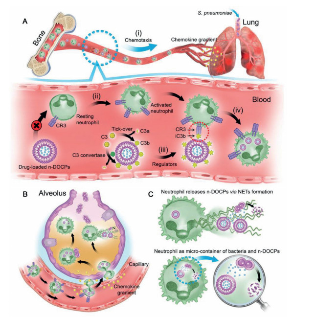
Macrophages/monocytes are widely recognized as having a remarkable tumor-homing capacity that is mediated by various chemoattractants that are secreted by the tumor cells. Taking advantage of their tumor-homing ability, macrophages/monocytes hitchhiker across the barrier to transport drug-loaded NPs, accumulating therapeutic molecules at the site of disease [152, 153]. Therefore, Tan et al. took advantage of the characteristic that apoptotic bodies are readily engulfed by phagocytic cell and used it as a biological carrier loaded with CpG, a Toll-like receptor 9 ligand, modified gold-silver nanorods for monocytes/macrophages-targeted cargo loading [152]. Intravenous administration of apoptotic body-encapsulated nanomedicine in C57BL/6 mice was rapidly phagocytic by circulating monocytes in the blood. Based on the homing behavior of monocytes/macrophages, nanomaterials can be efficiently transported to the inner region of the tumor (Fig. 5). With the photothermal effect of nanorods and the immune stimulation promoted by CpG, this cell-mediated delivery system can not only effectively ablate primary tumors, but also induce a potent immunity to prevent tumor metastasis and recurrence.
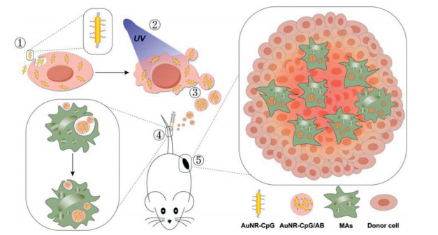
Nucleic acid therapy represents one of the most promising directions in drug development industry. Nucleic acid-based therapeutics, include RNA interference (RNAi) molecules (e.g., siRNA and miRNA), antisense oligonucleotide (ASO), mRNA modality (e.g., mRNA vaccine), CRISPR/Cas (clustered regularly interspaced short palindromic repeats–CRISPR-associated proteins) modules, aptamer, DNA-mediated gene therapy, small activating RNA (saRNA), Antagomir (anti-miR) and Ribozyme/DNAzyme, etc., have showed potent regulation capacity in the treatment of diverse diseases [154]. Small interfering RNA (siRNA) and microRNA (miRNA) can silence the expression of almost all genes of interest [155-157]. ASO regulates target gene expression by either recruiting RNase H to cleave target mRNA or modulating mRNA splicing [155]. DNA or mRNA (also including self-amplifying RNA and circle mRNA) can be used to express desired protein, thereby correcting gene expression defects or abnormalities, or expressing antigens to prepare vaccines [158, 159]. CRISPR/Cas modules can precisely change or modify target genes through efficient and stable knockout, knock-in or point mutation of specific genes, and realize the "close", "restoration" and "switching" of gene functions [160]. Therefore, CRISPR/Cas system was widely used for genome editing, gene detection, DNA and RNA imaging, regulation of gene transcription, establishment of disease model, etc. Aptamers can bind to target molecules (including proteins, small molecules, etc.), thereby potentially can be used to neutralize the target protein, or detect certain molecule, or achieve targeted drug delivery [161-163]. The sequence structure of small activating RNA (saRNA) is similar to that of siRNA, but it usually up-regulates the expression of the target gene by binding to the promoter region of the gene [164]. Antagomir is a single-stranded oligonucleotide that can bind to the target miRNA and inhibit the biological function of the miRNA [155]. Ribozyme or DNAzyme can be used to degrade the target nucleic acid molecules. However, one of critical and most challenging issue for broad application of these nucleic acid modalities is how to realize efficient and safe delivery in vivo.
For siRNA delivery, various strategies can be divided into three classicals according to the physiochemical properties of the materials or methods (Fig. 6): (1) Chemically-synthetic materials such as ionizable lipid/lipidoid nanoparticle or lipid-siRNA conjugate [165, 166], cationic polymer [167-169], inorganic nanoparticle [170], metal-covalent organic frameworks (MCOFs) [171]; (2) biological carriers including peptide [172], exosome (extracellular vesicle) [173], aptamer-siRNA conjugation [161], GalNAc-siRNA conjugation [154-156], three‐way junctions; (2) physical methods such as electroporation [174], microinjection, sonoporation, photoporation, magnetofection, poking devices [175]. Recently, Huang and colleagues designed and screened several panels of lipid-like materials. As a result, one leading structure termed A1-D1–5 was selected, and the optimized formulation called iLAND (ionizable lipid-assisted nucleic acid delivery system) efficiently deliver siRNA into hepatocytes and achieved excellent treatment effects in three hyperlipidemia disease models [166]. Huang and co-workers also engineered M1-macrophage-derived extracellular vesicles (EVs) by introducing vesicular stomatitis virus glycoprotein (VSV-G) and electroporating anti-PD-L1 siRNA (siPD-L1) into the EVs. Proposed EVs transported siRNA into the tumor cells via membrane fusion, successfully restored CD8+ T cell function and boosted macrophage phagocytosis in tumor microenvironment [173]. In addition, Huang's group fabricated a rolling microneedle electrode array (RoMEA) by utilizing parallel circular blades with microneedle arrays on edge as electrodes. RoMEA allowed low-damage and large-area siRNA transfection in vivo. By electroporating anti-PD-L1 siRNA, ideal immunotherapy effects were achieved in both B16-F10 and CT26 tumor models [174]. mRNA constitutes a promising nucleic acid modality that can be developed as therapeutic agent or therapeutic/prophylactic vaccine [176]. Lipids and lipid derivatives are the most widely-used and clinically-transformable delivery materials [175]. Two lipid-like structures termed SM-102 and ALC-0315 were employed in mRNA-1273 and BNT162b2, respectively (Fig. 7) [159]. These two COVID-19 protection vaccines have been approved and administered globally. N-[1-(2,3-dioleyloxy)propyl]-N,N,N-trimethylammonium chloride (DOTMA) and ATX lipid (Arcturus) were also used in clinical investigation of mRNA vaccines for controlling infectious disease or cancer. In addition, polymer or lipid-polymer hybrid were also employed to deliver mRNA. For example, nanoparticles prepared with poly(β amino ester) terpolymers (PBAEs) and PEG-lipid successfully transferred mRNA to the lungs after systemic administration in mice [177-179]. Zwitterionic phospholipidated polymers (ZPPs) were developed to deliver mRNA to spleen and lymph nodes in vivo [180]. Moreover, it was reported that zolitic imidazolate framework-8 (ZIF-8) confined with dendritic mesoporous organosilica nanoparticles (DMONs) enhanced mRNA transfection and translation efficiency than commercial products and toxic polymer-modified DMONs in vitro and in vivo [181].
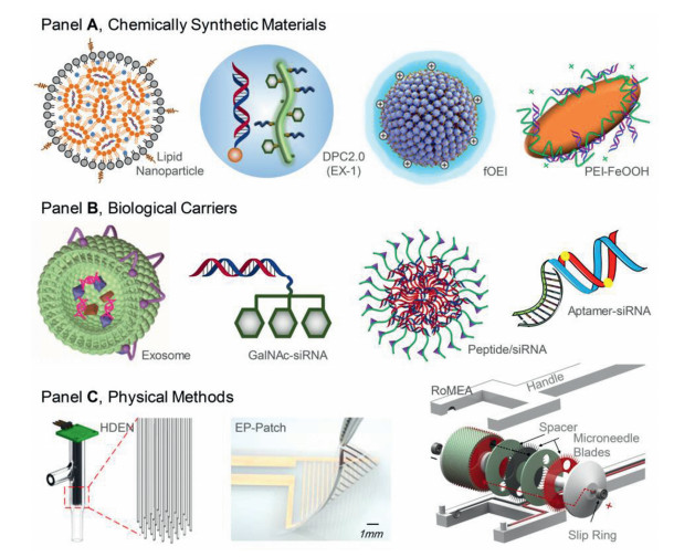
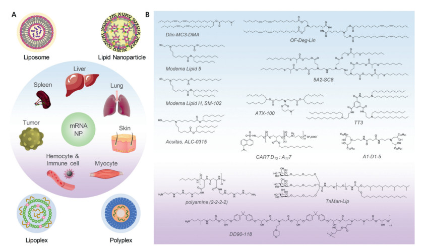
Plasmid DNA, mRNA and ribonucleoprotein (RNP) all can be utilized to achieve CRISPR/Cas-based genome editing (Fig. 8). Virus-based vectors, including adeno-associated viruses (AAVs), adenoviral vectors (AVs), lentiviral vectors (LVs) and phage, have been investigated in preclinical and clinical tests for genome editing and have shown excellent transfection efficacy [182]. However, potential immunogenicity of viral vectors limits its widespread application, although effective editing may be achieved with a single dose. In addition, non-viral synthetic materials are the more transformable choice for in vivo genome editing, because they can be controllably synthesized, easy to introduce desired chemical modifications, and show potent delivery efficiency in vivo. NTLA-2001, an in vivo gene-editing therapeutic agent for reduction of the concentration of transthyretin (TTR) in serum, was formulated with lipid nanoparticle (LNP). Phase 1 study showed that one dose of NTLA-2001 at 0.1 mg/kg and 0.3 mg/kg triggered 52% and 87% reduction of serum TTR protein from baseline, respectively [183]. iLP181, an ionizable LNP, could effectively load gene-editing plasmid and achieve ideal oncotherapy for liver cancer [160]. Daniel Siegwart and co-workers synthesized a series of multi-tailed ionizable phospholipids (iPhos) and the optimized formulation could deliver mRNA or mRNA/single-guide RNA for gene editing in vivo [69]. Moreover, cationic lipid-assisted polymeric nanoparticles (CLANs), PEI-based materials (PEI polyplexes, PEI-coated DNA nanoclews, etc.), cationic polypeptide, PAMAM dendrimers, cell-derived vesicles were also developed for CRISPR/Cas system delivery (Fig. 8) [182, 184].
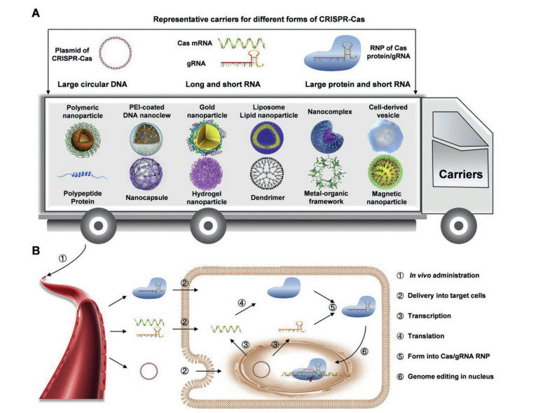
In summary, various nucleic acid modalities represent one of next-generation drug development platforms. Efficient and safe delivery is the bottleneck issue for clinical translation. Diverse strategies, including chemically-synthetic materials, biological carriers, and physical methods, were intensively investigated for in vitro and in vivo delivery of siRNA, mRNA, CRISPR/Cas system, etc.
The delivery of protein drugs at the living cell level is of great significance for chemical intervention in the life process, disease diagnosis and treatment research. After considerable advances in biotechnology and protein engineering, the emergence of therapies with peptides, proteins and antibodies has realized sustained clinical benefits for various diseases. However, the further popularity and the therapy potency of protein drugs in clinic are still hampered by the significant limitations, stemming from their severe vulnerability in biological circumstance, poor bioavailability, potential immunogenicity, high risk of infection, and generation of antidrug antibodies (ADAs). Protein drugs also suffer chemical instabilities (deamidation, racemization, oxidation, hydrolysis, etc.) and physical instabilities (denaturation, aggregation, surface adsorption, etc.). Therefore, it is highly desired to identify innovative protein delivery strategies. In this regard, we reviewed the latest advances in the delivery approach of therapeutic protein drugs.
Affected by the physiological environment of the human body, the parenteral injection is the most popular administration route of protein drugs, which greatly limits the wide application of protein drugs. The mentioned issues can be solved by optimizing the amino acid sequence of proteins to improve its stability, as well as chemical modification/attachment. For instance, the amino acid sequence of desmopressin (DDAVP) was optimized by a genetically engineering approach to reduce its immunogenicity and extend its half-life [185]. In order to reduce the renal clearance and extend plasma half-life of protein drugs, chemical modifications are also developed based on covalent attachment of polyethylene glycol (PEG) to protect protein immunogenic epitopes, reduce protein aggregation and protease digestion, and increase the hydrodynamic diameter of protein drugs [186]. Kyobum Kim et al. developed a series of mPEGylated PEADs with different PEG chain lengths and effectively encapsulated the VEGF-165 with high loading efficiency. This study provided a good paradigm to exploit PEG as potential growth factor delivery vehicles [187]. Nobuhiro Nishiyama et al. constructed a ternary a simple protein delivery system using TA and boronic acid-conjugated polymers. It showed significantly prolonged blood circulation and higher tumor accumulation than the conventional protein/TA complex in a subcutaneous tumor model [188].
In addition, the half-life of circulating target proteins can be prolonged by increasing the interaction between therapeutic proteins and serum components (such as albumin or immunoglobulin) [189, 190]. Niren Murthy et al. demonstrate that the coiled-coil forming peptide (KVSALKE)5 can act as a cell penetrating peptide (CPP) and interact with proteins containing the partner peptide E5. The study validated that the new type of CPP has great potential in improving the delivery of proteins towards specific cells and tissues in vivo [191]. In addition, the sequence of antibodies can be humanized to reduce the immunogenicity. Antibody-drug conjugates (ADCs) are developed from monoclonal antibodies that are conjugated with chemo drug payloads through a linker. ADCs have gained increasing attentions as an anti-cancer therapeutic agent [192, 193]. The latest Phase Ⅱ study shows that the ADC drug RC48-ADC that consists of a novel humanized anti-HER2 IgG1, a linker, and microtubule inhibitor, has shown promising antitumor activity in preclinical and early clinical studies [194].
Modifications of protein drugs can significantly change their pharmacokinetic behavior. However, the existing improvement methods cannot completely eliminate the inherent defects in the treatment of protein drugs. Related studies have found that the modification of protein drugs will lead to the loss of therapeutic effects, the increase of adverse effects, and generation of new immunogenicity [195, 196].
Lipid-based delivery systems including biocompatible polymers (PEGs and other synthetic or naturally derived macromolecules) and colloidal systems (liposomes and other lipid or polymer nanocarriers) have been utilized as protein nanocapsules to combat the formidable challenges related to the poor stability, rapid clearance, and immunogenicity limitations. Ulrich Lächelt et al. developed a novel delivery platform of Cas9 protein/sgRNA RNP complexes, which suggested great potential of lipid nanoparticles in genome editing applications in vivo [197]. Considering extracellular vesicles (EVs) are promising natural nanocarriers for delivery of proteins, Elena V. Batrakova et al. utilized macrophage-derived EVs to deliver soluble lysomal enzyme tripeptide peptidase-1 (TPP1) for the treatment of lysosomal storage disorder, neuronal ceroid lipofuscinoses 2 (CLN2) or Batten disease. In the in vitro model of CLN2, EV significantly improved the resistance of TPP1 to protease degradation and provided efficient TPP1 delivery to target cells [198].
Because of their excellent biocompatibility and structural and functional tunability, polymer carriers have received intense attentions in this field. The main challenge in polymer design lies on the low binding efficiency of polymer carriers to cargo proteins of different properties, especially under physiological conditions. Wang et al. carried out systematic research around the intelligent delivery of protein, and successively developed carriers such as cationic liposomes and metal organic frameworks [199, 200]. Due to the responsiveness of the carriers to the cell microenvironment and the intracellular degradation process, the as-constructed proterin delivery system realized functional regulation of protein drugs in vivo. In order to realize intracellular protein delivery, Cheng et al. utilized dynamic polymer super amphiphiles which can be co-assembled with proteins to form stable nanoparticles in water to release the bound protein in the cell [201]. This study integrated polymer chemistry and supramolecular engineering strategies to achieve intracellular delivery of superoxide dismutase for the treatment of ulcerative colitis in vivo, thus providing new opportunities for rational design and easy construction of robust intracellular protein delivery materials.
Numerous studies of drug delivery devices across biological barriers are also undergoing to address the challenges of non-invasive protein delivery (oral, transdermal, inhalation and mucosal administration). For non-invasive delivery systems, it remains a prominent challenge to realize spatiotemporal control of proteins. Drug delivery systems with biological responsiveness hold significant advantages to address the above issue. Guldberg et al. developed an injectable delivery system consisting of heparin microparticles and an alginate hydrogel, which was capable of delivering multiple growth factors in a tunable manner. Effective bone healing was achieved in composite injury models [202]. Gu et al. developed a glucose intelligent response to release insulin microneedle patch. The patch could quickly release the encapsulated insulin in response to the hyperglycemic state. This new type of smart insulin patch showed more rapid response reaction than the widely used pH-sensitive formulations, and thus can reduce the risk of hypoglycemia [203-205].
Hormonal drugs leuprorelin microspheres (Lupron Depot) and goserelin sustained-release implants can reduce the number of subcutaneous injections required and greatly reduce the side effects of the drug on the human body [206]. Afrezza is an artificial fast-acting inhaled insulin that uses Technosphere as an insulin delivery system to act through the lungs to control hyperglycemia in adult diabetic patients. It has been proven to help control blood sugar levels in patients with type 1 and type 2 diabetes [207]. Robert E. Guldberg et al. utilized the strong affinity interaction between heparin microparticles (HMP) and BMP-2 to improve protein delivery to bone defects. By evaluating HMP loaded with BMP-2 as a model for treating femoral defects in rats, the results show that HMP can effectively enhance the retention of BMP-2 in vivo, improve the spatial positioning of bone formation in large bone defects, and reduce heterotopic ossification [208].
Although parenteral administration of protein drugs is commonly used to bypass absorption barriers, it suffers from problems associated with repeated dosing, high cost, and pain, raising the need for alternatives to achieve non-invasive delivery system administration routes. However, protein administration methods face some absorption barriers, which significantly affect the bioavailability of protein drugs and drug treatment effects. Therefore, it is necessary to develop new alternatives to improve protein delivery efficiency. The development of non-invasive delivery systems for therapeutic protein drugs, especially the development of nanotechnology, provides a good opportunity, and can improve the delivery of protein drugs through non-invasive administration methods. In addition, the combined use of nano-formulations with complex medical equipment can maximize the efficiency of non-invasive drug delivery systems.
Oral administration with the advantage of high patient compliance remains one of the most common and convenient method of administration [209]. Recently, with the development of drug discovery technologies such as virtual screening, high throughput screening technology and high content screening, a large number of candidate drugs have been discovered. However, 40%−70% of these drugs are difficult to obtain sufficient therapeutic concentration due to their low solubility and poor oral absorption. In particular, protein and peptide drugs, with high molecular weight, are not easy to pass through the biological membrane and are susceptible to gastrointestinal microenvironment, as well as the first pass effect of liver [210]. Therefore, how to construct an ideal oral drug delivery system has become a significant problem to be solved in recent years.
Various nanocarriers including liposomes, polymer nanocapsules, microemulsions, polymer micelles, dendrimers, and nanomedical crystals have been emerged for the delivery of oral therapeutic drugs. Wang et al. designed engineered liposomes targeting the gut-CNS Axis for comprehensive therapy and realized comprehensive treatment of spinal cord injury [211]. Omar et al. found that oral delivery of controlled release polymer nanocapsules containing trypsin could target the small intestine and protect trypsin from the harsh condition during the process of preparation or the period of storage [212]. Akshay used the stable microemulsion to improve the oral bioavailability of felodipine, which can help to reduce the dose and its associated side effects [213]. Yang employed a delivery system based on L-carnitine (LC) conjugated chitosan (CS)-stearic acid polymeric micelles for improving the oral bioavailability of paclitaxel (PTX) through targeting intestinal organic cation/carnitine transporter 2 (OCTN2) [214]. Natalia et al. managed a fourth-generation polyamidoamine (PAMAM)-based nanovector for the efficient delivery of methotrexate to U87 glioma cells [215]. Although the above design strategies of delivery system are different, after entering the gastrointestinal tract, nanoparticles (NPs) can be absorbed mainly through the following three pathways: (1) cell bypass channel transport; (2) transcellular uptake of intestinal epithelial cells; (3) phagocytosis by microfold cells (M-cells) of Peyer's patches in ileum, which is the main absorption pathway of oral NPs. As the NPs enter gastrointestinal tract (GIT), partial drugs released and enter the bloodstream in the same way as free drugs are absorbed. After being phagocytosed by M-cells, the NPs are transported to the concave cavity of the basal surface of M-cells through cystic transport mode and released. Subsequently, the NPs pass through lymphatic vessels with a state of free or phagocytosed by macrophages and circulate from lymph into blood circulation.
However, challenges remain of delivering oral NPs site-specifically and efficiently. Researchers further modified the NPs based on the specific GIT microenvironment, which could greatly facilitate the interaction of NPs with tissues and cells in a particular section of GIT, increasing their residence time (Fig. 9). Specifically, for effective stomach targeting, NPs must overcome various physiological hurdles, including gastric motility, gastric pH and gastric mucus. Cai used the in situ pepsin-assisted needle assembly of magnetic-graphitic-nanocapsules for enhanced gastric retention and mucus penetration, which offers a robust strategy for effective oral drug delivery and site-selective therapy of gastric diseases [216]. And the small intestine employing nanotechnology was proved to be a more effective way for site-specific drug delivery [217]. Ma designed M-cell targeted polymeric lipid NPs containing a Toll-like receptor agonist to improve the cellular uptake of the lectin-functionalized NPs in the Peyer's patches and boost oral immunity [218]. Besides, colon targeting strategies can be conducive to maintaining the integrity of NPs and protecting the drug cargo during transit through the stomach and small intestines to maximize drug delivery to the colon [219]. Li developed an efficient enzyme-triggered controlled release system for colon targeting oral delivery to combat dextran sodium sulfate (DSS)-induced colitis in mice. They used curcumin-cyclodextrin (CD-Cur) inclusion complex as core and low molecular weight chitosan and unsaturated alginate resulting NPs (CANPs) as shell. The formed CD-Cur-CANPs have showed a narrow particle-size distribution and a compact structure with an efficient therapeutic efficacy, strong colonic biodistribution and accumulation, rapid macrophage uptake, as well as promoted colonic epithelial barrier integrity and modulated production of inflammatory cytokines, reshaped the gut microbiota in mice [220]. Notably, following the in-depth understanding of the organism microscopic world, intestinal microbiota, especially probiotics, has gradually attracted the attention of the researchers [221]. Liu et al. regulated the gut microbiota of mice by orally administering probiotics coated with lipid membrane via biointerfacial supramolecular self-assembly, which significantly ameliorate the severity of experimental colitis [222]. Wang et al. constructed an oral autonomous nanogenerator based on probiotic spores, which can simultaneously achieve the colonization of probiotics in the intestinal tract and the generation of therapeutic NPs, effectively overcoming multiple physiological barriers, showing a good application prospect in the treatment of colon cancer [223].
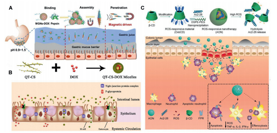
The "holy grails" of oral administration is to achieve efficient and effective administration, which is also the reason for NPs used for oral administration. Oral NPs could improve the solubility of insoluble drugs, enhance the adhesion of drug-loaded particles in the gastrointestinal tract, ensure the intestinal stability of drugs and protect drugs from enzymatic hydrolysis and pH influence, promote the lymphatic absorption and transport of drugs, and improve oral bioavailability. By further modification, some of them have achieved the advantages such as sustained release, controlled release, and targeted functions. With further research, the prospects for oral NPs will be significantly improved.
Gas therapy has emerged as a new class of therapeutic model to treat various diseases including tumor, inflammation and cardiovascular diseases [224]. As endogenous signal transduction molecules, gas molecules such as nitric oxide (NO), sulfur dioxide (SO2), carbon monoxide (CO), hydrogen (H2), hydrogen sulfide (H2S) and hydrogen selenide (H2Se) deeply involved in diverse physiological and pathological processes in living subjects [225]. The regulation of these gas molecules in lesion sites profoundly affect the occurrence and development the diseases [226]. To achieve the specific and on-demand release of gas molecules in disease sites, nanotechnology has been applied to integrate these gas molecules into prodrug which could be triggered by various stimulus such as light, X-rays, ultrasound, pH, glutathione, ROS [227-229].
As the first applied gas molecule in biomedical field, the discovery of the role of NO in the cardiovascular system leaded to the Nobel Prize in 1998. Since then, the function of NO in tumor or antibiotic therapy has been extensively exploited. For example, Gao et al. integrated near-infrared (NIR) laser-sensitive nitric oxide donor, prodrug of DOX and indocyanine green (ICG) into the hyaluronic acid shell. The obtained nanoparticles exhibited synergistic deep tumor penetration due to the enzymatically degradable hyaluronic acid shell and the vasodilatation by the NO release under the irradiation of NIR laser, leading to a much better anti-tumor efficiency with few side effects [230]. Moreover, Zhao et al. fabricated bismuth sulfide (Bi2S3) nanoparticles which was loaded with bis-N-nitroso compounds (BNN) (Fig. 10A). Upon the irradiation by 808 nm laser, the high photothermal conversion efficiency and on-demand NO release were realized simultaneously, resulted in the synergistic anti-tumor efficacy with mild PTT [231]. In recent years, SO2 is not only recognized as a polluting gas but can also be used for anti-tumor treatment. For example, Yang et al. developed a type of UV light responsive SO2 prodrug which was loaded into the rattle-structured upconversion@silica nanoparticles (RUCSNs) (Fig. 10B). The RUCSNs could convert NIR light into ultraviolet light so as to activate the prodrug for the controlled release of SO2, which leaded to the increase of intracellular reactive oxygen species levels and the damage of nuclear DNA [232]. To further improve the limited tumor penetration of current nanomedicine, Li et al. developed a type of SO2 prodrug (BTS) which was loaded into Au-Ag hollow nanotriangles. Under the irradiation by NIR laser, the nanocomposites converted light into heat for PTT, while the acidic environment in tumor lysosome resulted in rapid release of SO2 for deep tumor therapy, leading to the upregulation of apoptosis factor Bax and increase of caspase-3 expression to accelerate the apoptosis of tumor cells [91]. CO has been recognized as a double-edged sword, which protected tumor cells at low concentrations and killed them at high concentrations. Yang et al. prepared a multifunctional nanoplatform which was composed of mesoporous carbon nanoparticles (MCN) as drug carrier, DOX as chemotherapeutic drug and triiron dodecacarbonyl (FeCO) as thermo-sensitive CO prodrug. The nanoplatform could absorb NIR light and convert it into ample heat to trigger the rapid release of CO, which leaded to the enhanced cancer sensitivity to DOX by the ferroptosis pathway [233]. To increase the delivery efficiency of CO-based nanomedicine, He et al. developed a multistage assembly/disassembly strategy for mitochondria-targeting and mitochondrial microenvironment-responsive CO release (Fig. 10C) [234]. The obtained CO-based nanomedicine could achieve the sequential (ⅰ) the passive tumor targeting delivery, (ⅱ) the active tumor cell targeting delivery, (ⅲ) the acid-responsive CO prodrug release, (ⅳ) the mitochondria targeting prodrug delivery, and (ⅴ) the ROS responsive CO release by one single nanoplatform, which significantly augmented the anti-tumor efficiency [234]. H2 has been demonstrated to show the function of antioxidant, antiapoptosis and anti-tumor in recent years. He at al. integrated H2 into the Pd nanoparticles to fabricate a type of NIR laser-responsive H2 releasing nanoplatform for the first time (Fig. 10D). Under the irradiation of 808 nm laser, the Pd nanoparticles converted light into heat which triggered the rapid release of H2. The combination of PTT and H2 therapy exhibited cancer-selective synergistic therapy with negligible side effects [235]. To further broaden the H2 releasing nanoplatform, Yang et al. developed a type of Au-TiO2@ZnS: Cu, Co-A(Au-TiO2@ZnS) for X-ray triggered H2 release for synergistic H2-radiotherapy [236]. The H2-radiotherapy combined effect not only induced cell death through DNA damage caused by radiotherapy, but also induced cell death by mediating AMPK apoptosis pathway and caspase-3 apoptosis pathway, resulting in the complete inhibition of tumor growth. As a type of human endogenous gas, H2S has also been applied in anti-tumor therapy. Cai et al. synthesized a type of ferrous sulfide embedded bovine serum albumin nanoclusters via a self-assembly approach [129]. The obtained nanoclusters could achieve the controlled release of H2S and Fe2+ in tumor acidic environment. The accumulation of H2S gas in tumor cells resulted in the specific suppression effect to catalase activity and elevation of H2O2 concentration, which significantly enhanced the CDT effect by Fe2+. In addition, Chen et al. developed a kind of polyvinyl pyrrolidone modified multifunctional iron sulfide nanoparticles (Fe1-xS-PVP NPs) via a one-step hydrothermal method (Fig. 10E) [237]. Under tumor acidic environment, the Fe1-xS-PVP NPs produced H2S gas in situ, leading to the activity suppression of enzyme cytochrome c oxidase (COX Ⅳ) in cancer cells, which contributed to the combined inhibition of tumor growth with PTT. As the metabolic intermediates of selenium, hydrogen selenide (H2Se) played important roles in diverse physiological processes. Recently, Lu et al. developed a type of biocompatible ferrous selenide (FeSe2) nanoflowers which could achieve the on-demand H2Se release with the mild photothermal effect by the irradiation of 1064 nm laser (Fig. 10F) [238]. The accumulation of H2Se in tumor cells leaded to the down-regulated expression of high mobility group box 1 (HMGB1) protein, which could result in excessive cell autophagy via Akt/mTOR signaling pathway. The combined effect of PTT and H2Se effectively suppressed the tumor growth in a subcutaneous breast tumor-bearing mouse model and prevented the liver and lung metastasis by down-regulation of the metastasis-related proteins.
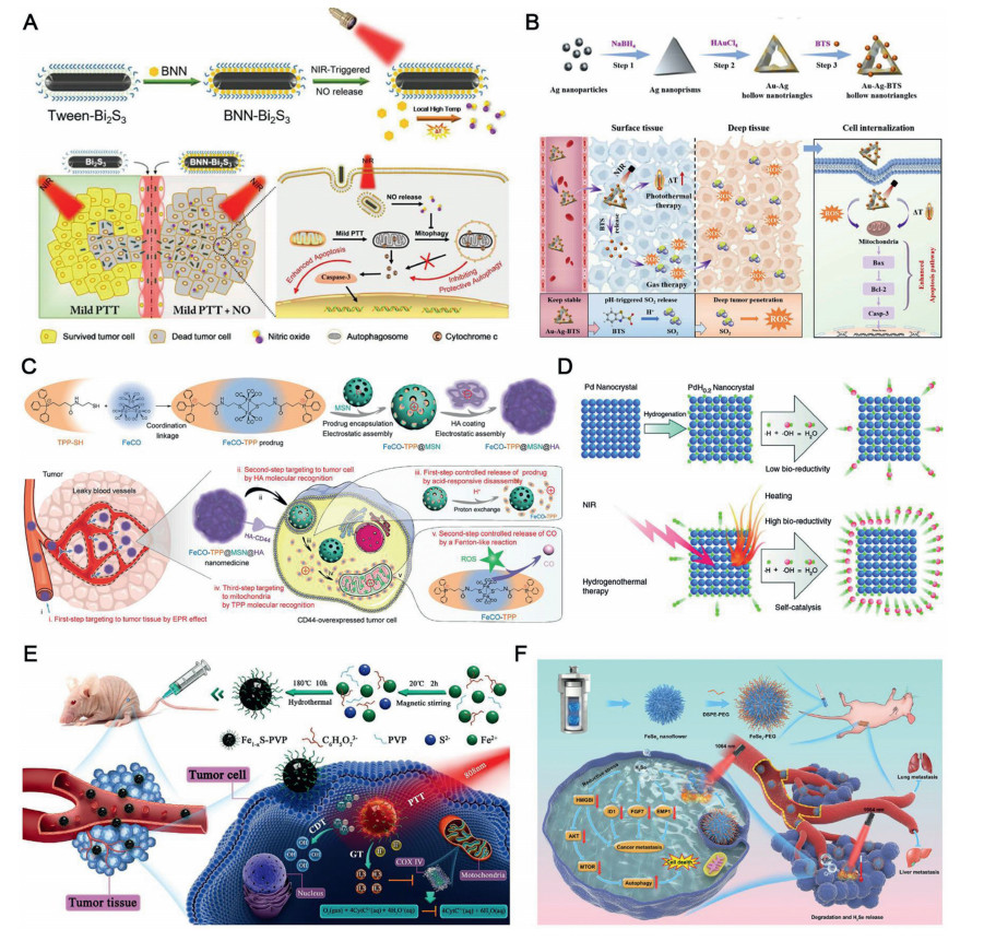
Inspired by the natural enzymes with critical roles in biology, significant efforts have been made to develop artificial enzymes to regulate biological process for disease management. Along with the progress of nanoscience, a special type of artificial catalyst call nanozyme has attracted particular attention owing to superiorities over nature enzymes, such as high-stability, low cost, ease of synthesis, tunable activity and selectivity. The term of "nanozyme" was introduced to describe the nanomaterials with intrinsic enzyme-like properties. While this concept is still under debate current [239], it is a rapidly growing field with more than 7500 publications since 2004, reporting around 300 different nanomaterials showing a broad range of catalytic activities [240]. The initial applications of nanozymes mainly focused on the industrial field for promoting the chemical synthesis efficiency. Later, the use of nanozymes have been expanded to biomedical field when some of them were attempted to initiate catalytic reactions in vivo for eliciting therapeutic effects, and the term of "nanocatalytic Medicine" was coined by Shi and co-worker to describe such application [241]. In addition, their nanomaterials' properties endow multifunctionalities such as magnetism, luminescence or UV–vis absorbance by rationally modulating the nanostructure, which is advantageous for multimodal diseases therapy as well as theranostic applications. Currently, various inorganic and organic nanomaterials have been demonstrated with enzyme-like properties, including noble metals, transition metal oxides, or carbon-based materials, which have been widely used for therapeutic applications.
Among various nanozymes, the subtype with oxidoreductase mimic activity is particular attractive for diseases treatment, and here we gave a brief introduction of several representative examples, including those with peroxidase (POD), oxidase (OXD), catalase (CAT) and superoxide dismutase (SOD) activities (Fig. 11). POD is the earliest defined activity of nanozyme. While the detailed mechanism is still elusive, its general reaction is to activate H2O2 to oxidize a substrate and produce water. During this process, reactive radicals (e.g., •OH and O2•−) can be generated to induce oxidative damage of biomacromolecules for diseases treatments. Similarly, OXD cause oxidative damage of the substrate using molecular oxygen as electron acceptor, producing H2O, H2O2 or O2•−. CAT, by contrast, displays antioxidant activity to catalyze H2O2 to produce O2 and water, which are commonly used to relief disease hypoxia by employing biological abundant H2O2. SOD is another antioxidant that catalyzes the disproportionation of superoxide anions (including HOO• and O2•−) into H2O2 and O2, thus protecting the body from oxidative damage. Interestingly, several nanozymes show distinct enzyme-like activities dependent of the reaction condition and properties of the nanomaterials (i.e., size, surface chemistry and morphology) [242]. Therefore, such nanozymes can be adapted to diverse biomedical applications by tunning the catalytic performance.
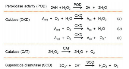
In contrast to many other nanomedicines, nanozymes can be applied for diseases therapy without need of additional drugs loading, which is achieved by catalytic scavenging or generating of specific molecules in pathological environment of diseases. For example, various oxidoreductase mimic nanozymes have been demonstrated with excellent therapeutic efficacy owing to their activities to modulate reactive oxygen and nitrogen species (RONS). RONS are important signaling molecules to regulate abundant physiological functions. However, excessive RONS are produced in many inflammatory diseases, such as rheumatoid arthritis, acute lung/kidney injury, and RONS scavenging nanozymes are benefit for inflammation alleviation and anti-oxidative protection. For this purpose, the CeO2 nanozymes have been extensively explored owing to their SOD and CAT mimicking activities. Huang group recently reported citric acid modified CeO2 nanozymes as antioxidants to treat acute kidney injury, which showed good efficacy in vivo due to multi-enzymatic properties and efficient accumulation in the kidneys of the ultrasmall size nanozymes [243]. Notably, most of anti-oxidant nanozymes mainly have ability to eliminate oxygen radicals, while the capability to scavenge nitrogen radicals is relatively weak. To this end, a multifunctional rhodium nanozyme was developed with a broad RONS scavenging properties and photothermal activities, which found application for colon diseases therapy [244]. The RONS generating nanozymes, by contrast, are commonly employed to treat cancer and bacterial infection via oxidative damage to cause cell death [245, 246]. Cancer microenvironment is pathologically featured with mild acidity and overproduction of H2O2, which benefits Fenton reaction catalyzed by POD for ROS generation [247]. For such application, iron-based nanozymes are most widely explored.
To enhance the treatment efficacy, several attempts have been made to develop hybrid nanozymes with multiple catalysis for synergistic effect. For instance, Fan and coworkers fabricated an OXD- and POD-dual active nanozyme, in which OXD can consume GSH and convert O2 into H2O2 to supplied POD activity for self-cascade anti-tumor therapy [248]. In a similar design, the CAT and SOD nanozymes were incorporated into a single nanosystem to synergistically scavenge ROS and produce O2 for reducing pro-inflammatory macrophage levels and inducing anti-inflammatory macrophages to treat rheumatoid arthritis [249]. Besides efficacy, another important concern of nanozymes is the biocompatibility. Since most nanozymes are metal-based materials, potential metal poisoning may occur after high-lose or frequent administration. To address this issue, targeting delivery of nanozymes to diseased tissue would be a good choice by surface ligands modification. However, since the chemical catalysis mainly happen on the surface of nanozymes, surface modification may have complicated impact on the catalytic activity [250]. Therefore, rational design of post-modification of nanozymes' surface should be carefully explored. For example, we recently systematically studied the adsorption of DNA on CuO nanozyme surface, based on which a polarity control of DNA adsorption was achieved by designing a di-block DNA sequence for targeting tumor therapy [251].
Although huge amount of efforts and resources have been utilized in research and development of drug delivery nanoparticles, there are still a long way to the clinical use of these novel-designed nanoparticles, with many impedances in the way. Over hundred clinical researches have been made on nanoparticles, with almost 200 clinical researches undergoing, the low efficacy is the major problem that results failures in phase Ⅱ and Ⅲ (overall success lowers than 10%) [252, 253]. The substandard preclinical outcome, potential toxicity of nanomaterials, low quality control and difficult production amplification of nanoparticles should take the responsibility. There remains lack of accurate in vitro and in vivo models to evaluate real patient treatment, resulting in the disparity between clinical and preclinical outcomes. Therefore, future preclinical investigations should develop and utilize innovative techniques to assist evaluation, such as accurately-simulated organoid, organ on the chip and patient-derived xenografts. More important, regarding nanoparticles' limited elevation and remaining insufficient delivery in many disease, there remain continuous demand of new delivery materials, strategies and techniques. On the other hand, clarifying the mechanism of diseases and interactions between diseases and nanoparticles would strongly promote the efficacy and future researches on nanoparticles. Besides, when administrated for patient, nanoparticles' potential toxicity strongly hinders the clinical translation. This situation is also contributed by the inaccurate simulation and evaluation at preclinical stage. On the other hand, the exogenous materials derived nanoparticles commonly do more harm to the progress, while some engineering endogenous nanoparticles also face concerns like immunogenicity and unavoidable effects on normal physiological processes. However, most researchers focus on the efficacy but ignore potential toxicity of nanoparticles in preclinical study, and lack of information about toxicity always makes clinical research hard. Therefore, it is urgent demand to call for more evaluations in these aspects when design, optimize and develop a nanoparticle at preclinical stage. Besides, more biocompatible materials used to compose nanoparticles are required, and there also need some more efficient and convenient methods to evaluate nanoparticles' toxicity. Additionally, the quality control in expandable production of nanoparticles is an important concern. Many novel and intelligent nanoparticles are complicated in compositions and functions, and excess in materials, which makes it hard to ensure the quality. Future nanoparticles need optimization in both compositions, preparations and efficacy, aiming at retaining functions the most and simplifying materials to meet production demands. Excellent alternatives of compositions are also of values and require exploration. There remain other aspects in which future drug delivery nanoparticles need optimization, such as off-target distribution, unexpected in vivo interaction and drug disability, resolving these concerns would strongly promote the success of clinical research. In conclusion, future investigation and development of nanoparticles should integrate and optimize nanoparticles' composition, evaluation methods and accurate efficacy, toxicity and potential interaction, instead of focusing on the efficacy. A comprehensive research of nanoparticles is essential and significant, and could promote the clinical translation efficiently.
The authors declare no conflict of interest.
This work was supported by National Natural Science Foundation of China (No. 81961138009), 111 Project (No. B18035), and the Key Research and Development Program of Science and Technology Department of Sichuan Province (No. 2020YFS0570).
Y.C. Barenholz, J. Control. Release 160 (2012) 117–134. doi: 10.1016/j.jconrel.2012.03.020
C.M. Dawidczyk, C. Kim, J.H. Park, et al., J. Control. Release 187 (2014) 133–144. doi: 10.1016/j.jconrel.2014.05.036
M.R. Green, G.M. Manikhas, S. Orlov, et al., Ann. Oncol. 17 (2006) 1263–1268. doi: 10.1093/annonc/mdl104
D. Stevens, J. Infect. 28 (1994) 45–49. doi: 10.1016/S0163-4453(94)95971-4
J. Geng, K. Li, D. Ding, et al., Small 8 (2012) 3655–3663. doi: 10.1002/smll.201200814
K.Y. Choi, H. Chung, K.H. Min, Y.Y. Hong, S.Y. Jeong, Biomaterials 31 (2009) 106–114. doi: 10.1016/j.biomaterials.2009.09.030
Z. Luo, Y. Dai, H. Gao, Acta Pharm. Sin. B 9 (2019) 1099–1112. doi: 10.1016/j.apsb.2019.06.004
J.V. Jokerst, Z. Miao, C. Zavaleta, C. Zhen, S.S. Gambhir, Small 7 (2015) 625–633. doi: 10.1002/smll.201002291
M. Lundqvist, J. Stigler, T. Cedervall, et al., ACS Nano 5 (2011) 7503–7509. doi: 10.1021/nn202458g
W. Xiao, H. Gao, Int. J. Pharm. 552 (2018) 328–339. doi: 10.1016/j.ijpharm.2018.10.011
L. Yu, M. Xu, W. Xu, W. Xiao, H. Gao, Nano Lett. 20 (2020) 8903–8911. doi: 10.1021/acs.nanolett.0c03982
S. Acharya, S.K. Sahoo, Adv. Drug Deliv. Rev. 63 (2011) 170–183. doi: 10.1016/j.addr.2010.10.008
S. Sindhwani, A.M. Syed, J. Ngai, B.R. Kingston, W. Chan, Nat. Mater. 19 (2020) 566–575. doi: 10.1038/s41563-019-0566-2
B. Ouyang, W. Poon, Y.N. Zhang, et al., Nat. Mater. 19 (2020) 1362–1371. doi: 10.1038/s41563-020-0755-z
O. Trédan, C.M. Galmarini, K. Patel, I.F. Tannock, J. Natl. Cancer Inst. 99 (2007) 1441–1454. doi: 10.1093/jnci/djm135
S. Yang, H. Gao, Pharmacol. Res. 126 (2017) 97–108. doi: 10.1016/j.phrs.2017.05.004
Y. Zhou, X. Chen, J. Cao, H. Gao, J. Mater. Chem. B 8 (2020) 6765–6781. doi: 10.1039/D0TB00649A
J.E. Lee, D.J. Lee, N. Lee, et al., J. Mater. Chem. 21 (2011) 16869–16872. doi: 10.1039/c1jm11869b
S. Ruan, X. Cao, X. Cun, et al., Biomaterials 60 (2015) 100–110. doi: 10.1016/j.biomaterials.2015.05.006
W. Yu, M. Shevtsov, X. Chen, H. Gao, Chin. Chem. Lett. 31 (2020) 1366–1374. doi: 10.1016/j.cclet.2020.02.036
Q. Lv, L. Cheng, Y. Lu, X. Zhang, J. Liu, Adv. Sci. 7 (2020) 2000515. doi: 10.1002/advs.202000515
F. Yang, Z. Zhao, B. Sun, Q. Chen, C. Luo, Trends Cancer 6 (2020) 645–659. doi: 10.1016/j.trecan.2020.05.001
C. Luo, J. Sun, D. Liu, et al., Nano Lett. 16 (2016) 5401–5408. doi: 10.1021/acs.nanolett.6b01632
H. Mei, S. Cai, D. Huang, et al., Bioact. Mater. 8 (2022) 220–240. doi: 10.1016/j.bioactmat.2021.06.035
C. Luo, B. Sun, C. Wang, et al., J. Control. Release 302 (2019) 79–89. doi: 10.1016/j.jconrel.2019.04.001
S. Zhang, Z. Wang, Z. Kong, Y. Wang, J. Sun, Theranostics 11 (2021) 6019–6032. doi: 10.7150/thno.59065
S. Zhang, Y. Wang, Z. Kong, et al., Acta Pharm. Sin. B 11 (2021) 3636–3647. doi: 10.1016/j.apsb.2021.04.005
X. Shan, X. Zhang, C. Wang, et al., J. Nanobiotechnol. 19 (2021) 282. doi: 10.1186/s12951-021-01037-6
S. Li, F. Yang, X. Sun, et al., Chem. Eng. J. 426 (2021) 130838. doi: 10.1016/j.cej.2021.130838
M. Li, Z. Xu, L. Zhang, et al., ACS Nano 15 (2021) 9808–9819. doi: 10.1021/acsnano.1c00680
X. Zhang, J. Xiong, K. Wang, et al., Bioact. Mater. 6 (2021) 2291–2302. doi: 10.1016/j.bioactmat.2021.01.004
Z. Zhao, X. Zhang, H. Zhang, et al., Adv. Sci. 9 (2021) 2104264. doi: 10.1002/advs.202104264
Y. Yan, B. Chen, Z. Wang, et al., Adv. Sci. 8 (2021) 2002253. doi: 10.1002/advs.202002253
C.M. Hu, L. Zhang, S. Aryal, C. Cheung, R.H. Fang, Proc. Natl. Acad. Sci. U.S.A. 108 (2011) 10980–10985. doi: 10.1073/pnas.1106634108
R. Li, Y. He, Y. Zhu, et al., Nano Lett. 19 (2019) 124–134. doi: 10.1021/acs.nanolett.8b03439
Q. Jiang, Y. Liu, R. Guo, et al., Biomaterials 192 (2019) 292–308. doi: 10.1016/j.biomaterials.2018.11.021
Y. He, R. Li, H. Li, et al., ACS Nano 13 (2019) 4148–4159. doi: 10.1021/acsnano.8b08964
L. Zhang, Y. Zhu, X. Wei, et al., Acta Pharm. Sin. B 22 (2022) 3427–3447.
Y. Song, N. Zhang, Q. Lia, et al., Chem. Eng. J. 408 (2021) 127296. doi: 10.1016/j.cej.2020.127296
H. Tan, Y. Song, J. Chen, et al., Adv. Sci. 8 (2021) e2100787. doi: 10.1002/advs.202100787
T. Yong, X. Zhang, N. Bie, et al., Nat. Commun. 10 (2019) 3838. doi: 10.1038/s41467-019-11718-4
T. Tan, H. Hu, H. Wang, et al., Nat. Commun. 10 (2019) 3322. doi: 10.1038/s41467-019-11235-4
T.G. Barclay, C.M. Day, N. Petrovsky, S. Garg, Carbohyd. Polym. 221 (2019) 94–112. doi: 10.1016/j.carbpol.2019.05.067
A. Varanko, S. Saha, A. Chilkoti, Adv. Drug Deliv. Rev. 156 (2020) 133–187. doi: 10.1016/j.addr.2020.08.008
A.O. Elzoghby, M.A. Abdelmoneem, I.A. Hassanin, et al., Biomaterials 263 (2020) 120355. doi: 10.1016/j.biomaterials.2020.120355
M. Zu, Y. Ma, B. Cannup, et al., Adv. Drug Deliv. Rev. 176 (2021) 113887. doi: 10.1016/j.addr.2021.113887
Q. Cheng, L. Liu, M. Xie, et al., Adv. Healthc. Mater. 10 (2021) e2001953. doi: 10.1002/adhm.202001953
Y. Wang, J. Yu, Z. Luo, et al., Adv. Mater. 33 (2021) 2103497. doi: 10.1002/adma.202103497
H. Liu, Z. Cai, F. Wang, et al., Adv. Sci. 8 (2021) e2101619. doi: 10.1002/advs.202101619
N. Dabholkar, S. Gorantla, T. Waghule, et al., Int. J. Biol. Macromol. 170 (2021) 602–621. doi: 10.1016/j.ijbiomac.2020.12.177
E. Kim, G. Erdos, S. Huang, et al., EBioMedicine 55 (2020) 102743. doi: 10.1016/j.ebiom.2020.102743
R. Dimatteo, N.J. Darling, T. Segura, Adv. Drug Deliv. Rev. 127 (2018) 167–184. doi: 10.1016/j.addr.2018.03.007
R. Liu, Y. An, W. Jia, et al., J. Control. Release 321 (2020) 589–601. doi: 10.1016/j.jconrel.2020.02.043
Y. Luo, J. Li, Y. Hu, et al., Acta Pharm. Sin. B 10 (2020) 2227–2245. doi: 10.1016/j.apsb.2020.05.011
V. Taghipour-Sabzevar, T. Sharifi, M.M. Moghaddam, Ther. Deliv. 10 (2019) 527–550. doi: 10.4155/tde-2019-0044
A. Zielinska, F. Carreiro, A.M. Oliveira, et al., Molecules 25 (2020) 3731. doi: 10.3390/molecules25163731
X. Zheng, J.Z. Xie, X. Zhang, et al., Chin. Chem. Lett. 32 (2021) 243–257. doi: 10.1016/j.cclet.2020.11.029
J.A. Kulkarni, D. Witzigmann, S.B. Thomson, et al., Nat. Nanotechnol. 16 (2021) 630–643. doi: 10.1038/s41565-021-00898-0
A.S. Piotrowski-Daspit, A.C. Kauffman, L.G. Bracaglia, W.M. Saltzman, Adv. Drug Deliv. Rev. 156 (2020) 119–132. doi: 10.1016/j.addr.2020.06.014
N.D. Sonawane, F.C. Szoka Jr., A.S. Verkman, J. Biol. Chem. 278 (2003) 44826–44831. doi: 10.1074/jbc.M308643200
S. Taranejoo, J. Liu, P. Verma, K. Hourigan, J. Appl. Polym. Sci. 132 (2015) 42096.
S.H. Lee, S.H. Choi, S.H. Kim, T.G. Park, J. Control. Release 125 (2008) 25–32. doi: 10.1016/j.jconrel.2007.09.011
I. Lostale-Seijo, J. Montenegro, Nat. Rev. Chem. 2 (2018) 258–277. doi: 10.1038/s41570-018-0039-1
X. Fu, L. Hosta-Rigau, R. Chandrawati, J.W. Cui, Chem-US 4 (2018) 2084–2107. doi: 10.1016/j.chempr.2018.07.002
X. Hou, T. Zaks, R. Langer, Y. Dong, Nat. Rev. Mater. 6 (2021) 1078–1094. doi: 10.1038/s41578-021-00358-0
R. Tenchov, R. Bird, A.E. Curtze, Q. Zhou, ACS Nano 15 (2021) 16982–17015. doi: 10.1021/acsnano.1c04996
H. Park, A. Otte, K. Park, J. Control. Release 342 (2021) 53–65. doi: 10.1016/j.jconrel.2021.12.030
M.A. Maier, M. Jayaraman, S. Matsuda, et al., Mol. Ther. 21 (2013) 1570–1578. doi: 10.1038/mt.2013.124
S. Liu, Q. Cheng, T. Wei, et al., Nat. Mater. 20 (2021) 701–710. doi: 10.1038/s41563-020-00886-0
L. Miao, L. Li, Y. Huang, et al., Nat. Biotechnol. 37 (2019) 1174–1185. doi: 10.1038/s41587-019-0247-3
A.J. Wilson, D. Devasia, P.K. Jain, Chem. Soc. Rev. 49 (2020) 6087–6112. doi: 10.1039/D0CS00338G
T. Sun, G. Zhang, T. Ning, et al., Adv. Sci. 8 (2021) 2102256. doi: 10.1002/advs.202102256
N. Rohaizad, C.C. Mayorga-Martinez, M. Fojtu, N.M. Latiff, M. Pumera, Chem. Soc. Rev. 50 (2021) 619–657. doi: 10.1039/D0CS00150C
L. Jin, P. Hu, Y. Wang, et al., Adv. Mater. 32 (2020) e1906050. doi: 10.1002/adma.201906050
M. Wan, Q. Wang, R. Wang, et al., Sci. Adv. 6 (2020) eaaz9014. doi: 10.1126/sciadv.aaz9014
X. Qin, C. Wu, D. Niu, et al., Nat. Commun. 12 (2021) 5243. doi: 10.1038/s41467-021-25561-z
J. Shen, Z. Lu, J. Wang, et al., Adv. Mater. 33 (2021) 2101993. doi: 10.1002/adma.202101993
H. Pan, M. Zheng, A. Ma, L. Liu, L. Cai, Adv. Mater. 33 (2021) 2100241. doi: 10.1002/adma.202100241
Q. Song, C. Zheng, J. Jia, et al., Adv. Mater. 31 (2019) 1903793. doi: 10.1002/adma.201903793
H. Kim, S. Beack, S. Han, et al., Adv. Mater. 30 (2018) 1701460. doi: 10.1002/adma.201701460
Z. Shen, A. Wu, X. Chen, Mol. Pharm. 14 (2017) 1352–1364. doi: 10.1021/acs.molpharmaceut.6b00839
G. Chen, H. Qiu, P.N. Prasad, X. Chen, Chem. Rev. 114 (2014) 5161–5214. doi: 10.1021/cr400425h
L. Zeng, L. Huang, Z. Wang, et al., Angew. Chem. Int. Ed. 60 (2021) 23569–23573. doi: 10.1002/anie.202108076
Z. Yang, J.H. Lee, H.M. Jeon, et al., J. Am. Chem. Soc. 135 (2013) 11657–11662. doi: 10.1021/ja405372k
T. Sun, G. Zhang, Q. Wang, et al., Biomaterials 183 (2018) 268–279. doi: 10.1016/j.biomaterials.2018.04.016
W. Chen, Z. Sun, L. Lu, Angew. Chem. Int. Ed. 60 (2021) 5626–5643. doi: 10.1002/anie.201914511
M.A. Rahim, N. Jan, S. Khan, et al., Cancers 13 (2021) 670. doi: 10.3390/cancers13040670
M. Merino, T. Lozano, N. Casares, H. Lana, M.J. Garrido, J. Nanobiotechnol. 19 (2021) 102. doi: 10.1186/s12951-021-00846-z
Q. Ji, J. Hou, X. Yong, et al., Adv. Mater. 33 (2021) 2007798. doi: 10.1002/adma.202007798
S. Zou, B. Wang, C. Wang, Q. Wang, L. Zhang, Nanomedicine 15 (2020) 625–641. doi: 10.2217/nnm-2019-0388
M. Xu, Q. Lu, Y. Song, et al., Biomaterials 250 (2020) 120076. doi: 10.1016/j.biomaterials.2020.120076
J. Ouyang, L. Sun, J. Pan, et al., Small 17 (2021) 2102598. doi: 10.1002/smll.202102598
W. Jia, Y. Wang, R. Liu, X. Yu, H. Gao, Adv. Funct. Mater. 31 (2021) 2009765. doi: 10.1002/adfm.202009765
M. Ayer, M. Schuster, I. Gruber, et al., Adv. Healthc. Mater. 10 (2021) e2001375. doi: 10.1002/adhm.202001375
Z. Xie, Y. Su, G.B. Kim, et al., Small 13 (2017) 1603121. doi: 10.1002/smll.201603121cale||9||16024|2017|||
A. Xie, S. Hanif, J. Ouyang, et al., EBioMedicine 56 (2020) 102821. doi: 10.1016/j.ebiom.2020.102821
B. Chen, W. Dai, B. He, et al., Theranostics 7 (2017) 538–558. doi: 10.7150/thno.16684
A. Raza, T. Rasheed, F. Nabeel, et al., Molecules 24 (2019) 1117. doi: 10.3390/molecules24061117
G. Liu, J.F. Lovell, L. Zhang, Y. Zhang, Int. J. Mol. Sci. 21 (2020) 6380. doi: 10.3390/ijms21176380
J. Hu, X. Yuan, F. Wang, et al., Chin. Chem. Lett. 32 (2021) 1341–1347. doi: 10.1016/j.cclet.2020.11.006
M.T. Zhu, G.J. Nie, H. Meng, et al., Acc. Chem. Res. 46 (2013) 622–631. doi: 10.1021/ar300031y
S. Zhang, G. Deng, F. Liu, et al., Adv. Funct. Mater. 30 (2020) 1910651. doi: 10.1002/adfm.201910651
L. Chu, R. McPhee, W. Huang, et al., Vaccine 39 (2021) 2791–2799. doi: 10.1016/j.vaccine.2021.02.007
S. Ruan, C. Hu, X. Tang, et al., ACS Nano 10 (2016) 10086–10098. doi: 10.1021/acsnano.6b05070
S. Ruan, L. Qin, W. Xiao, et al., Adv. Funct. Mater. 28 (2018) 1802227. doi: 10.1002/adfm.201802227
X. Gao, Q. Yue, Z. Liu, et al., Adv. Mater. 29 (2017) 1603917. doi: 10.1002/adma.201603917
M. Karimi, A. Ghasemi, P. Sahandi Zangabad, et al., Chem. Soc. Rev. 45 (2016) 1457–1501. doi: 10.1039/C5CS00798D
R. Xie, S. Ruan, J. Liu, et al., Biomaterials 275 (2021) 120891. doi: 10.1016/j.biomaterials.2021.120891
F. Danhier, J. Control. Release 244 (2016) 108–121. doi: 10.1016/j.jconrel.2016.11.015
L. Tang, X. Yang, Q. Yin, et al., Proc. Natl. Acad. Sci. 111 (2014) 15344–15349. doi: 10.1073/pnas.1411499111
Z. Popovic, W. Liu, V.P. Chauhan, et al., Angew. Chem. Int. Ed. 49 (2010) 8649–8652. doi: 10.1002/anie.201003142
H. Wang, D. Mao, Y. Wang, et al., Sci. Rep. 5 (2015) 16680. doi: 10.1038/srep16680
P.P. Yang, Q. Luo, G.B. Qi, et al., Adv. Mater. 29 (2017) 1605869. doi: 10.1002/adma.201605869
X.X. Hu, P.P. He, G.B. Qi, et al., ACS Nano 11 (2017) 4086–4096. doi: 10.1021/acsnano.7b00781
W. Yu, R. Liu, Y. Zhou, H. Gao, ACS Central Sci. 6 (2020) 100–116. doi: 10.1021/acscentsci.9b01139
R. Xie, S. Ruan, J. Liu, L. Qin, Y. Qin, Biomaterials 275 (2021) 120891. doi: 10.1016/j.biomaterials.2021.120891
Y. Wang, X. Li, P. Chen, Y. Dong, Y. Yu, Nanoscale 12 (2020) 1886–1893. doi: 10.1039/C9NR09235H
W. Yu, X. He, Z. Yang, et al., Biomaterials 217 (2019) 119309. doi: 10.1016/j.biomaterials.2019.119309
R. Liu, W. Xiao, C. Hu, R. Xie, H. Gao, J. Control. Release 278 (2018) 127–139. doi: 10.1016/j.jconrel.2018.04.005
R. Liu, C. Hu, Y. Yang, J. Zhang, H. Gao, Acta Pharm. Sin. B 9 (2019) 410–420. doi: 10.1016/j.apsb.2018.09.001
C. Hu, X. Cun, S. Ruan, et al., Biomaterials 168 (2018) 64–75. doi: 10.1016/j.biomaterials.2018.03.046
Z. Cong, L. Zhang, S.Q. Ma, et al., ACS Nano 14 (2020) 1958–1970. doi: 10.1021/acsnano.9b08434
C. Hu, X. He, Y. Chen, et al., Adv. Funct. Mater. 31 (2021) 2007149. doi: 10.1002/adfm.202007149
H. Wang, X. Han, Z. Dong, et al., Adv. Funct. Mater. 29 (2019) 1902440. doi: 10.1002/adfm.201902440
C.N. Loynachan, A.P. Soleimany, J.S. Dudani, et al., Nat. Nanotechnol. 14 (2019) 883–890. doi: 10.1038/s41565-019-0527-6
L. Zhang, D. Jing, N. Jiang, et al., Nat. Nanotechnol. 15 (2020) 145–153. doi: 10.1038/s41565-019-0626-4
Y. Qin, F. Tong, W. Zhang, et al., Adv. Funct. Mater. 31 (2021) 2104645. doi: 10.1002/adfm.202104645
C. Xu, Y. Yu, Y. Sun, et al., Adv. Funct. Mater. 29 (2019) 1905213. doi: 10.1002/adfm.201905213
C. Lin, F. Tong, R. Liu, et al., Acta Pharm. Sin. B 10 (2020) 2348–2361. doi: 10.1016/j.apsb.2020.10.009
R. Liu, M. Yu, X. Yang, et al., Adv. Funct. Mater. 29 (2019) 1808462. doi: 10.1002/adfm.201808462
H. Yang, X. Zheng, Z. Zheng, et al., ACS Appl. Mater. Inter. 13 (2021) 54715–54726. doi: 10.1021/acsami.1c15858
R. Lin, W. Yu, X. Chen, H. Gao, Adv. Healthc. Mater. 10 (2021) e2001212. doi: 10.1002/adhm.202001212
A. Joseph, C. Contini, D. Cecchin, et al., Sci. Adv. 3 (2017) e1700362. doi: 10.1126/sciadv.1700362
C. Gao, Y. Wang, Z. Ye, et al., Adv. Mater. 33 (2021) e2000512. doi: 10.1002/adma.202000512
W. Yu, R. Lin, X. He, et al., Acta Pharm. Sin. B 11 (2021) 2924–2936. doi: 10.1016/j.apsb.2021.04.006
H.Y. Huang, L.Q. Chen, W. Sun, et al., Theranostics 11 (2021) 906–924. doi: 10.7150/thno.47446
Q. Guo, Q. Zhu, T. Miao, et al., J. Control. Release 303 (2019) 117–129. doi: 10.1016/j.jconrel.2019.04.031
N.U. Khan, J. Ni, X. Ju, et al., Acta Pharm. Sin. B 11 (2021) 1341–1354. doi: 10.1016/j.apsb.2020.10.015
X. Ju, T. Miao, H. Chen, J. Ni, L. Han, Adv. Healthc. Mater. 10 (2021) e2001997. doi: 10.1002/adhm.202001997
X. Ju, H. Chen, T. Miao, J. Ni, L. Han, Mol. Pharm. 18 (2021) 2694–2702. doi: 10.1021/acs.molpharmaceut.1c00224
L. Han, C. Jiang, Acta Pharm. Sin. B 11 (2021) 2306–2325. doi: 10.1016/j.apsb.2020.11.023
J. Ni, T. Miao, M. Su, et al., J. Control. Release 329 (2021) 934–947. doi: 10.1016/j.jconrel.2020.10.023
T.T. Miao, X.F. Ju, Q.N. Zhu, et al., Adv. Funct. Mater. 29 (2019) 1900259.
J. Che, A. Najer, A.K. Blakney, et al., Adv. Mater. 32 (2020) e2003598. doi: 10.1002/adma.202003598
M. Li, S. Li, H. Zhou, et al., Nat. Commun. 11 (2020) 1126. doi: 10.1038/s41467-020-14963-0
L. Tang, Z. Wang, Q. Mu, et al., Adv. Mater. 32 (2020) e2002739. doi: 10.1002/adma.202002739
C. Gao, Q. Cheng, J. Li, et al., Adv. Funct. Mater. 31 (2021) 2102440. doi: 10.1002/adfm.202102440
X. Xu, G. Deng, Z. Sun, et al., Adv. Mater. 33 (2021) e2102322. doi: 10.1002/adma.202102322
D. Chu, X. Dong, Q. Zhao, J. Gu, Z. Wang, Adv. Mater. 29 (2017) 1701021. doi: 10.1002/adma.201701021
G.L. Burn, A. Foti, G. Marsman, D.F. Patel, A. Zychlinsky, Immunity 54 (2021) 1377–1391. doi: 10.1016/j.immuni.2021.06.006
S. Li, M. Li, S. Huo, et al., Adv. Mater. 33 (2021) e2006160. doi: 10.1002/adma.202006160
L. Zheng, X. Hu, H. Wu, et al., J. Am. Chem. Soc. 142 (2020) 382–391. doi: 10.1021/jacs.9b11046
Y.B. Miao, K.H. Chen, C.T. Chen, et al., Adv. Mater. 33 (2021) e2100701. doi: 10.1002/adma.202100701
B. Hu, L. Zhong, Y. Weng, et al., Signal Transduct. Target. Ther. 5 (2020) 101. doi: 10.1038/s41392-020-0207-x
Y. Weng, H. Xiao, J. Zhang, X.J. Liang, Y. Huang, Biotechnol. Adv. 37 (2019) 801–825. doi: 10.1016/j.biotechadv.2019.04.012
Y. Huang, Mol. Ther. Nucleic Acids 6 (2017) 116–132. doi: 10.1016/j.omtn.2016.12.003
B. Hu, Y. Weng, X.H. Xia, X.J. Liang, Y. Huang, J. Gene Med. 21 (2019) e3097. doi: 10.1002/jgm.3097
T. Yang, C. Li, X. Wang, et al., Bioact. Mater. 5 (2020) 1053–1061. doi: 10.1016/j.bioactmat.2020.07.003
Y. Weng, Y. Huang, Asian J. Pharm. Sci. 16 (2021) 263–264. doi: 10.1016/j.ajps.2021.02.005
C. Li, T. Yang, Y. Weng, et al., Bioact. Mater. 9 (2022) 590–601. doi: 10.1016/j.bioactmat.2021.05.051
D. Zhao, G. Yang, Q. Liu, et al., Nanoscale 12 (2020) 10939–10943. doi: 10.1039/D0NR00301H
G. Yang, Z. Li, I. Mohammed, et al., Signal Transduct. Target. Ther. 6 (2021) 227. doi: 10.1038/s41392-021-00649-6
G. Yang, C. Zhu, L. Zhao, et al., Chin. Chem. Lett. 32 (2021) 218–220. doi: 10.1016/j.cclet.2020.10.018
L.C. Li, Adv. Exp. Med. Biol. 983 (2017) 1–20. doi: 10.1007/978-981-10-4310-9_1
S. Guo, K. Li, B. Hu, et al., Exploration 1 (2021) 35–49. doi: 10.1002/EXP.20210008
B. Hu, B. Li, K. Li, et al., Sci. Adv. 8 (2022) eabm1418. doi: 10.1126/sciadv.abm1418
K. Li, M. Lu, X. Xia, Y. Huang, Chin. Chem. Lett. 32 (2021) 1010–1016. doi: 10.1016/j.cclet.2020.09.010
C. Li, J. Zhou, Y. Wu, et al., Nano Lett. 21 (2021) 3680–3689. doi: 10.1021/acs.nanolett.0c04468
M. Zhang, Y. Weng, Z. Cao, et al., ACS Appl. Mater. Inter. 12 (2020) 32289–32300. doi: 10.1021/acsami.0c06614
S. Guo, B. Liu, M. Zhang, et al., Chin. Chem. Lett. 32 (2021) 102–106. doi: 10.1016/j.cclet.2020.11.024
J. Zhuang, H. Gong, J. Zhou, et al., Sci. Adv. 6 (2020) eaaz6108. doi: 10.1126/sciadv.aaz6108
D. Yin, M. Zhang, J. Chen, Y. Huang, D. Liang, Chin. Chem. Lett. 32 (2021) 1731–1736. doi: 10.1016/j.cclet.2020.12.005
H. Liu, L. Huang, M. Mao, et al., Adv. Funct. Mater. 30 (2020) 2006515. doi: 10.1002/adfm.202006515
T. Yang, D. Huang, C. Li, et al., Nano Today 36 (2021) 101017. doi: 10.1016/j.nantod.2020.101017
Y. Zhang, C. Sun, C. Wang, K.E. Jankovic, Y. Dong, Chem. Rev. 121 (2021) 12181–12277. doi: 10.1021/acs.chemrev.1c00244
Y. Weng, C. Li, T. Yang, et al., Biotechnol. Adv. 40 (2020) 107534. doi: 10.1016/j.biotechadv.2020.107534
J.C. Kaczmarek, A.K. Patel, K.J. Kauffman, et al., Angew. Chem. Int. Ed. 55 (2016) 13808–13812. doi: 10.1002/anie.201608450
J.C. Kaczmarek, A.K. Patel, L.H. Rhym, et al., Biomaterials 275 (2021) 120966. doi: 10.1016/j.biomaterials.2021.120966
A.K. Patel, J.C. Kaczmarek, S. Bose, et al., Adv. Mater. 31 (2019) e1805116. doi: 10.1002/adma.201805116
S. Liu, X. Wang, X. Yu, et al., J. Am. Chem. Soc. 143 (2021) 21321–21330. doi: 10.1021/jacs.1c09822
Y. Wang, H. Song, C. Liu, et al., Natl. Sci. Rev. 8 (2021) nwaa268. doi: 10.1093/nsr/nwaa268
D. Wilbie, J. Walther, E. Mastrobattista, Acc. Chem. Res. 52 (2019) 1555–1564. doi: 10.1021/acs.accounts.9b00106
J.D. Gillmore, E. Gane, J. Taubel, et al., N. Engl. J. Med. 385 (2021) 493–502. doi: 10.1056/NEJMoa2107454
C.F. Xu, G.J. Chen, Y.L. Luo, et al., Adv. Drug Deliv. Rev. 168 (2021) 3–29. doi: 10.1016/j.addr.2019.11.005
J.L. Lau, M.K. Dunn, Med. Chem. 26 (2018) 2700–2707. doi: 10.1016/j.bmc.2017.06.052
F.M. Veronese, G. Pasut, Drug Discov. Today 10 (2005) 1451–1458. doi: 10.1016/S1359-6446(05)03575-0
H. Jo, M. Gajendiran, K. Kim, J. Ind. Eng. Chem. 82 (2020) 234–242. doi: 10.1016/j.jiec.2019.10.018
Y. Honda, T. Nomoto, M. Matsui, et al., Biomacromolecules 21 (2020) 3826–3835. doi: 10.1021/acs.biomac.0c00903
D.C. Roopenian, S. Akilesh, Nat. Rev. Immunol. 7 (2007) 715–725. doi: 10.1038/nri2155
Y. Zong, X. Tan, J. Xiao, et al., Protein Expres. Purif. 153 (2019) 53–58. doi: 10.1016/j.pep.2018.08.012
J. Li, J. Tuma, H. Han, et al., Adv. Healthc. Mater. 11 (2022) 2102118. doi: 10.1002/adhm.202102118
C.H. Chau, P.S. Steeg, W.D. Figg, Lancet 394 (2019) 793–804. doi: 10.1016/S0140-6736(19)31774-X
A. Beck, L. Goetsch, C. Dumontet, N. Corvaïa, Nat. Rev. Drug Discov. 16 (2017) 315–337. doi: 10.1038/nrd.2016.268
Z. Peng, T. Liu, J. Wei, et al., J. Clin. Oncol. 38 (2020) 4560-4560. doi: 10.1200/JCO.2020.38.15_suppl.4560
N.E. Elsadek, A.S. Abu Lila, T. Ishida, Immunological responses to PEGylated proteins: anti-PEG antibodies, in: G Pasut, S Zalipsky (Eds.), Polymer-Protein Conjugates, Elsevier, 2020, pp. 103–123.
T.J. Povsic, M.G. Lawrence, A.M. Lincoff, et al., J. Allergy Clin. Immun. 138 (2016) 1712–1715. doi: 10.1016/j.jaci.2016.04.058
J. Kuhn, Y. Lin, A.K. Levacic, et al., Bioconjugate Chem. 31 (2020) 729–742. doi: 10.1021/acs.bioconjchem.9b00853
M.J. Haney, N.L. Klyachko, E.B. Harrison, et al., Adv. Healthc. Mater. 8 (2019) 1801271. doi: 10.1002/adhm.201801271
J. Chang, X. Chen, Z. Glass, et al., Acc. Chem. Res. 52 (2019) 665–675. doi: 10.1021/acs.accounts.8b00493
J. Liu, J. Sheng, L. Shao, et al., Angew. Chem. Int. Ed. 60 (2021) 26740–26746. doi: 10.1002/anie.202111213
J. Xu, Z. Li, Q. Fan, et al., Adv. Mater. 33 (2021) 2104355. doi: 10.1002/adma.202104355
R. Subbiah, A. Cheng, M.A. Ruehle, et al., Acta Biomater 114 (2020) 63–75. doi: 10.1016/j.actbio.2020.07.026
J. Yu, Y. Zhang, Y. Ye, et al., Proc. Natl. Acad. Sci 112 (2015) 8260. doi: 10.1073/pnas.1505405112
Z. Wang, J. Wang, H. Li, et al., Proc. Natl. Acad. Sci. U.S.A. 117 (2020) 29512. doi: 10.1073/pnas.2011099117
J. Yu, J. Wang, Y. Zhang, et al., Nat. Biomed. Eng. 4 (2020) 499–506. doi: 10.1038/s41551-019-0508-y
A.M. Dlugi, J.D. Miller, J. Knittle, Fertil. Steril. 54 (1990) 419–427. doi: 10.1016/S0015-0282(16)53755-8
A. Pfützner, A.E. Mann, S.S. Steiner, Diabetes Technol. Ther. 4 (2002) 589–594. doi: 10.1089/152091502320798204
H.H. Marian, L. Krishnan, T. Rouse, et al., Sci. Adv. 6 (2020) eaay1240. doi: 10.1126/sciadv.aay1240
Z. Vinarov, B. Abrahamsson, P. Artursson, et al., Adv. Drug Deliv. Rev. 171 (2021) 289–331. doi: 10.1016/j.addr.2021.02.001
P. Lundquist, P. Artursson, Adv. Drug Deliv. Rev. 106 (2016) 256–276. doi: 10.1016/j.addr.2016.07.007
X. Wang, J. Wu, X. Liu, et al., J. Control. Release 331 (2021) 390–403. doi: 10.1016/j.jconrel.2021.01.032
O.S. Abu Abed, C.S. Chaw, L. Williams, A.A. Elkordy, Int. J. pharm. 592 (2021) 120094. doi: 10.1016/j.ijpharm.2020.120094
A.R. Koli, K.M. Ranch, H.P. Patel, et al., Int. J. pharm. 596 (2021) 120202. doi: 10.1016/j.ijpharm.2021.120202
T. Yang, J. Feng, Q. Zhang, et al., Drug Deliv. 27 (2020) 575–584. doi: 10.1080/10717544.2020.1748762
N. Ortiz, P.A. Vasquez, F. Vidal, et al., Nanomedicine 15 (2020) 2771–2784. doi: 10.2217/nnm-2020-0305
X. Cai, Y. Xu, L. Zhao, et al., Nano Today 36 (2021) 101032. doi: 10.1016/j.nantod.2020.101032
Y. Mu, Y. Fu, J. Li, et al., Carbohyd. Polym. 203 (2019) 10–18. doi: 10.1016/j.carbpol.2018.09.020
T. Ma, L. Wang, T. Yang, G. Ma, S. Wang, Int. J. pharm. 473 (2014) 296–303. doi: 10.1016/j.ijpharm.2014.06.052
C. Li, Y. Zhao, J. Cheng, et al., Adv. Sci. 6 (2019) 1900610. doi: 10.1002/advs.201900610
S. Li, M. Jin, Y. Wu, et al., Drug Deliv. 28 (2021) 1120–1131. doi: 10.1080/10717544.2021.1934189
Q.W. Chen, J.Y. Qiao, X.H. Liu, C. Zhang, X.Z. Zhang, Chem. Soc. Rev. 50 (2021) 12576–12615. doi: 10.1039/D0CS01571G
Z. Cao, X. Wang, Y. Pang, S. Cheng, J. Liu, Nat. Commun. 10 (2019) 5783. doi: 10.1038/s41467-019-13727-9
Q. Song, C. Zheng, J. Jia, et al., Adv. Mater. 31 (2019) e1903793. doi: 10.1002/adma.201903793
L. Yu, P. Hu, Y. Chen, Adv. Mater. 30 (2018) 1801964. doi: 10.1002/adma.201801964
Y. Zhao, X. Ouyang, Y. Peng, S. Peng, Pharmaceutics 13 (2021) 1917. doi: 10.3390/pharmaceutics13111917
Y. Wang, T. Yang, Q. He, Natl. Sci. Rev. 7 (2020) 1485–1512. doi: 10.1093/nsr/nwaa034
L. Chen, S.F. Zhou, L. Su, J. Song, ACS Nano 13 (2019) 10887–10917. doi: 10.1021/acsnano.9b04954
L. Qin, H. Gao, Asian J. Pharm. Sci. 14 (2019) 380–390. doi: 10.1016/j.ajps.2018.10.005
Y. Zhou, W. Yu, J. Cao, H. Gao, Biomaterials 255 (2020) 120193. doi: 10.1016/j.biomaterials.2020.120193
C. Hu, X. Cun, S. Ruan, et al., Biomaterials 168 (2018) 64–75. doi: 10.1016/j.biomaterials.2018.03.046
X. Zhang, J. Du, Z. Guo, et al., Adv. Sci. 6 (2019) 1801122. doi: 10.1002/advs.201801122
A. Akinc, M.A. Maier, M. Manoharan, et al., Nat. Nanotechnol. 14 (2019) 1084–1087. doi: 10.1038/s41565-019-0591-y
X. Yao, P. Yang, Z. Jin, et al., Biomaterials 197 (2019) 268–283. doi: 10.1016/j.biomaterials.2019.01.026
J. Meng, Z. Jin, P. Zhao, et al., Sci. Adv. 6 (2020) eaba1362. doi: 10.1126/sciadv.aba1362
P. Zhao, Z. Jin, Q. Chen, et al., Nat. Commun. 9 (2018) 4241. doi: 10.1038/s41467-018-06630-2
Y. Wu, L. Su, M. Yuan, et al., Angew. Chem. Int. Ed. 60 (2021) 12868–12875. doi: 10.1002/anie.202100002
Z. Yang, Y. Luo, Y. Hu, et al., Adv. Funct. Mater. 31 (2021) 2007991. doi: 10.1002/adfm.202007991
S. Peng, H. Wang, Y. Xin, et al., Nano Today 40 (2021) 101240. doi: 10.1016/j.nantod.2021.101240
S. Scott, H. Zhao, A. Dey, T.B. Gunnoe, ACS Catal. 10 (2020) 14315–14317. doi: 10.1021/acscatal.0c05047
H. Wei, L.Z. Gao, K.L. Fan, et al., Nano Today 40 (2021) 101269. doi: 10.1016/j.nantod.2021.101269
B.W. Yang, Y. Chen, J.L. Shi, Adv. Mater. 31 (2019) 1901778. doi: 10.1002/adma.201901778
M. Wei, J. Lee, F. Xia, et al., Acta Biomater. 126 (2021) 15–30. doi: 10.1016/j.actbio.2021.02.036
D.Y. Zhang, H. Liu, C. Li, et al., ACS Appl. Mater. Inter. 12 (2020) 56830–56838. doi: 10.1021/acsami.0c17579
Z. Miao, S. Jiang, M. Ding, et al., Nano Lett. 20 (2020) 3079–3089. doi: 10.1021/acs.nanolett.9b05035
D.Z. Yang, Z.Z. Chen, Z. Gao, S.K. Tammina, Y.L. Yang, Colloids Surf. B: Biointerfaces 195 (2020) 111252. doi: 10.1016/j.colsurfb.2020.111252
W.P. Yang, X. Yang, L.J. Zhu, et al., Coord. Chem. Rev. 448 (2021) 214170. doi: 10.1016/j.ccr.2021.214170
J. Ma, J.J. Qiu, S.R. Wang, ACS. Appl. Nano Mater. 3 (2020) 4925–4943. doi: 10.1021/acsanm.0c00396
X. Meng, D. Li, L. Chen, et al., ACS Nano 15 (2021) 5735–5751. doi: 10.1021/acsnano.1c01248
J. Kim, H.Y. Kim, S.Y. Song, et al., ACS Nano 13 (2019) 3206–3217. doi: 10.1021/acsnano.8b08785
B.W. Liu, J.W. Liu, Nano Res. 10 (2017) 1125–1148. doi: 10.1007/s12274-017-1426-5
Y.C. Meng, Y. Chen, J.J. Zhu, et al., Mater. Horiz. 8 (2021) 972–986. doi: 10.1039/D0MH01372B
Y. Jiang, Z. Jiang, M. Wang, L. Ma, Adv. Drug Deliv. Rev. 180 (2022) 114034. doi: 10.1016/j.addr.2021.114034
H. He, L. Liu, E.E. Morin, M. Liu, A. Schwendeman, Acc. Chem. Res. 52 (2019) 2445–2461. doi: 10.1021/acs.accounts.9b00228

Figure 2 Schematic diagram of the synthesis and application of engineered exosomes-thermosensitive liposomes hybrid NPs. Copied with permission [21]. Copyright 2020, Wiley Publishing Group.

Figure 3 Schematic illustration of hybrid cell-membrane-cloaked biomimetic nanoparticles designed for noninvasive targeted treatment of laser-induced CNV. (A) Preparation process of [RBC-REC]NPs by enclosing polymeric cores with fused RBC-REC membranes. (B) [RBC-REC]NPs administered intravenously absorb proangiogenic factors, resulting in the blocking of their effects on host neovascular endothelial cells. Copied with permission [93]. Copyright 2021, American Chemical Society.

Figure 4 Schematic illustration of neutrophils infiltrate into tumor tissue after hijacking nanoparticles in the blood. Reproduced with permission [151]. Copyright 2021, Wiley Publishing Group.

Figure 5 Tumor homing after uptake by monocytes of nanodrugs encapsulated by apoptotic bodies in blood of C57BL/6 mice. Reproduced with permission [152]. Copyright 2020, American Chemical Society.

Figure 6 Representative siRNA delivery strategies. (Panel A) Synthetic materials including lipid nanoparticle [166], DPCiv™, fluorinated oligoethylenimine (fOEI) nanoassembly, and polyethylenimine (PEI)-functionalized FeOOH [170]. (Panel B) Several biological vectors including exosome, GalNAc-siRNA conjugate, etc. [161, 173]. (Panel C) Some siRNA electroporation devices such as high-density distributed electrode network (HDEN), flexible electroporation patch, and rolling microneedle electrode array (RoMEA) [174]. Reproduced with permission [155]. Copyright 2019, Elsevier.

Figure 7 Representative mRNA delivery platforms. (A) Lipid or polymer-based nanoparticles for mRNA delivery, and the targeted organs or cells that mRNA nanoparticles could reach. (B) Representative chemical structures of lipids or polymers that have been investigated for mRNA delivery in vivo. Reproduced with permission [176]. Copyright 2020, Elsevier.

Figure 8 Non-viral delivery carriers of CRISPR-Cas system. (A) DNA, RNA and RNP-based CRISPR-Cas gene-editing elements and various delivery vectors. (B) Illustration of the working mechanism of CRISPR-Cas system in vivo. Copied with permission [184]. Copyright 2021, Elsevier.

Figure 9 (A) Schematic illustration of gastric retention and penetration in vivo. MGNs-DOX orally administered to BALB/c mice formed MNAs with the help of in situ pepsin and stomach-targeted MF, then DOX-loaded MNAs penetrated the mucus with DOX release under the gastric acid conditions. Reproduced with permission [216]. Copyright 2021, Elsevier. (B) Scheme of the effect of QT-CS Micelles on improving the oral absorption of DOX. Reproduced with permission [217]. Copyright 2019, Elsevier. (C) Schematic illustration of oral targeted therapy of AON through the leaky intestine and the intrinsic adhesiveness. AON exerts multiple biological effects in the colon. Reproduced with permission [219]. Copyright 2019, Wiley Publishing Group.

Figure 10 Schematic diagram of diverse gas releasing nanoplatform. (A) Near-infrared (NIR) laser triggered NO release for mild photothermal augmented anti-tumor therapy. Copied with permission [231]. Copyright 2019, Wiley Publishing Group. (B) Tumor acidic environment triggered SO2 release for deep anti-tumor therapy. Copied with permission [91]. Copyright 2020, Elsevier. (C) A multistage assembly/disassembly strategy for tumor-targeted CO delivery. Copied with permission [234]. Copyright 2020, Wiley Publishing Group. (D) NIR laser triggered H2 release for cancer-selective therapy. Copied with permission [235]. Copyright 2018, Nature Publishing Group. (E) Tumor acidic environment triggered H2S release for the augmented nanocatalysis therapy. Copied with permission [237]. Copyright 2021, Wiley Publishing Group. (F) Second near-infrared photo-activatable H2Se nanogenerators for metastasis-inhibited cancer therapy. Copied with permission [238]. Copyright 2021, Elsevier.

 扫一扫看文章

扫一扫关注我们
 DownLoad:

 下载:
下载:
