Figure 1.
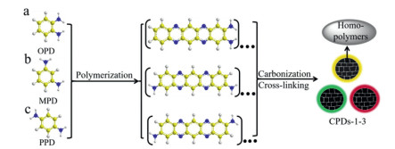
Schematic diagram of synthesis of CPDs-1–3.
Polymer types regulation strategy toward the synthesis of carbonized polymer dots with excitation-wavelength dependent or independent fluorescence
Jianliang Bai , Xinyu Wang , Yaqing Zhu , Guojun Yuan , Shuang Wu , Fu Qin , Xu Yu , Lili Ren

Carbon-based nanomaterials have been fascinating scientists in recent years because of multitudinous source of precursors and outstanding mechanical, optical and electrical properties [1-9]. Different from fullerenes, nanotubes, nanodiamonds and graphene, carbon dots (CDs) is a new member of the family of carbon nano-materials [10, 11]. Since it was discovered in 2004, as a class of photoluminescent carbon-based nanomaterials, CDs have since emerged in bioengineering, chemical engineering and energy applications due to desirable properties such as high photo-stability against photobleaching [12, 13], low toxicity [14-16], biocompatibility [17] and easy surface functionalization [17-19]. Applications include drug delivery [20-22], bioimaging [23-28], fluorescence (FL) sensing [29-31], optoelectronic device [32-34], photovoltaics [35-37] and information encryption [38]. Compared with the trend of CDs in full swing, the diversity of CDs and the complexity of chemical structure of CDs undermine the understanding of the underlying general FL phenomena and make the optical properties of the CDs elusive [39-41].
Among these optical properties, one of the most interesting properties is the excitation-wavelength dependent (a feature in stark contrast to the traditional inorganic semiconductor quantum dots) and the excitation-wavelength independent characteristics of emission for CDs [11, 42-44]. Up to now, the optical properties and corresponding luminescence mechanism of CDs have been studied extensively [45-51]. Some studies have thought that excitation-dependent FL is associated with the inhomogeneous ultrafine size. The wide particle size distribution can appear multiple energy gaps and result in multiple transition modes. Hence, under different excitation-wavelengths, it shows excitation-wavelength dependence characteristic [52-54]. More views have suggested that the various surface states can effectively introduce new energy levels for electron transitions and lead to continuously tunable excitation-wavelength dependent emission [42, 47, 55]. In addition, some researchers have believed that the ratio of core-state and molecular fluorophore contained in CDs will affect the excitation-wavelength dependence [56]. The above viewpoints can explain many optical properties of CDs, however, some of these problems have not been resolved, such as the actual composition and the accurate structure of CDs have not been expounded [11]. Therefore, obvious model structures are urgently needed to investigate the luminescence origin of CDs.
Previous studies have shown that the vast majority of CDs synthesized by the "bottom-up" process should strictly be considered as carbonized polymer dots (CPDs) [57-60]. The "bottom-up" strategy usually involves the process polymerization, cross-linking and carbonization of polymers or organic small molecules at high temperatures in microwave-assisted or solvothermal reactions [61]. Obviously, the formation of polymer structure in the bottom-up reaction process will affect the physical and chemical properties of the CPDs. It is well known that polymers can usually be divided into homopolymers and copolymers, and copolymers include block copolymers, random copolymers, alternating copolymers and gradient copolymers (Fig. S1 in Supporting information) [62]. According to previous reports, if the nanoparticle contains homogeneous polymer as the luminescence unit, the CPDs show excitation-wavelength independent FL [39]. Correspondingly, if the nanoparticle contains a variety of random copolymers as luminescence units, the CPDs show excitation-wavelength dependent FL [63]. Nevertheless, the precise nature of the polymer subfluorophores and their role in the FL emission of CPDs remain unclear, mainly due to the lack of studies focusing on the formation process and precise accurate structure of CPDs.
Based on the above considerations, in this work we have tried to shed light on this problem by controlling the precursor types and reaction conditions, and by developing a model system designed to imitate the FL properties of CPDs. With this hypothesis here we have designed a model to describe the polymer structures contained in the CPDs and adequately display transition from excitation-wavelength independent FL to excitation-wavelength dependent FL of CPDs.
In order to provide a striking contrast, phenylenediamine isomers were used as precursors to synthesize "carbonized homopolymer dots", and CA and DTEA as precursors to synthesize "carbonized copolymer dots". As shown in Fig. 1, CPDs-1–3 with excitation-wavelength independent FL characteristic were synthesized using o-phenylenediamine (OPD), m-phenylenediamine (MPD) and p-phenylenediamine (PPD) through hydrothermal or solvothermal reactions. However, polymer gel with excitation-wavelength independent FL characteristic and CPDs-4 and CPDs-5 with excitation-wavelength dependent FL characteristic were synthesized using CA and DTEA through hydrothermal reactions (Fig. 2).
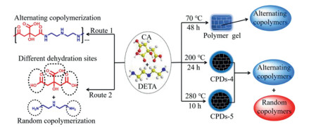
Fig. 3 shows the optical properties of CPDs-1–3. The UV–vis absorption spectra for yellow, green, and red CPDs are shown in Figs. 3a-c. Normally, obvious π-π* transitions appear ~250 nm region which correspond to sp2-hybridized carbon. For CPDs-1–3, low energy bands can be observed in the 400–550 nm region. These low energy bands are caused by n-π* transitions of the extended conjugation system including C=N and C=O [64]. FL spectra illustrates that CPDs-1–3 all own a single emission center, and under 360 nm excitation, the yellow emission, green emission and red emission are located at 540 nm, 509 nm and 617 nm, respectively (Figs. 3a-c). In addition, although the excitation-wavelength increased from 320 nm to 460 nm, the emission maxima of CPDs-1 are fixed at 540 nm, indicating that CPDs-1 has excitation-wavelength independent FL characteristics (Fig. 3d). CPDs-2 and CPDs-3 show similar excitation-wavelength independence FL characteristics like CPDs-1 (Figs. 3e and f), indicating that these three CPDs have similar structures. It should be emphasized that the choice of solvent has an important influence on the structure of the final CPDs, because the solvent may influence the reaction process [65]. In addition, the FL emission of CPDs-3 can be tuned by adjusting the polarity of the solvent, which is named a solvent-dependent characteristic (Fig. S2 in Supporting information) [24].
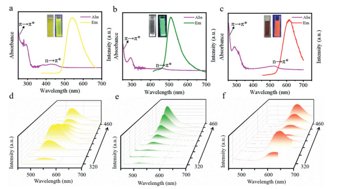
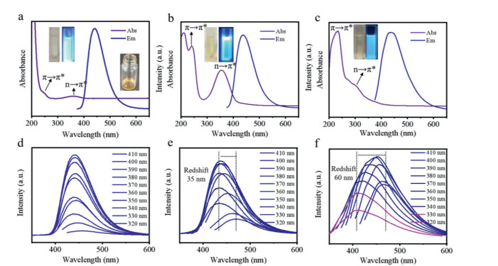
Fig. 4 shows the optical properties of polymer gel and CPDs-4 and CPDs-5. In the UV–vis absorption spectra of polymer gel and CPDs-4 and CPDs-5, both the polymer gel and two CPDs display absorption peaks at ~250 nm in the UV region, which are assigned to the π-π* transitions originate from sp2 carbon. The n-π* transitions of C=O at ~350 nm are usually related to doping and surface functional groups of CPDs (Figs. 4a-c) [64]. As shown in Fig. 4d, when the excitation-wavelength increased from 320 nm to 410 nm, the emission maxima of polymer gel are fixed at 442 nm, indicating that polymer gel has excitation-wavelength independent FL characteristics. On the contrary, when the excitation-wavelength increased from 320 nm to 410 nm, the emission-wavelength of CPDs-4 also redshifted from 435 nm to 470 nm (redshift 35 nm), indicating that CPDs-4 has obvious excitation-wavelength dependent FL characteristics (Fig. 4e). Even more surprising, CPDs-5 has shown more obvious and higher excitation-wavelength dependent FL (redshift 60 nm) than CPDs-4 under the same excitation-wavelength variation (Figs. 4e and f).
In order to study the effect of the structure and composition of two types of CPDs on the FL behaviors, the CPDs are characterized by transmission electron microscopy (TEM), Fourier transform infrared spectra (FT-IR), X-ray photoelectron spectroscopy (XPS) and Raman spectroscopy technologies. CPDs-3 is taken as a typical example to analyze the "carbonized homopolymer-dots". As shown in Fig. S3a (Supporting information), the CPDs-3 are quasi-spherical particles and the averaged particle size are determined to be 1.93 nm. The Raman spectrum of CPDs-3 shows a typical CDs structure, with D band at 1349 cm−1 indicating disordered carbon structure and G band at 1537 cm−1 indicating graphitized carbon structure (Fig. S3b in Supporting information). For detailed information of structure, FT-IR spectrum is obtained (Fig. S3c in Supporting information). CPDs-3 display the characteristic peaks for O—H/N—H stretching vibration (3407 cm−1), C—H stretching vibration (2930 cm−1), C=N stretching vibration (1631 cm−1), C=C stretching vibration (1603 cm−1), C—N stretching vibration (1511 cm−1) and C—O stretching vibration (1120 cm−1), indicating that CPDs-3 are composed of aromatic structure, and surface are rich in -NH2 and -OH. Then the functional groups of the CPDs-3 are further identified by XPS (Figs. S3d-f in Supporting information). Fig. S3d shows three fitting peaks for the high resolution C 1s spectrum of CPDs-3: C—C/C=C (284.3 eV), C—N (285.9 eV) and C—O (288.2 eV) [28]. Deconvolution of the N 1 s spectrum reveals the presence of pyridinic N at 398.6 eV, amino N at 400.0 eV and pyrrolic N at 401.1 eV (Fig. S3e) [28]. O 1s spectrum has only one peak at 533.67 eV, which is attributed to C—O-C and C—OH bonds (Fig. S3f) [28]. The above results indicate that CPDs-3 are partially graphitized nano particles with surface rich in –NH2 and –OH functional groups.
CPDs-4 and CPDs-5 are taken as typical examples to analyze the "carbonized copolymer-dots". CPDs-4 and CPDs-5 are uniformly dispersed with an average particle size of 2.55 and 5.61 nm, respectively (Figs. S4a and g in Supporting information). The Raman spectra of both CPDs show two peaks at 1342 and 1585 cm−1 (Figs. S4b and h in Supporting information), corresponding to the disordered structures (D band) and the graphitic carbon domains (G band) [24]. The intensity ratio of ID/IG is found to decrease from 1.08 to 0.80 for CPDs-4 to CPDs-5, indicating the increase of reaction temperature is beneficial to the increase of graphitization degree of CPDs. To further verify the surface structure and composition of CPDs-4 and CPDs-5, the FT-IR spectroscopy and XPS are measured. The same bonds are observed in the FT-IR spectra of two CPDs, such as O—H/N—H (3445 cm−1), C—H (2917 cm−1), C=O (1664 cm−1) and C=N (1638 cm−1), which indicates that both CPDs contained oxygen- and nitrogen- functional groups (Figs. S4c and i in Supporting information). Fig. S4d (Supporting information) shows three fitting peaks for the high resolution C 1s spectrum of CPDs-4: C—C/C=C (284.5 eV), C—N/C—O (285.7 eV) and C=O/C=N (287.6 eV) [24]. Deconvolution of the N 1s spectrum reveals the presence of C—N=C at 399.5 eV and amino N at 400.3 eV (Fig. S4e in Supporting information) [66]. In the O 1s spectrum of CPDs-4, three peaks are corresponding to C=O (530.5 eV), C—O/O—H (531.4 eV) and O—C=O (532.4 eV) (Fig. S4f in Supporting information) [24]. XPS analysis of the CPDs-5 shows that is similar to CPDs-4 in composition (Figs. S4j-l in Supporting information). By combining all these above characterization data and analyses, the CPDs-4 and CPDs-5 are carbon particles with varying degrees of graphitization, and surface rich in -NH2 and -OH functional groups.
We speculate that the prepared CPDs have different polymer structures (including homogeneous polymer chains, alternating copolymers and random copolymers), which may lead to different FL origins. In order to prove this hypothesis, first, we have studied the properties of "carbonized homopolymer-dots" and selected CPDs-3 as a typical example. 1H NMR spectrum of PPD shows two major signal peaks at 6.35 and 4.17 ppm (Fig. S5 in Supporting information, top). After polymerization, cross-linking and carbonization, PPD forms CPDs-3 with uniform polymer structure (Fig. 1). The 1H NMR spectrum of the CPDs-3 shows five distinct peaks at 7.08, 6.64, 5.76, 4.51 and 4.46 ppm, respectively (Fig. S5, bottom), corresponding to the homogeneous polymers contain some repeating units (Fig. S6 in Supporting information) contained in carbon nanoparticles. Therefore, CPDs show excitation-wavelength independence FL characteristics because the main FL origin is the homogeneous polymer chain. Raman spectrum shows that the carbon-core associated with excitation-wavelength dependent FL produced by high temperature graphitization does not become the luminescence center [56], which further indicates that the luminescence origin of the CPDs is polymer fluorophore.
Next, we have explored the luminescence mechanism of polymer gel and "carbonized copolymer dots". The 1H NMR spectrum of the polymer gel with excitation-wavelength independence shows clear structure and no obvious "noisy" signal peak (Fig. S7 in Supporting information, top), indicating that the two precursors formed homogeneous alternating copolymers under low temperature reaction conditions (Fig. 2, route 1) [39]. However, when the reaction temperature rises to 200 ℃, the diversity of dehydration sites (formation of amide) tends to form random copolymers, resulting in excitation-wavelength dependent characteristics of CPDs-4 (Fig. 2, route 2). Therefore, the signal peaks in the 1H NMR are "noisy" (Fig. S7, bottom). In fact, CPDs-4 produced at 200 ℃ contains a mixture of alternating copolymers and random copolymers. Therefore, when the reaction temperature raised to 280 ℃, the proportion of random copolymers contained in CPDs-5 continued to increase, resulting in more obvious and higher degree excitation-wavelength dependent characteristics. It can be seen that the polymer structures contained in CPDs is the main origin of their luminescence, and the uniform polymer structures contributes to produce excitation-wavelength independent FL, and the random copolymer structures contributes to produce excitation-wavelength dependent FL.
In conclusion, we have prepared five kinds of CPDs by using simple solvothermal or hydrothermal methods, and studied the structures of CPDs in order to obtain insight into interesting optical properties of these fluorescent emitters. Detailed structural characterization and optical properties reveal that the homogeneous polymer structures contained in CPDs are the main origin of excitation-wavelength independent FL, and the random copolymer structures contained in CPDs are the main origin of excitation-wavelength dependent FL. More importantly, we have achieved controllable preparation of excitation-wavelength independent, excitation-wavelength dependent and higher degree excitation-wavelength dependent fluorescent materials by simple tuning the reaction temperature. Looking toward the future, it is very meaningful to closely correlate the FL properties of CPDs with the accurate polymer structures.
The authors declare no conflict of interest.
We gratefully acknowledge the Priority Academic Program Development of Jiangsu Higher Education Institutions (No. 1107047002), the Key Research and Development Plan (Modern Agriculture) of Jiangsu Province (No. BE2018385) and Innovation Platform Project supported by Jiangsu Province (No. 6907041203). The authors would like to thank the shiyanjia lab (www.shiyanjia.com) for the XPS and Raman tests.
Supplementary material associated with this article can be found, in the online version, at doi:
W. Li, Y. Liu, B. Wang, et al., Chin. Chem. Lett. 30 (2019) 2323–2327. doi: 10.1016/j.cclet.2019.06.040
X. Niu, T. Song, H. Xiong, et al., Chin. Chem. Lett. 32 (2021) 1953–1956. doi: 10.1016/j.cclet.2021.01.006
A.K. Geim, K.S. Novoselov, Nat. Mater. 6 (2007) 183–191. doi: 10.1038/nmat1849
J. Zhou, C. Booker, R. Li, et al., J. Am. Chem. Soc. 129 (2007) 744–745. doi: 10.1021/ja0669070
B. Wang, J. Yu, L. Sui, et al., Adv. Sci. 8 (2021) 2001453. doi: 10.1002/advs.202001453
O.A. Shenderova, V.V. Zhirnov, D.W. Brenner, Solid State Mater. Sci. 27 (2002) 227–356.
G. Hong, S. Diao, A.L. Antaris, et al., Chem. Rev. 115 (2015) 10816–10906. doi: 10.1021/acs.chemrev.5b00008
O. Kozák, M. Sudolská, G. Pramanik, et al., Chem. Mater. 28 (2016) 4085–4128. doi: 10.1021/acs.chemmater.6b01372
C. Cha, S.R. Shin, N. Annabi, et al., ACS Nano 7 (2013) 2891–2897. doi: 10.1021/nn401196a
K. Hola, Y. Zhang, Y. Wang, et al., Nano Today 9 (2014) 590–603. doi: 10.1016/j.nantod.2014.09.004
M. Fu, F. Ehrat, Y. Wang, et al., Nano Lett. 15 (2015) 6030–6035. doi: 10.1021/acs.nanolett.5b02215
L. Wang, Y. Wang, T. Xu, et al., Nat. Commun. 5 (2014) 5357–5366. doi: 10.1038/ncomms6357
H. Jia, Z. Wang, T. Yuan, et al., Adv. Sci. 6 (2019) 1900397. doi: 10.1002/advs.201900397
Y. Ding, J. Yu, X. Chen, et al., Adv. Sci. 8 (2021) 2002404. doi: 10.1002/advs.202002404
C. Ding, A. Zhu, Y. Tian, Acc. Chem. Res. 47 (2014) 20–30. doi: 10.1021/ar400023s
Z. Zhu, R. Cheng, L. Ling, et al., Angew. Chem. Int. Ed. 59 (2020) 3099–3105. doi: 10.1002/anie.201914331
Z.T. Rosenkrans, T. Sun, D. Jiang, et al., Adv. Sci. 7 (2020) 2000420. doi: 10.1002/advs.202000420
X. Liu, H. Jiang, J. Ye, et al., Adv. Funct. Mater. 26 (2016) 8694–8706. doi: 10.1002/adfm.201603084
Y. Yang, X. Lin, W. Li, et al., ACS Appl. Mater. Interfaces 9 (2017) 14953–14959. doi: 10.1021/acsami.7b00282
Y. Sun, W. Cao, S. Li, et al., Sci. Rep. 3 (2013) 3036. doi: 10.1038/srep03036
G. Zuo, A. Xie, X. Pan, et al., ACS Appl. Nano Mater. 1 (2018) 2376–2385. doi: 10.1021/acsanm.8b00521
M. Zheng, S. Liu, J. Liu, et al., Adv. Mater. 26 (2014) 3554–3560. doi: 10.1002/adma.201306192
J. Kim, S. Jung, M. Shin, Opt. Mater. 72 (2017) 45–51. doi: 10.1016/j.optmat.2017.05.041
J. Bai, Y. Ma, G. Yuan, et al., J. Mater. Chem. C 7 (2019) 9709–9718. doi: 10.1039/C9TC02422K
A. Kundu, J. Lee, B. Park, et al., J. Colloid Interface Sci. 513 (2018) 505–514. doi: 10.1016/j.jcis.2017.10.095
K. Sun, Y. Tang, Q. Li, et al., ACS Nano 10 (2016) 6769–6781. doi: 10.1021/acsnano.6b02386
Q. Xiao, Y. Liang, F. Zhu, et al., Microchim. Acta 184 (2017) 2429–2438. doi: 10.1007/s00604-017-2242-z
K. Jiang, S. Sun, L. Zhang, et al., Angew. Chem. Int. Ed. 54 (2015) 5360–5363. doi: 10.1002/anie.201501193
R. Guo, B. Chen, F. Li, et al., Sens. Actuators B 264 (2018) 193–201. doi: 10.1016/j.snb.2018.02.175
X. Chen, J. Bai, Y. Ma, et al., Microchem. J. 149 (2019) 103981. doi: 10.1016/j.microc.2019.103981
Y. Jiao, Y. Gao, Y. Meng, et al., ACS Appl. Mater. Interfaces 11 (2019) 16822–16829. doi: 10.1021/acsami.9b01319
L. Wang, W. Li, L. Yin, et al., Sci. Adv. 6 (2020) eabb6772. doi: 10.1126/sciadv.abb6772
B. Zhao, Z. Tan, Adv. Sci. 8 (2021) 2001977. doi: 10.1002/advs.202001977
X. Yang, L. Sui, B. Wang, et al., Sci. China Chem. 64 (2021) 1547–1553. doi: 10.1007/s11426-021-1033-6
J. Liu, Y. Liu, N. Liu, et al., Science 347 (2015) 970–974. doi: 10.1126/science.aaa3145
M. Han, S. Zhu, S. Lu, et al., Nano Today 19 (2018) 201–218. doi: 10.1016/j.nantod.2018.02.008
Q. Chang, X. Han, C. Xue, et al., Chem. Commun. 53 (2017) 2343–2346. doi: 10.1039/C6CC09508A
Y. Ding, X. Wang, M. Tang, et al., Adv. Sci. (2021) 2103833.
L. Vallan, E.P. Urriolabeitia, F. Ruipérez, et al., J. Am. Chem. Soc. 140 (2018) 12862–12869. doi: 10.1021/jacs.8b06051
B. Wang, S. Lu, Matter 5 (2022) 110–149. doi: 10.1016/j.matt.2021.10.016
J. Yu, X. Yong, Z. Tang, et al., J. Phys. Chem. Lett. 12 (2021) 7671–7687. doi: 10.1021/acs.jpclett.1c01856
H. Nie, M. Li, Q. Li, et al., Chem. Mater. 26 (2014) 3104–3112. doi: 10.1021/cm5003669
Y. Dong, H. Pang, H. Yang, et al., Angew. Chem. 125 (2013) 7954–7958. doi: 10.1002/ange.201301114
H. Li, X. He, Z. Kang, et al., Angew. Chem. Int. Ed. 49 (2010) 4430–4434. doi: 10.1002/anie.200906154
M.J. Krysmann, A. Kelarakis, P. Dallas, et al., J. Am. Chem. Soc. 134 (2012) 747–750. doi: 10.1021/ja204661r
Y. Fang, S. Guo, D. Li, et al., ACS Nano 6 (2012) 400–409. doi: 10.1021/nn2046373
P. Yu, X. Wen, Y. Toh, et al., J. Phys. Chem. C 116 (2012) 25552–25557. doi: 10.1021/jp307308z
L. Tang, R. Ji, X. Cao, et al., ACS Nano 6 (2012) 5102–5110. doi: 10.1021/nn300760g
H. Ding, S. Yu, J. Wei, et al., ACS Nano 10 (2016) 484–491. doi: 10.1021/acsnano.5b05406
H.A. Nguyen, I. Srivastava, D. Pan, et al., ACS Nano 14 (2020) 6127–6137. doi: 10.1021/acsnano.0c01924
H.A. Nguyen, I. Srivastava, D. Pan, et al., Proc. Natl. Acad. Sci. U. S. A. 118 (2021) e2023083118. doi: 10.1073/pnas.2023083118
J. Wang, C. Cheng, Y. Huang, et al., J. Mater. Chem. C 2 (2014) 5028–5035. doi: 10.1039/C3TC32131B
X. Yan, X. Cui, L. Li, J. Am. Chem. Soc. 132 (2010) 5944–5945. doi: 10.1021/ja1009376
Y. Wang, Y. Li, Y. Yan, et al., Chem. Commun. 49 (2013) 9006–9008. doi: 10.1039/c3cc43375g
S.K. Das, Y. Liu, S. Yeom, et al., Nano Lett. 14 (2014) 620–625. doi: 10.1021/nl403820m
Y. Song, S. Zhu, S. Zhang, et al., J. Mater. Chem. C 3 (2015) 5976–5984. doi: 10.1039/C5TC00813A
S. Tao, T. Feng, C. Zheng, et al., J. Phys. Chem. Lett. 10 (2019) 5182–5188. doi: 10.1021/acs.jpclett.9b01384
C. Xia, S. Zhu, T. Feng, et al., Adv. Sci. 6 (2019) 1901316. doi: 10.1002/advs.201901316
Y. Ru, L. Ai, T. Jia, et al., Nano Today 34 (2020) 100953. doi: 10.1016/j.nantod.2020.100953
Z. Wang, Y. Liu, S. Zhen, et al., Adv. Sci. 7 (2020) 1902688. doi: 10.1002/advs.201902688
J. Bai, G. Yuan, Y. Zhu, et al., J. Phys. Chem. C 125 (2021) 18543–18551. doi: 10.1021/acs.jpcc.1c06137
M. Ouchi, T. Terashima, M. Sawamoto, Chem. Rev. 109 (2009) 4963–5050. doi: 10.1021/cr900234b
S. Hu, A. Trinchi, P. Atkin, et al., Angew. Chem. Int. Ed. 54 (2015) 2970–2974. doi: 10.1002/anie.201411004
B. Zhi, X. Yao, M. Wu, et al., Chem. Sci. 12 (2021) 2441–2455. doi: 10.1039/D0SC05743F
H. Liu, X. Lv, C. Li, et al., Nanoscale 12 (2020) 10956–10963. doi: 10.1039/D0NR01903H
K. Jiang, S. Hu, Y. Wang, et al., Small 16 (2020) 2001909. doi: 10.1002/smll.202001909

Figure 3 (a-c) UV–vis absorption and FL spectra for CPDs-1–3. Insets in (a-c): The images of the CPDs-1–3 in ethanol solvents under daylight and UV lamp (365 nm), respectively; (d-f) The FL spectra of CPDs-1–3 in ethanol solvents excited by different wavelength lights.

Figure 4 (a-c) UV–vis absorption and FL spectra for polymer gel and CPDs-4 and CPDs-5. Insets in (a-c): The images of polymer gel and CPDs-4 and CPDs-5 in ethanol solvents under daylight and UV lamp (365 nm), respectively; (d-f) The FL spectra of polymer gel and CPDs-4 and CPDs-5 in ethanol solvents excited by different wavelength lights in ethanol.

 扫一扫看文章

扫一扫关注我们
 DownLoad:

 下载:
下载:
