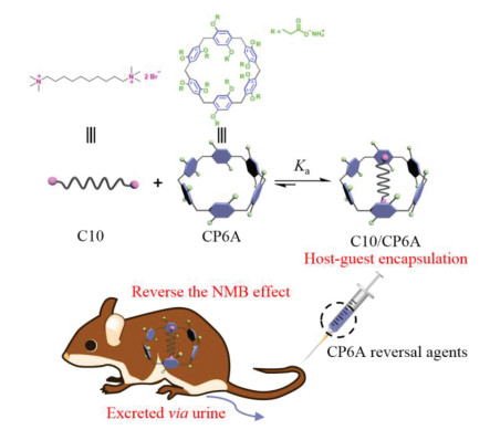
Figure 1.
Schematic illustration of host-guest encapsulation strategy based on CP6A to reverse the NMB effect of C10.
Reversing neuromuscular blocking agent decamethonium by carboxylatopillar[6]arene based on host-guest encapsulation
Yao Chai , Longming Chen , Yahan Zhang , Liang Zhao , Zhao Meng , Junyi Chen , Chunju Li , Qingbin Meng

Neuromuscular blocking agents (NMBAs) known as skeletal muscle relaxants are widely administered by anesthesiologists in anesthesia practice [1, 2], which prevent patients from moving in the surgical table and provide muscle relaxation to meet clinical requirements [3-9]. Annually more than 400 million patients receive NMBAs during anesthesia in operating rooms emergency or medicine departments [10]. Rapid reversal of neuromuscular blocker (NMB) is desirable to recover the muscular activity and improve patient safety at the end of surgery [11-13]. NMBAs are nicotinic acetylcholine receptor (nAChR) blocking drugs, achieving their effect by blocking the transmission of nerves impulses to skeletal muscle [14, 15]. According to their various mechanisms, NMBAs generally can be divided into two categories, including non-polarizing and depolarizing muscular relaxants [16-18]. Non-polarizing muscular relaxants act as competitive antagonists that bind with nAChR to induce muscle relaxation and their efficacies can be antagonized by anticholinesterase [19]. Depolarizing muscular relaxants exhibit agonist behavior upon binding to nAChR [16]. Persistent depolarizing of muscle cell membrane bring about inactivation of sodium transport mechanism, and consequently cause desensitization of end-plate membrane [20, 21]. Contrary to non-polarizing muscular relaxants, the NMB effect of depolarizing muscular relaxants cannot be inhibited by anticholinesterase. With respect to the foregoing, it is urgent to synthesize specific reversal agent of depolarizing muscular relaxants.
This research aimed at developing a supramolecular strategy to reverse NMB effect of depolarizing muscular relaxants by an artificial receptor. Sugammadex, a major advance in clinical anesthesia, is made by introduction of a modified γ-cyclodextrin molecular container which can bind with rocuronium and vecuronium with high affinities and rapidly reverse their NMB effect in vivo [22-25]. In addition, Issacs and co-workers developed acyclic analog of cucurbit[n]uril type molecular containers to efficiently reverse NMB effect of non-polarizing steroidal NMBAs [10, 12]. Given that, we propose that water-soluble macrocycles can function as reversal agents to reverse NMB effect of depolarizing muscular relaxants via host-guest interactions.
As a proof of concept, decamethonium (C10), the first depolarizing muscular relaxant which can avert to metabolically convert and reduce arterial blood pressure for patients with myasthenia gravis, served as model NMBAs [26-29]. C10 possess two potential binding sites, a central hydrophobic fatty chain and two flanking ammonium heads. Referring to the structural features of C10, carboxylatopillar[6]arene (CP6A), a popular water-soluble pillararene derivative with good water solubility and excellent biocompatibility was chosen as macrocyclic receptor [30-38]. CP6A possesses highly symmetrical prism-structure, satisfactory cavity size and anionic charge substituents at both rims [39, 40]. The alkyl chain of C10 was engulfed in CP6A cavity by hydrophobic interaction, accompanying with electrostatic interaction between ammonium heads and carboxylate groups. We found that CP6A could bind to C10 with a high affinity about 107 L/mol in phosphate buffer solution (PBS) and efficiently accelerate recovery from muscle relaxation in model mice, originating from rapid excretion of C10 via urine. Therefore, we suggested that CP6A could be employed as a reversal agent of C10 to reverse the NMB effect in vivo by direct host-guest encapsulation to meet clinical requirement (Fig. 1).
Initially 1H NMR spectroscopy was carried out to verify the host-guest complexation behaviors between CP6A and C10. Figs. 2a–c showed the spectra of C10 in D2O in absence and presence of CP6A. Upon addition of 1 equiv. of CP6A, the proton signals of C10, especially Hd, He and Hf exhibited substantial upfield shifts and broadening effects comparing to free C10, revealing that Hd, He and Hf were located in the cavity of CP6A and shielded by the electron-rich cyclic structure. Meanwhile the protons signals of CP6A (H1, H3) underwent downfield shift due to complexation-induced deshielding effects. For comparison, controlled 1H NMR experiment of mixtures of monomer (M, 6 equiv.) and C10 was also performed, showing that C10 could not interact with M (Fig. S1 in Supporting information).

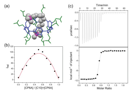
To further support the existence of 1:1 host-guest complex and guest binding geometry within host cavity, an MM2-minimized molecular model of C10/CP6A complex was conducted. As shown in Fig. 3a, the fatty chain of C10 was engulfed in the cavity of CP6A and its two flanking ammonium heads were close to carboxylate groups of CP6A to produce electrostatic interaction, which was in consistent with above 1H NMR spectroscopy results. The 1:1 binding stoichiometry between CP6A and C10 was also determined by continuous variation method (Job's plot) using the fluorescence emission values at 290 nm (Fig. 3b). Then isothermal titration calorimetry (ITC) experiment was employed to measure the association constant (Ka) of C10/CP6A. As shown in Fig. 3c, the binding between CP6A and C10 was dominantly governed by enthalpic gains (ΔH = −48.31 ± 0.42 kJ/mol) and the unfavorable entropic loss (-TΔS = 8.08 ± 0.54 kJ/mol), yielding a Ka value of (1.07 ± 0.14) × 107 L/mol. Such a high binding affinity promoted to further investigate whether CP6A might serve as a reversal agent of C10 to reverse its NMB effect in vivo.
Prior to biological activity analysis, the cytotoxicity of CP6A was assessed in mouse fibroblast cells (L929) by Cell Counting Kit-8 (CCK-8) assay. The results indicated that even at relatively high concentrations, CP6A displayed minimal cytotoxicity against this cell line (Fig. S2 in Supporting information), well matching previous results [41, 42]. Further supports for low system cytotoxicity from tissue sections, including heart, lung, spleen, liver, and kidney of mice at 7 days after intravenous injection of CP6A were also investigated. As shown in Fig. S4 (Supporting information), compared to PBS group, no inflammatory or toxicological responses were seen in CP6A administration groups. These results led us to consider that CP6A has excellent biocompatibility and low system cytotoxicity.
Next, a mouse model for muscle relaxation of C10 according to previous reports [43, 44], was set up to test reversal activity of CP6A. Initially, the cytotoxicity of C10/CP6A complex was evaluated in L929 cell line by CCK-8 assay. C10/CP6A complex displayed negligibly cytotoxicity against this cell line, with about 90% cell viability even at relatively high concentrations (Fig. S3 in Supporting information). In this study, KM mice (~20 g body weight) were intravenously administered with 0.69 mg/kg of C10. Within 1 min, all the mice showed muscle relaxation which was characterized by loss of balance and falling from the rotor. One minute after injection of C10, the mice were administrated with PBS, 2.7 mg/kg M (6 equiv. of C10), 2.7 mg/kg CP6A (equal equivalent of C10), respectively and immediately monitored locomotion behaviors. The time was recorded when mice could continuously move on the rotor for 30 s, which was regarded as recovery from muscle relaxation. As shown in Fig. 4, that mice in PBS group completely recovered from muscle relaxation exceed 8 min. Similarly, administration with M (negative control) was invalid in recovery time. Distinctively it was noted that a statistical significant in recovery time was seen for CP6A group (108 ± 10 s), which was about 78% than PBS group (499 ± 57 s). These results proved that locomotion recovery from muscle relaxation was efficiently accelerated by CP6A.
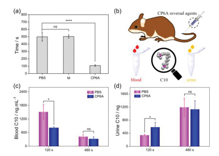
Furthermore, possible mechanism of reversal agent CP6A was explored and the calibration curves of C10 in mice blood and urine were first derived respectively (Figs. S5 and S6 in Supporting information). Previous studies reported that C10 excreted primarily via urine as a prototype [44]. We thus quantitatively detected C10 concentration in blood and its cumulative excretion amount in urine to verify possible mechanism of reversal agent CP6A. 60 s after intravenous injection of C10, the KM mice were randomly divided into two groups and administrated with PBS and CP6A. At predetermined intervals, blood was collected from ocular venous plexus and subsequently concentration of C10 was determined by LC-MS/MS. The results revealed that administration of CP6A could efficiently decrease C10 concentration within examination range. For example, at a time interval of 120 s, C10 concentration of PBS group (1517 ± 322 ng/mL) was about 85% higher than that of CP6A group (819 ± 32) ng/mL). A similar trend was also observed at 480 s after treatment (Fig. 4c). Meanwhile the cumulative excretion amount of C10 in urine was also monitored at same time points. C10 was found to be excreted via urine more quickly with assistance of CP6A. For example, its cumulative excretion amount of CP6A group at 120 s (590 ± 136 ng) was about 70% higher than PBS group (346 ± 20 ng) (Fig. 4d). Taken together, the above results suggested that CP6A could lower the concentration of C10 in blood of mice in vivo and reverse the muscle relaxation by capturing C10 and excreting it via urinary clearance.
In summary, we have provided a feasible supramolecular method that could reverse NMB effect of C10 via direct host-guest encapsulation. CP6A possessed strong binding ability towards C10 with a Ka value in the vicinity of 107 L/mol. Intravenous injection of CP6A could contribute to reverse NMB effect in a mouse model of muscle relaxation. The mechanism of reversal agent CP6A indicated that reducing the duration of muscle relaxation was attributed to accelerating the metabolism of C10 from blood to urine. These favorable results revealed that CP6A might function as a potential antidote to fill the clinical gap of C10.
The authors report no declarations of interest.
We acknowledge the Natural Science Foundation of Beijing Municipality (No. 7204285), the National Natural Science Foundation of China (Nos. 81573354, 21772118, 21971192). We are grateful to Prof. Xiaomei Zhuang for LC-MS/MS analyses.
Supplementary material associated with this article can be found, in the online version, at doi:10.1016/j.cclet.2021.11.087.
J.M. Adam, D.J. Bennett, A. Bom, et al., J. Med. Chem. 45 (2002) 1806-1816. doi: 10.1021/jm011107f
A. Bom, M. Bradley, K. Cameron, et al., Angew. Chem. Int. Ed. 41 (2002) 266-270.
J.M. Hunter, N. Engl. J. Med. 332 (1995) 1691-1699. doi: 10.1056/NEJM199506223322507
E.N. Brown, R. Lydic, N.D. Schiff, N. Engl. J. Med. 363 (2010) 2638-2650. doi: 10.1056/NEJMra0808281
C. Claroni, M. Covotta, G. Torregiani, et al., J. Clin. Med. 8 (2019) 1774-1783. doi: 10.3390/jcm8111774
E. Kirmeier, L.I. Eriksson, H. Lewald, et al., Lancet. Respir. Med. 7 (2019) 129-140. doi: 10.1016/S2213-2600(18)30294-7
A.S. Slutsky, N. Engl. J. Med. 363 (2010) 1176-1180. doi: 10.1056/NEJMe1007136
C.L. Morales-Perez, C.M. Noviello, R.E. Hibbs, Nature 538 (2016) 411. doi: 10.1038/nature19785
A.G. Engel, X.M. Shen, D. Selcen, et al., Lancet Neurol. 14 (2015) 420-434. doi: 10.1016/S1474-4422(14)70201-7
D. Ma, B. Zhang, U. Hoffmann, et al., Angew. Chem. Int. Ed. 51 (2012) 11358-11362. doi: 10.1002/anie.201206031
M. Grosse-Sundrup, J.P. Henneman, W.S. Sandberg, et al., BMJ 345 (2012) e6329. doi: 10.1136/bmj.e6329
X. Zhang, Q. Cheng, L. Li, et al., Theranostics 9 (2019) 3107-3121. doi: 10.7150/thno.34947
F. Haerter, J.C. Simons, U. Foerster, et al., Anesthesiology 123 (2015) 1337-1349. doi: 10.1097/ALN.0000000000000868
L.D. Gray, C. Morris, Anaesthesia 68 (2013) 14-29. doi: 10.1111/anae.12057
H. Yin, X. Zhang, J. Wei, et al., Theranostics 11 (2021) 1513-1526. doi: 10.7150/thno.53459
R.K. Jones, J.E. Caldwell, S.J. Brull, et al., Anesthesiology 109 (2008) 816-824. doi: 10.1097/ALN.0b013e31818a3fee
C.L. Deng, S.L. Murkli, L.D. Isaacs, Chem. Soc. Rev. 49 (2020) 7516-7532. doi: 10.1039/D0CS00454E
X.Y. Wu, Y.L. Wang, Y. Hai, et al., Chin. Chem. Lett. 28 (2017) 487-492. doi: 10.1016/j.cclet.2016.10.026
G.M. Keating, Drugs 76 (2016) 1041-1052. doi: 10.1007/s40265-016-0604-1
S. Thesleff, D.M. Quastel, Annu. Rev. Pharmacol. 5 (1965) 263-284. doi: 10.1146/annurev.pa.05.040165.001403
A.I. Lazar, F. Biedermann, K.R. Mustafina, et al., J. Am. Chem. Soc. 138 (2016) 13022-13029. doi: 10.1021/jacs.6b07655
H. Yin, D. Bardelang, R. Wang, Trends Chem. 3 (2021) 1-4. doi: 10.1016/j.trechm.2020.08.008
K. Wang, D.S. Guo, H.Q. Zhang, et al., J. Med. Chem. 52 (2009) 6402-6412. doi: 10.1021/jm900811z
S. Kheterpal, M.T. Vaughn, T.Z. Dubovoy, et al., Anaesthesiology 132 (2020) 1371-1381. doi: 10.1097/ALN.0000000000003256
K.S. Khuenl-Brady, M. Wattwil, B.F. Vanacker, et al., Anesth. Analg. 110 (2010) 64-73. doi: 10.1213/ane.0b013e3181ac53c3
C. Ball, R. Westhorpe, Anaesth. Intensive Care 33 (2005) 709. doi: 10.1177/0310057X0503300601
N.T. Campkin, J.R. Hood, S.A. Feldman, Anesth. Analg. 77 (1993) 78-80.
M.H. Davison, Lancet 1 (1950) 252.
A.J. Gray, Lancet 1 (1950) 253-255.
T. Ogoshi, T. Yamagishi, Y. Nakamoto, Chem. Rev. 116 (2016) 7937-8002. doi: 10.1021/acs.chemrev.5b00765
J. Zhou, G.C. Yu, F.H. Huang, Chem. Soc. Rev. 46 (2017) 7021-7053. doi: 10.1039/C6CS00898D
H.C. Zhang, Z.N. Liu, Y.L. Zhao, Chem. Soc. Rev. 47 (2018) 5491-5528. doi: 10.1039/C8CS00037A
T.X. Xiao, L. Zhou, L.X. Xu, et al., Chin. Chem. Lett. 30 (2019) 271-276. doi: 10.1016/j.cclet.2018.05.039
J.Y. Chen, Y.H. Zhang, Y.D. Zhang, et al., Chin. Chem. Lett. 32 (2021) 3034-3038. doi: 10.1016/j.cclet.2021.03.079
T.X. Xiao, W.W. Zhong, L. Zhou, et al., Chin. Chem. Lett. 30 (2019) 31-36. doi: 10.1016/j.cclet.2018.05.034
X. Zhang, X.Y. Wang, B. Wang, et al., Chin. Chem. Lett. 31 (2020) 3230-3232. doi: 10.1016/j.cclet.2020.02.037
D.R. Cao, M. Herbert, Chin. Chem. Lett. 30 (2019) 1758-1766. doi: 10.1016/j.cclet.2019.06.026
G.C. Yu, M. Xue, Z.B. Zhang, et al., J. Am. Chem. Soc. 134 (2012) 13248-13251. doi: 10.1021/ja306399f
G.C. Yu, M. Xue, Z.B. Zhang, et al., J. Am. Chem. Soc. 134 (2012) 13248-13251. doi: 10.1021/ja306399f
B. Li, Z. Meng, Q.Q. Li, et al., Chem. Sci. 8 (2017) 4458-4464. doi: 10.1039/C7SC01438D
J.Y. Chen, Y.D. Zhang, Z. Meng, et al., Chem. Sci. 11 (2020) 6275-6282. doi: 10.1039/D0SC01756F
J.Y. Chen, Y.D. Zhang, Y. Chai, et al., Chem. Sci. 12 (2021) 5202-5208. doi: 10.1039/D1SC00426C
W.D. Paton, E.J. Zaimis, Br. J. Pharmacol. 120 (1997) 60-79.
B. Christensen, J. Holm, Acta Pharmacol. Toxicol. 27 (1969) 17-26.

Figure 1 Schematic illustration of host-guest encapsulation strategy based on CP6A to reverse the NMB effect of C10.

Figure 2 1H NMR spectra (400 MHz, D2O) of (a) C10 (2 mmol/L), (b) C10 (2 mmol/L) + CP6A (2 mmol/L), and (c) CP6A (2 mmol/L).

Figure 3 (a) Optimized structure of MM2 energy-minimized model of C10/CP6A complex. (b) Job's plot for CP6A with C10 in 10 mmol/L PBS buffer at pH 7.4 (λex = 290 nm [CP6A] + [C10] = 10 µmol/L). (c) ITC titration of CP6A and C10 in 10 mmol/L PBS buffer at 298 K. One binding site model was utilized to fit the data. (ΔH: −48.31 ± 0.42 kJ/mol, -TΔS: 8.08 ± 0.54 kJ/mol and ΔG: −40.09 ± 0.21 kJ/mol).

Figure 4 (a) Diagram of the mice recovery time. Mice were intravenously administered with PBS, M and CP6A at 60 s after 0.69 mg/kg of C10 administration (mean ± SD, n = 10). (b) Schematic illustration of the clearance of C10 by CP6A in vivo. The concentration of C10 in (c) blood and its cumulative excretion amount in (d) urine after intravenous injection of PBS and CP6A within 8 min (mean ± SD, n = 6). Significant differences were assessed using t-test. ns, not significant. P < 0.05, ****P < 0.0001.

 扫一扫看文章

扫一扫关注我们
 DownLoad:
 下载:
下载: