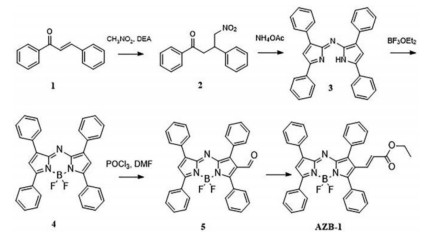
Scheme 1.
Synthetic route of the probe AZB-1.
Aza-BODIPY based probe for photoacoustic imaging of ONOO− in vivo
Danghui Ma , Shumin Hou , Chaeeon Bae , Thanh Chung Pham , Songyi Lee , Xin Zhou

As a typical reactive nitrogen specie, peroxynitrite (ONOO−) has drew wide attention due to its unusually potent oxidizing ability and strongly nucleophilic character [1-5]. The normal endogenous ONOO− is essential for maintaining cellular processes. Its role has been clearly proved in a mass of biophysiological events, including signal transduction pathways, forming nitrotyrosine residues and nitro fatty acid [6-10]. In contrast, the abnormal high concentration of ONOO− is harmful for the normal growth of cells, leading to the emergence of various diseases, such as diabetes, Alzheimer's disease, cardiovascular disease, and neurodegenerative diseases [11-14]. Therefore, it is of great significance to develop powerful analysis method for detecting ONOO− levels in organisms, in order to better understand complex pathophysiological processes.
Recently, the fluorescent probes have made significant progress in detecting ONOO−, due to their unique advantages of high temporal and spatial resolution, as well as high sensitivity [15, 16]. Moreover, some of them have realized the nondestructive and real-time imaging ONOO− in living cells and in vivo [17-22]. However, a major challenge with these fluorescent probes is the low resolution of imaging depth less than 1 mm due to their limited tissue penetration, which hampered the further monitoring ONOO− in vivo. Alternatively, photoacoustic molecular imaging (PA) is an emerging non-invasive biological imaging and sensing technology based on photoacoustic effect [23]. When biological tissue is irradiated by laser, photosensitive substances in the tissue absorb laser energy, generate instantaneous thermal expansion, and then radiate ultrasonic waves and stimulate photoacoustic signals. Photoacoustic imaging technology has the advantages of strong optical absorption, high spatial resolution, high contrast, and the ability to image in deep tissue [24]. Moreover, as a non-ionizing radiation imaging technology, photoacoustic imaging showed considerable tissue safety [25-28]. For this reason, some PA probes have been developed for a variety of applications, such as cancer detection [29], arthritis imaging [30], metal ions detection [31, 32], imaging of reducing agents [33, 34] and reactive oxygen species [35]. Although numbers of PA probes have been reported, only a few examples regarding ONOO− photoacoustic probes have been achieved to data [36, 37].
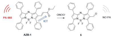
Herein, we reported a photoacoustic probe AZB-1 based on an aza-BODIPY derivative for detecting ONOO−. The probe showed a strongest absorption peak at 660 nm and gave a strong photoacoustic signal. When reacted with ONOO−, the conjugated group at 2-position of aza-BODIPY core can be readily oxidized, resulting in the shutting the ICT effect and the photoacoustic signal of the probe correspondingly. Wisely utilized this strategy may serve as powerful platforms for the preparation of novel photoacoustic chemosensors. Moreover, the probe was successfully employed to imaging ONOO− variations in inflammatory mice models.
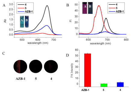
To obtain an ideal probe with initial strong photoacoustic signal, tetraphenyl substituted aza-BODIPY dye was employed as the core, due to its NIR absorbance and excellent optical properties, as well as easy modification at 2-position [38]. The probe AZB-1 was then synthesized by introduction a conjugated, unsaturated ketone group at 2-position of aza-BODIPY core (Scheme 1). As shown in Fig. 1, the optical and photoacoustic properties were first investigated. All of them showed typical absorbance peaks around 660 nm. Upon excitation, the parent compound 4 gave an emission peak at 690 nm, and the precursor 5 showed a hyperchromatic emission at 650 nm. Both of compounds 4 and 5 displayed ignorable photoacoustic signals as expected (Fig. 1c). In contrast, the probe AZB-1 showed almost no fluorescence, but with a strong photoacoustic signal. It is believed that the strong ICT effect occurred between the parent core and the conjugated α, β-unsaturated ketone group, relaxed the excited energy through a non-radiative pathway, resulting in switching its fluorescence into photoacoustic signal. Once reacting with ONOO−, the double bond could be oxidized, breaking the extending conjugated system, leading a turn-off photoacoustic response. These results indicated that the design strategy of photoacoustic probe could be realized by modifying aza-BODIPY with an election withdrawing group.
The solubility and aggregation behaviors of AZB-1 were firstly examined. As showed in Figs. S1-S3 (Supporting information), AZB-1 features a typical dose-dependent aggregation behavior in aqueous solutions due to its hydrophobic nature. The absorption intensity of the probe varies with the ratio of PBS buffer solution. AZB-1 showed a stable response to ONOO− in a mixture aqueous solution (PBS: DMSO = 1:1) under the concentration of 3 µmol/L. In addition, we tried to increase the solubility by preparing nanoparticles. As showed in Fig. S4 (Supporting information), probe AZB-1-F127 was assembled by the amphiphilic polyether F127 and formed a uniformed nanoparticle with an average diameter of 200 nm. However, the nanoparticles AZB-1-F127 showed ignorable response after the reaction with ONOO−. The results indicated that the polyether-coated probe cannot react with the ONOO−, which may be because the ONOO− could not break through the outer polyether to enter the interior of the nanoparticle. Therefore, the subsequent tests were performed in mixture aqueous solution with AZB-1.
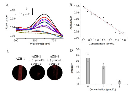
The response of AZB-1 to ONOO− was then launched. As shown in the Fig. 2, in a mixture aqueous solution (DMSO: PBS = 1:1), the absorption intensity of the probe AZB-1 at 660 nm gradually decreases with the increase of the concentration of ONOO−, and the decrease of the absorption value has a good linear relationship with the concentration of ONOO−. Meanwhile, the value of photoacoustic signal at 660 nm was significantly reduced, after the addition of ONOO− with different concentrations. These data indicated that this probe could detect ONOO− in solution by a photoacoustic way. In addition, the photoacoustic signal value of the probe AZB-1 reduced obviously over the change of the concentration of ONOO−, even 2 µmol/L ONOO− has quenched the photoacoustic signal.
The proposed sensing mechanism was documented in Scheme 2. According to the absorbance spectra (Figs. 2A and 1A), the product of AZB-1 with ONOO− showed similar absorbance peak with compound 5. Consequently, we assumed that the double bond of α, β-unsaturated ketone group could be readily oxidized, resulting in the quenching the photoacoustic signal. To confirm this proposed reaction mechanism, high resolution mass spectra were used to investigate oxidation of AZB-1 by ONOO−. When 10 equiv. of ONOO− was added to the solution of AZB-1, a peak corresponding to [5−H]− (Fig. S7 in Supporting information) appeared at m/z 566.8860, demonstrating that the compound 5 was generated.
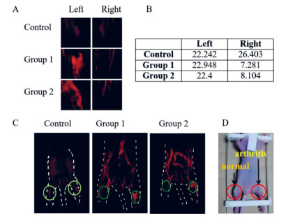
Based on abovementioned data, we further investigated the actual application of AZB-1 for photoacoustic imaging of ONOO− in vivo. All animal experiments were approved by the Institutional Animal Ethics Committee at Qingdao University, and the experiments were performed in compliance with the Care and Use of Laboratory Animal of Qingdao University. BALB/c nude mice (4−5 weeks) were used for establishment of mouse rheumatoid arthritis models by injecting λ-carrageenan (5 mg/mL, 50 µL in PBS) into the right hind limbs of mice. The left limbs were injected with normal saline (50 µL) as control. Four hours later, the right leg of the experimental mice was found to be arthritic and inflamed. It is known from previous reports that arthritis causes a rapid increase in the concentration of ONOO− [39]. Photoacoustic imaging was performed after intravenous injection of AZB-1. As shown in Fig. 3, there was no significant difference in photoacoustic signal values between the left and right limbs of the control group. But the photoacoustic signal of the right limb was significantly reduced in comparison with that of the left limb. These data demonstrated that probe AZB-1 can be used as a reliable and high-resolution imaging tool for the detection of ONOO− variations in mice.
To conclude, we have developed a new photoacoustic probe AZB-1, which exhibits good sensitivity in monitoring ONOO− oxidation under solution and living mice. The fluorescence signal of the probe itself is very weak, but it has a very strong photoacoustic signal. When it reacted with ONOO− in solution system, the value of photoacoustic signal is significantly reduced. The same phenomenon can also be verified in vivo. When the probe was injected into mice, the photoacoustic signal of the right arthritis limb is very weak, while the photoacoustic signal of the left normal limb was obvious. The value of the photoacoustic signal of the left limb is about 3 times than that of the right limb. The results demonstrated the probe AZB-1 can be used to detect ONOO− in vivo. Wisely utilized this strategy may serve as powerful platforms for the preparation of novel PA chemosensors in the future.
The authors declared that they have no conflicts of interest to this work.
Dr. Xin Zhou acknowledged the National Natural Science Foundation of China (Nos. 21662037, 21762045, 21911540466), Shandong Provincial Natural Science Foundation (No. ZR2019YQ12), China Postdoctoral Science Foundation (No. 219M652306), and Taishan Scholar Project (No. tsqn 201812049) for supporting this work. This research was also supported through the Basic Science Research Program of the National Research Foundation of Korea (NRF) funded by the Ministry of Education (No. 2017R1A6A3A04004954) to S. Lee.
Supplementary material associated with this article can be found, in the online version, at doi:10.1016/j.cclet.2021.05.048.
F.J. Corpas, J.B. Barroso, Ann. Bot. 113(2014) 87-96. doi: 10.1093/aob/mct260
H. Li, X. Li, X. Wu, W. Shi, H. Ma, Anal. Chem. 89(2017) 5519-5525. doi: 10.1021/acs.analchem.7b00503
J. Liu, Y. Dong, Y. Ma, et al., Nanoscale 10(2018) 13589-13598. doi: 10.1039/C8NR04596H
Y. Deng, G. Feng, Anal. Chem. 92(2020) 14667-14675. doi: 10.1021/acs.analchem.0c03199
J.H.M. Tsai, J.G. Harrison, J.C. Martin, et al., J. Am. Chem. Soc. 116(1994) 4115-4116. doi: 10.1021/ja00088a072
G. Ferrer-Sueta, R. Radi, ACS Chem. Biol. 4(2009) 161-177. doi: 10.1021/cb800279q
C. Ducrocq, B. Blanchard, B. Pignatellib, H. Ohshima, Cell. Mol. Life Sci. 55(1999) 1068-1077. doi: 10.1007/s000180050357
Y. Li, X. Xie, X. Yang, et al., Chem. Sci. 8(2017) 4006-4011. doi: 10.1039/C7SC00303J
P. Pacher, J.S. Beckman, L. Liaudet, Physiol. Rev. 87(2007) 315-424. doi: 10.1152/physrev.00029.2006
T. Sawa, M.H. Zaki, T. Okamoto, et al., Nat. Chem. Biol. 3(2007) 727-735. doi: 10.1038/nchembio.2007.33
T. Koppal, J. Drake, S. Yatin, et al., J. Neurochem. 72(1999) 310-317. doi: 10.1046/j.1471-4159.1999.0720310.x
D. Lee, C.S. Lim, G. Ko, et al., Anal. Chem. 90(2018) 9347-9352. doi: 10.1021/acs.analchem.8b01960
V.L. Dawson, T.M. Dawson, J. Chem. Neuroanat. 10(1996) 179-190. doi: 10.1016/0891-0618(96)00148-2
T. Franc, S. Salman-Tabcheh, M.C. Guérin, J. Torreilles, Res. Rev. 30(1999) 153-163. doi: 10.1016/S0165-0173(99)00014-4
S. Wang, L. Chen, P. Jangili, et al., Coord. Chem. Rev. 374(2018) 36-54. doi: 10.1016/j.ccr.2018.06.013
B. Li, Z. He, H. Zhou, H. Zhang, T. Cheng, Chin. Chem. Lett. 28(2017) 1929-1934. doi: 10.1016/j.cclet.2017.08.055
X. Li, X. Gao, W. Shi, H. Ma, Chem. Rev. 114(2014) 590-659. doi: 10.1021/cr300508p
J.F. Zhang, Y. Zhou, J. Yoon, J.S. Kim, Chem. Soc. Rev. 40(2011) 3416-3429. doi: 10.1039/c1cs15028f
X. Chen, X. Tian, I. Shin, J. Yoon, Chem. Soc. Rev. 40(2011) 4783-4804. doi: 10.1039/c1cs15037e
J. Lu, Y. Song, W. Shi, X. Li, H. Ma, Sens. Actuators B: Chem. 161(2012) 615-620. doi: 10.1016/j.snb.2011.11.009
H. Maeda, H. Matsuno, M. Ushida, et al., Angew. Chem. Int. Ed. 44(2005) 2922-2925. doi: 10.1002/anie.200500114
C. Yin, F. Huo, J. Zhang, et al., Chem. Soc. Rev. 42(2013) 6032-6059. doi: 10.1039/c3cs60055f
M. Xu, L.V. Wang, Rev. Sci. Instrum. 77(2006) 041101. doi: 10.1063/1.2195024
Q. Fu, R. Zhu, J. Song, H. Yang, X. Chen, Adv. Mater. 31(2019) 1805875.
L.V. Wang, Nat. Photonics 3(2009) 503-509. doi: 10.1038/nphoton.2009.157
J. Weber, P.C. Beard, S.E. Bohndiek, Nat. Methods 13(2016) 639-650. doi: 10.1038/nmeth.3929
K.J. Cash, C. Li, J. Xia, L.V. Wang, H.A. Clark, ACS Nano 9(2015) 1692-1698. doi: 10.1021/nn5064858
P. Beard, Interface Focus 1(2011) 602-631. doi: 10.1098/rsfs.2011.0028
S. Mallidi, G.P. Luke, S. Emelianov, Trends Biotechnol. 29(2011) 213-221. doi: 10.1016/j.tibtech.2011.01.006
J. Jo, C. Tian, G. Xu, et al., Photoacoustics 12(2018) 82-89. doi: 10.1016/j.pacs.2018.07.004
H. Li, P. Zhang, L.P. Smaga, R.A. Hoffman, J. Chan, J. Am. Chem. Soc. 137(2015) 15628-15631. doi: 10.1021/jacs.5b10504
Y. Liu, S. Wang, Y. Ma, et al., Adv. Mater. 29(2017) 1606129. doi: 10.1002/adma.201606129
X. Li, Y. Tang, J. Li, et al., Chem. Commun. 55(2019) 5934-5937. doi: 10.1039/C9CC02224D
J. Zheng, Q. Zeng, R. Zhang, D. Xing, T. Zhang, J. Am. Chem. Soc. 141(2019) 19226-19230. doi: 10.1021/jacs.9b10353
C.J. Reinhardt, E.Y. Zhou, M.D. Jorgensen, G. Partipilo, J. Chan, J. Am. Chem. Soc. 140(2018) 1011-1018. doi: 10.1021/jacs.7b10783
J. Zhang, X. Zhen, J. Zeng, K. Pu, Anal. Chem. 90(2018) 9301-9307. doi: 10.1021/acs.analchem.8b01879
X. Qin, F. Li, Y. Zhang, et al., Anal. Chem. 90(2018) 9381-9385. doi: 10.1021/acs.analchem.8b01992
W. Zhao, E.M. Carreira, Angew. Chem. Int. Ed. 44(2005) 1677-1679. doi: 10.1002/anie.200461868
A.H. Panchal, N.J. Patel, N.G. Patel, et al., Pharma. Sci. Monit. 1(2010) 79-94.

Figure 1 (A) Absorption and (B) fluorescence spectra of the AZB-1 (10 µmol/L), and its precursor 5 (10 µmol/L), and 4 (10 µmol/L), in DMSO solution. Inset: The color of the three compounds under natural light and UV lamp excited at 365 nm (A represents compound 5, B represents AZB-1). (C) Photoacoustic signal of compounds AZB-1, 5, 4. (D) Photoacoustic signal value of AZB-1, 5, 4. The excitation wavelengths were 660 nm.

Figure 2 (A) Absorption spectra of AZB-1 probe after reaction with different concentrations of ONOO−. (B) Absorption value of probe AZB-1 at 660 nm after reaction with different concentrations of ONOO−. (C) Photoacoustic signal of AZB-1 reacting with different concentrations of ONOO−. (D) Photoacoustic signal value of AZB-1 reacting with different concentrations of ONOO−. The excitation wavelengths were all at 660 nm.

Figure 3 (A) Photoacoustic imaging of left and right limbs of mice in control group and experimental group. The mice in the control group were injected with normal saline, while the mice in the experimental group had arthritis in the right limb and normal left limb. (B) Photoacoustic signal values of left and right limbs of mice in control group and experimental group. (C) Photoacoustic signal images of the whole body of mice in control group and experimental group. (D) Photograph of the experimental group mice (excitation wavelength is 660 nm).

 扫一扫看文章

扫一扫关注我们
 DownLoad:

 下载:
下载:
