Received Date:
13 May 2019 Accepted Date:
24 July 2019 Revised Date:
13 July 2019 Available Online:
22 February 2020
Abstract:
Host-guest supramolecular gels were developed via the self-assembly of inclusion complexes (ICs) of β-cyclodextrins/phenylboronic acid gelator (PBA). Salts and current were involved in the self-assembly to stabilize the host-guest gels. The stability of the gels was greatly improved after salts were added. The stable time of gels was extended from 2.5 h to 120 h with the addition of NH4NO3 at the concentration of 2.5×10⁻² g/mL. The morphology of the gel was affected by the concentrations of NH4NO3. SEM images revealed that the gels were three-dimensional nanofibrous networks, the sizes of fibers decreased with decreasing NH4NO3 concentrations, which affected the stability of gels, further proved by the rheological properties of gels. More stable gels were obtained with current stimulation, the stable time of the gel was increased from 2.5 h to 55 h with current by adding NaBF4. The current also exhibited significant influence on the aggregation as the voltage varied (0-500 mV) with a constant concentration of salts. The result showed the self-assembly process of host-guest gel could be well controlled via the addition of salts and current to desired morphology and stability.
Low-molecular-weight gels (LMWG) are important materials for biomedical applications. The gels are formed via the selfassembly of gelators driven by noncovalent interactions including H-bonding, π-π stacking, van der Waals forces, electrostatic and coordination interactions [1-3], which are reversible and sensitive to surrounding environment [3, 4]. LMWGs have attracted great interest to biomaterials scientists for drug administration (e.g., oral, transdermal, or parenteral) [5-7].
LMWGs based on urea derivatives, amino acid derivatives, polysaccharide derivatives, cholesterol derivatives, and complex organic compounds were fabricated only in organic solvents, pursuing from organogels to biocompatible gels made them possible for the applications in drug delivery [8-10]. Phenylboronic acid gelator (PBA) based LMWGs were biocompatible and sensitive to both glucose concentration and pH value, which evoked stimulisensitivity in drug delivery, however, the gelation occurred in toxic solvents such as cyclohexane, chloroform, toluene, and so on, the gelation conditions retarded the medical applications of PBAbased LMWGs [11, 12]. Moreover, the stability of LMWGs was another concern as the poor stability directly affected the gel's application [13, 14].
Cyclodextrins have been used as pharmaceutical materials with a long history [15, 16]. The inclusion complex (IC) supramolecular gel with β-CD as the host molecule and PBA as the guest molecule could improve the gelation and biocompatibility of gels. This fabrication would provide a strategy to control and optimize the structure as well as property of LMWGs [17, 18]. In this study, a β-CD/PBA inclusion complex based LMWG was prepared, the stability of the gel triggered by salt and current was further investigated to offer an effective way in controlling morphology, stability and properties of the host-guest LMWG as a potential drug delivery vehicle.
The gelation behavior of the ICs was examined in nineteen polar and non-polar solvents (Table S1 in Supporting information). A translucent gel as shown in Fig. 1e was only obtained in ethanol; however, neither β-CD nor the PBA could gelate individually in ethanol. The critical gelation concentration (CGC) of the ICs was 2.7 mg/mL (0.34 wt%), the ICs exhibited strong capability in gelling as the lowest CGC of single PBA gelator was 24.4 mg/mL in many solvents.
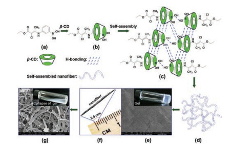
Figure 1
Figure 1.
The formation of gel via self-assembly. (a) Molecular structure of the gelator;
(b) inclusion complex of β-CD/gelator; (c) self-assembly of IC; (d) gel network;
(e) SEM image of gel, the insert image was the picture of gel; (f) nanofiber in the gel;
(g) SEM image of gel.
Fig. 1 presented the formation of gel. PBA based gelators (Fig. 1a) were threaded into the cage of β-CDs to form ICs (Fig. 1b). Driven by the van der Waals force, hydrogen bonding between β-CDs and π-interaction between the conjugation moieties of gelators (Fig. 1c) [19], the ICs self-assembled and rearranged fibrils, the fibrils were tangled to form 3-dimensional networks (Fig. 1d) and the gels were produced (Fig. 1e). The rearrangement of selfassembled fibrils was dynamic, once the fibrils grew into fibers (Fig. 1f), the 3-dimensional networks were destroyed and the fibers collapsed and precipitated (Fig. 1g). The lifetime of networks were considered as the stability of gels, which was tested as 2.5 h.
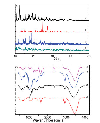
XRD is a powerful tool to characterize the crystal structure of molecules. XRD patterns of β-CD, PBA based gelator, IC and gel were presented in Fig. 2A. It was clear that the characteristic peaks of ICs appeared, it revealed the host-guest interaction between β-CDs and PBA based gelators. All the three samples of β-CD, gelator and IC exhibited strong crystallization capability, the gel showed weak crystallization due to its porous 3-dimensional network architecture. The formation of the inclusion complex gel was further characterized by FTIR spectrum (Fig. 2B), where the characteristic absorption peaks of the gelator, β-CD, and inclusion complex were shifted due to the enhanced intermolecular hydrogen bonding during the gelation [16]. For instance, the characteristic absorption peaks of the gelator at 3455.8 cm-1 (NH, OH) and at 1641.1 cm-1 (C=O, C=C) were shifted to 3451.7 cm-1 and 1635.3 cm-1 for the xerogel.
Figure 2
Figure 2.
(A) The XRD patterns of β-CD (a), gelator (b), IC (c) and gel (d). (B) FTIR spectra of gelator (a), β-CD (b), IC (c) (gelator: β-CD = 1.0 mol:1.0 mol), xerogel of IC (d).
The inclusion complex could form a supramolecular gel and exhibited rapid fiber formation in 18 min, however, the inclusion complex gel displayed poor stability and it was fractured in less than 150 min due to the aggregation of the nanofibers. Different kinds of salts were used to control the lifetime of gels, the effects of both inorganic and organic salts on the aggregation behavior of ICs were investigated (Table S2 in Supporting information).
In ethanol solutions containing various salts, the stability of the gel was greatly improved. For example, the stability time of the gel increased from the original time of 2.5 h to 4.7 h in a NaCl ethanol solution with the concentration of 7.9 × 10⁻⁴ g/mL, to 6.0 h in a NaHCO3 ethanol solution with the concentration of 4.7 × 10⁻³ g/mL, and to 27.0 h in a NaBF4 ethanol solution with the concentration of 4.9 × 10⁻⁴ g/mL. With the addition of inorganic salts, the electrostatic repulsion among the gelators was a key factor to influence the aggregation and result in the precipitation of fibers [20, 21]. As a Lewis acid, the boric acid moiety in the gelator was a good anion receptor, which combined with the anions from the inorganic salts. The increased electrostatic repulsion between these boric acid-anion complexes led to slower aggregation and sedimentation of ICs.
With anionic and cationic surfactants, the IC gels showed rapid self-assembly and good stability. The stability time of the gels was much longer in the cationic surfactants than that in anionic surfactants. For instance, the gel existed stably for 27 h with the addition of the cationic surfactant N-benzyl-N, N, N-trimethylammonium tribromide, further stability of 45 h was obtained with the addition of 1-decyl-3-methylimidazolium bromide. These two salts were ionic liquids. The effect of ionic liquids on the aggregation behaviors of supermolecules in aqueous solution was previously reported [22]. The imidazole rings in organic salts were probably penetrated into nanofibers to strengthen the intermolecular π-π stacking interaction within the gelators, leading to tight packing of gelators into nanofibers and less breakage of nanofibers [23, 24].
We also found that some salts did not increase the stability of the gels, but inhibited gel formation. No gel formation was observed with the addition of salts such as LiCl, CaCl2, FeCl3, CuCl2, NaBr, KI, CsF, CsI, sodium stearate, potassium tert-butanoate, sodium 4-nitrobenzenesulfonate, and sodium 2-naphthalenesulfonate. These results demonstrated that the inorganic and organic salts presented the ability to either prevent gel formation or improve the stability of the gel.
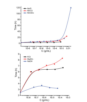
The aggregation behavior of ICs was also affected by the salt concentrations. Higher salt concentration resulted in longer gel lifetime. The lifetimes (stability) of gels incubated with KCl, NaCl, NaOH, NH4Cl, NH4NO3 and MgSO4 were investigated (Fig. 3). NaCl showed the weakest influence on gelation as the lifetime of gel was 4.7 h even when the salt concentration was the saturated 7.9 × 10⁻⁴ g/mL in ethanol, it was nearly the same as that with the salt concentration less than 7.9 × 10⁻⁵ g/mL. The other five salts exhibited greater influence on the gelation, with the addition of KCl (2.4 × 10⁻⁸ g/mL) and MgSO4 (2.0 × 10⁻⁸ g/mL) with very low concentrations, the lifetime of the gel was increased from 2.5 h to 6.0 h, it implied that the gel was more sensitive to these two salts. However, little stabilization effect was observed when the concentration of KCl was increased from 2.4 × 10⁻⁸ g/mL to the saturated 2.4 × 10⁻⁴ g/mL. The stability of gel depended on the concentration of NH4Cl and NH4NO3. The gel was stable for 24 h with NH4Cl (6.0 × 10⁻³ g/mL) and for 120 h with NH4NO3 (2.5 × 10⁻² g/mL). As for NaOH, the concentration less than 5.0 × 10⁻⁵ g/mL was conducive to form stable gel, no gel was obtained when the concentration was higher than 5.0 × 10⁻⁵ g/mL.
Figure 3
Figure 3.
The effect of salts concentration on the lifetime of gels.
To further understand the effect of the concentration of inorganic salts on the gelation, SEM images of the gels with various concentrations of NH4NO3 (2.5 ×10⁻⁶ – 2.5 ×10⁻² g/mL) were observed (Fig. 4). At low NH4NO3 concentrations, the gel maintained fiber network morphology, the fibers were significantly different from those of the gel without NH4NO3 in Fig. 1e. For instance, nanofibers and microbelts with width of 100–1400 nm were observed with NH4NO3 concentration of 2.5 × 10⁻⁶ g/mL (Fig. 4d), and the width was 80–1100 nm with NH4NO3 concentration of 2.5 × 10⁻⁵ g/mL (Fig. 4c). The width distribution of nanofibers and microbelts for these two gels were relatively wide. As the concentration of NH4NO3 increased to 2.5 × 10⁻⁴ g/mL, substantial changes in morphology happened. The nano-and microfibers disappeared, mainly short fiber sections were obtained (Fig. 4b). Micron flakes were predominant when the concentration of NH4NO3 was further increased to 2.5 ×10⁻³ g/mL (Fig. 4a). Although it was not easy for these microflakes to form a threedimensional network, the overlapping and aggregation of the microflakes were easier and tighter, which resulted in long lifetime of gel [25].
Figure 4
Figure 4.
SEM images of gels with different NH4NO3 concentrations, (a) 2.5 ×10⁻³ g/mL; (b) 2.5 ×10⁻⁴ g/mL; (c) 2.5 ×10⁻⁵ g/mL; (d) 2.5 ×10⁻⁶ g/mL. (e) The rheological properties of gels with different concentration of 10 mg/mL and 3 mg/L, storage and loss modulus as a function of angular frequency for the gels, all the gels were measured at 25 ℃.
The stability was further interpreted by the rheological properties of the gels (Fig. 4e). The frequency dependence of storage modulus (G') of the gels with different concentration of NH4NO3 was higher than their corresponding loss modulus (G"), it demonstrated that real gels were formed [26, 27]. The mechanical strength of this gel was improved with increasing the concentration of salt, the G' increased from 1.22 × 10² Pa to 2.14 × 10³ Pa as the concentration of NH4NO3 increased from 2.5 ×10⁻⁶ g/mL to 2.5 × 10⁻³ g/mL. This result demonstrated the mechanical strength of this gel was depended on the concentration of salt, it further proved that the salt induced long lifetime of gels.
Besides the effect on the stability of gels, salts also affect the size and morphology of the self-assemblies in the gels [21]. Significant morphology changes of self-assemblies in gels were observed in the presence of different salts (NH4NO3, KCl, MgSO4 and NH4Cl) (Fig. S1 in Supporting information). Interpenetrated porous and network structures were observed in the gel with NH4NO3 (2.5 × 10⁻⁴ g/mL) (Fig. S1a), and micro-flakes with particle width of 2.6–13.5 μm appeared with the addition of MgSO4 (2.0 × 10⁻⁴ g/mL) (Fig. S1c). Uniform micro-rods were obtained with addition of KCl (2.4 × 10⁻⁴ g/mL) (Fig. S1b), and fragments of fibers with varying lengths and widths were observed in the organogel with NH4Cl (6.0 × 10⁻⁴ g/mL) (Fig. S1d).
Inorganic and organic salts were used as electrolytes in the ICs solutions (Table S3 in Supporting information) with ethanol as the solvent. Interestingly, the application of current triggered the formation of gel with extremely high stability. The stability time of the gel with NaBF4 was greatly increased from 27 h to 55 h with current. The stability time for the gel was 17 h (LiF, saturation), 12 h (KCl, 2.4 × 10⁻⁴ g/mL), and 8 h (NaCl, 7.9 × 10⁻⁴ g/mL) when the current was applied, and the corresponding stability time without current was 10 h, 6.7 h, and 4.7 h, respectively. However, the stability of the gel did not change in the presence of organic salts such as AgOOCCF3, EDTA, tetrabutylammonium iodide, tetrabutylammonium bromide, and N-benzyl-N, N, N-trimethylammonium tribromide. This result might be related to the ionic conductivities of these electrolyte solutions.
Moreover, the current also influenced the aggregation of the ICs, even in the same electrolyte solution with different current intensities. For example, the uniform micro-rods were obtained in the presence of KCl (2.4 × 10⁻⁴ g/mL) and without current (Fig. 5a). With a low voltage stimulation (100 mV), mainly square or hexagonal flakes were observed (Fig. 5b). When the voltage was further increased to 300 mV, three-dimensional nanofibrous networks containing flake nodular structures were obtained (Fig. 5c). At the maximum voltage studied of 500 mV (Fig. 5d), fan-shaped microaggregates were formed. The current conduction was transferred through the electrolyte, which affected the morphology of the self-assembly. Interestingly, the formation of fan-shaped aggregates was widespread in the gels trigged by the application of a voltage of 500 mV for many different types of salts, including tetrabutylammonium iodide, N-benzyl-N, N, N-trimethylammonium tribromide, NaBF4, CsI, and KCl (Fig. S2 in Supporting information). These segmented aggregates were composed of microneedle shapes, microrods, and micro-sheet-like particles.
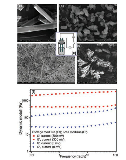
Figure 5
Figure 5.
SEM images of xerogels of inclusion complex with varying current intensity of KCl (2.4 ×10⁻⁴ g/mL), (a) 0 mV; (b) 100 mV; (c) 300 mV; (d) 500 mV; (e) diagram of current-trigged gelation. (f) the rheological properties of gels with different concentration of 10 mg/mL and 3 mg/L, storage and loss modulus as a function of angular frequency for the gels, all the gels were measured at 25 ℃.
The rheological properties of the gels were tested (Fig. 5f). The G' of the gel without current was 1.2 × 10² Pa in the presence of KCl (2.4 ×10⁻⁴ g/mL), and the G' was 1.99 × 10³ Pa as the current increased to 300 mV. The mechanical strength of the gel was improved with increasing the current. This result demonstrated the mechanical strength and the lifetime of the gel could be induced by current.
We prepared β-CD/PBA gelator inclusion complexes based supramolecular gels. Translucent gels consisted of macroscopic nanofibers or nanobelts were obtained in ethanol with a CGC of 0.34 wt%. The stability of the gel was greatly improved with the addition of different salts and current. The gelation was affected by salt types and concentrations. The lifetime of gels was extended from 2.5 h to 120 h with the addition of 2.5 ×10⁻² g/mL NH4NO3. The mechanical strength of this gel was improved with increasing the concentration of salt, the G' increased from 1.22 × 10² Pa to 2.14 ×10³ Pa as the concentration of NH4NO3 increased from 2.5 ×10⁻⁶ g/mL to 2.5 ×10⁻³ g/mL. The SEM images of the gels revealed that the gels were three-dimensional nanofibrous networks, the sizes of fibers decreased with decreasing NH4NO3 concentrations. Both the rheological properties and micromorphology demonstrated the salt-induced long lifetime of gels. Current influenced the aggregation of ICs as a trigger. The storage modulus of the gel increased from 1.2 × 10² Pa to 1.99 × 10³ Pa as the current varied from 0 mV to 300 mV.
Acknowledgments
The authors thank for the financial support from the National Natural Science Foundation of China (No. 21672164), Wenzhou Science and Technology Bureau (No. Y20170162), and Graduate Innovation Fund of Wenzhou University (No. 3162018031).
A. Roy, S. Comesse, M. Grisel, et al., Biomacromolecules 15 (2014) 1160-1170. doi: 10.1021/bm4017034
Figure 1
The formation of gel via self-assembly. (a) Molecular structure of the gelator;
(b) inclusion complex of β-CD/gelator; (c) self-assembly of IC; (d) gel network;
(e) SEM image of gel, the insert image was the picture of gel; (f) nanofiber in the gel;
(g) SEM image of gel.
Figure 2
(A) The XRD patterns of β-CD (a), gelator (b), IC (c) and gel (d). (B) FTIR spectra of gelator (a), β-CD (b), IC (c) (gelator: β-CD = 1.0 mol:1.0 mol), xerogel of IC (d).
Figure 4
SEM images of gels with different NH4NO3 concentrations, (a) 2.5 ×10⁻³ g/mL; (b) 2.5 ×10⁻⁴ g/mL; (c) 2.5 ×10⁻⁵ g/mL; (d) 2.5 ×10⁻⁶ g/mL. (e) The rheological properties of gels with different concentration of 10 mg/mL and 3 mg/L, storage and loss modulus as a function of angular frequency for the gels, all the gels were measured at 25 ℃.
Figure 5
SEM images of xerogels of inclusion complex with varying current intensity of KCl (2.4 ×10⁻⁴ g/mL), (a) 0 mV; (b) 100 mV; (c) 300 mV; (d) 500 mV; (e) diagram of current-trigged gelation. (f) the rheological properties of gels with different concentration of 10 mg/mL and 3 mg/L, storage and loss modulus as a function of angular frequency for the gels, all the gels were measured at 25 ℃.

 DownLoad:


 下载:
下载:


