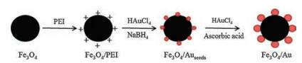
Scheme 1.
Synthetic process of Fe3O4/Au nanocomposites.
Cancer cell detection and imaging: MRI-SERS bimodal splat-shaped Fe3O4/Au nanocomposites
Xinmei Zhao , Leyong Zeng , Narayan Hosmane , Yan Gong , Aiguo Wu

Cancer, unrestrained development of abnormal cells in the body, is a serious health threatening disease which has been defeating many important lives every year. Early diagnosis with appropriate treatment can overcome this fatal disease to increase the survival rate of the patients. Presently, nanotechnology produces several multifunctional contrast agents in the form of nanoparticles (NPs) for imaging, such as magnetic resonance imaging/computed tomography (MRI/CT) [1], MRI/photo-acoustic [2], MRI/near infrared ray (NIR) [3] and MRI/optical/thermal imaging [4]. However, the control synthesis of solution based multimode nanoparticles with enhanced MRI and biocompatibility are exiting challenges. To improve the sensitivity in cancer diagnosis, surface enhanced Raman scattering (SERS) is one of the best for tumor detection, because (1) the detection limit is lower than the fluorescence method, (2) the effect by autofluorescence of the tumor can be avoided, and (3) SERS has high selectivity for the Raman reporter, so that the impurities in complex samples do not have any influence on the signal collection. Thus, the SERS technology is one of important analytical methods in biomolecular identification, and, therefore, it has been widely utilized in tumor detection and resection [5, 6]. Thus, combination of SERS and MRI techniques improves the accuracy of cancer diagnosis and therapy. Nowdays, researchers have been showing interest to fabricate the novel nanomaterials and nanostructures with dual MRI/SERS imaging applications.
The composites combined with MRI-active super-paramagnetic iron oxide nanoparticles and SERS-active gold substrates were prepared in 2011, in order for in vivo magnetic resonance imaging and Raman spectroscopy [7]. In 2012, a distinct triple-modality Au@SiO2@DOTA-Gd composite was synthesized and explored in brain tumor imaging and resection [8]. But, the smaller size and aggregation problems related to super-paramagnetic iron oxide nanoparticles restrict their usage in nanomedicine. The toxicity and nephrogenic systemic fibrosis (NSF) are also associated with commercially available Gd complex. For SERS application, silver nanoparticles are good SERS substrates, but they are toxic and easy to be oxidized. Gold nanoparticles have better stability and biocompatibility, therefore, gold nanopartilcles are often used as SERS substrate [9, 10]. The gold nanoparticles has excellent SERS property in biodetection [11, 12]. To resolve these issues, we designed new MRI/SERS probe consisting of Fe3O4/Au nano-composites. Recently, gold-coated Fe3O4 nanorose clusters were synthesized for cancer cell targeting, imaging and therapy [13, 14]. But these studies were not focused on the fields of MRI/SERS applications. In 2014, a new type of magneto-plasmonic Au-Fe alloy nanoparticles was designed for multimode SERS/MRI/CT imaging [15]. However, the studies about MRI/SERS properties of Fe3O4/Au were infancy.
In this work, Fe3O4 microspheres with an average size of ~130 nm were used to design splat-shaped Fe3O4/Au nanocomposites for MRI/SERS multimode imaging applications. There are some advantages of this prepared material such as the Fe3O4 are composed of tiny nanoparticles with super-paramagnetic nature and the gold nanoparticles can be grown on the modified surfaces of these microspheres uniformly. The uniform gold nanoparticles have more SERS "hot pot" to enhance the SERS property. The results of this investigation demonstrate the effectiveness of these unique splat-shaped nanocomposites as an excellent combined MRI and SERS agent for cancer detection.
In the experiments, two steps are employed to fabricate the splat-like Fe3O4/Au nanocomposites. Firstly, simple hydrothermal method is used to prepare the spherical Fe3O4 NPs in which FeCl3·6H2O, sodium 2-hydroxypropane tricarboxylate, and NaOAc·3H2O were mixed in ethylene glycol (EG) and resulting mixture was heated at 200 ℃ for 10 h [16]. To enhance the solubility of as-synthesized NPs, the surfaces of Fe3O4 microspheres are modified with polyetherimide (PEI) containing lots of amino group, which was improved by zeta potential study of Fe3O4 and Fe3O4/PEI microsphere (Fig. S1 in Supporting information). Finally, the seed mediated growth method is used to grow the gold nanoparticles onto the surface of the modified Fe3O4 microspheres [17]. The synthesis procedure for Fe3O4/Au composites is shown in Scheme 1.
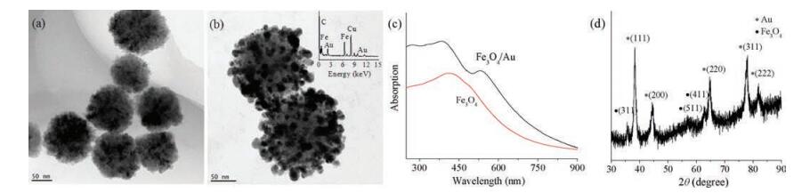
Fig. 1 shows the transmission electron microscopy (TEM) images and UV–vis absorption characteristics of the Fe3O4 and Fe3O4/Au nanostructures. It is clear from the TEM images that the microspheres are composed of tiny nanoparticles, and the average size is about 130 nm (Fig. 1a). Fig. 1b illustrates the gold nanoparticles with an average size of 10 nm that is grown on the surface of Fe3O4 microsphere uniformly shaped NPs in the form of splat-like structure, which can be indicated by energydispersive X-ray spectroscopy (EDS) spectra inserted in Fig. 1b. Compared with the UV–vis absorption spectra of Fe3O4 microspheres in Fig. 1c, an additional absorption peak is observed at 532 nm for Fe3O4/Au nanocomposites, which is attributed to the gold plasmonic oscillations indicating the formation of gold nanoparticles [18]. All the X-ray powder diffraction (XRD) peaks of the Fe3O4/Au composites (Fig. 1d) are well indexed to the structure of gold nanoparticles or Fe3O4 [19].
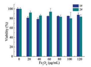
To examine the feasibility of the composites in the bioapplications, the toxicity of the nanoparticles was assessed with human breast cancer cells (MCF-7) by using the 3-(4, 5-dimethyliazol-2-yl)-2, 5-diphenyltetrazolium bromide (MTT) method as described before [20]. A 2.0 mL sample of MCF-7 in complete Dulbecco's modified eagle medium (DMEM) were seeded into each well at 6 × 10⁵ cells per well and allowed to adhere at 37 ℃. After 24 h, the DMEM medium was then changed to a fresh medium containing different amounts of the nanoparticles (0.115, 0.229, 0.343 mol of Fe) in 2 mL of the DMEM medium, and cells were incubated for 24 h. As shown in Fig. 2, no significant cell death or proliferation defects are recorded with different concentrations (corresponding to the weight concentration of Fe3O4/Au composites). This result suggests that Fe3O4/Au nanocomposites do not pose any considerable toxicity problem to the cells.
The magnetic hysteresis (Fig. S2 in Supporting information) and MRI relaxavities values (Fig. S3 in Supporting information) of splatlike Fe3O4/Au nanostructures are shown in Supporting information. The magnetic properties of the samples were measured at 298 K with the magnetic field sweeping from -30 kOe to +30 kOe. The magnetization saturation values are about 69.6, 58.7 and 33.6 emu/g for Fe3O4, Fe3O4/PEI and Fe3O4/Au, respectively. Compared to Fe3O4, the PEI and gold are the nonmagnetic materials. Therefore, the magnetization saturation value decreases with the addition of nonmagnetic materials [21]. The slope of the Fe3O4/Au is 64 and 1 L mmol-1 s-1 for r2 and r1, respectively, which is better than that reported [22]. This result suggested that synthesized material can be an excellent T2 contrast agent for MRI application.
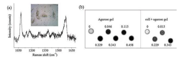
The in vitro MRI and SERS detection in the MCF-7 cells were studied (Fig. 3). For the SERS in vitro experiment, 0.229 mol (corresponding to the Fe concentration) of the Fe3O4/Au nanocomposites was dispersed in 2 mL of DMEM and incubated with the cells for 4 h at 37 ℃. Two peaks were observed in Fig. 3a at 1078 cm-1 and 1583 cm-1, which are attributed to the C—S and C—C band shift of the 4-ATP, respectively [23]. The good SERS property was also studied without cells, which is shown in Fig. S4 (Supporting information). For MRI detection, the 1% low-melting point agarose gels were prepared to simulate the human living tissue. The cells were removed from the wells, washed with phosphate buffer saline (PBS) twice and detached with trypsinEDTA. The cells were centrifuged twice and were dispersed in 1% low-melting agarose gel at a density of 5 ×10⁶ cells/mL. A significant dark contrast is obtained which is also increasing with Fe concentrations (Fig. 3b). All the images were obtained at the same clinical magnetic resonance imager with the same sequence. These results revealed that the Fe3O4/Au nanocomposites can be used as SERS and T2-weighted MRI cancer detection.
In conclusion, unique splat-shaped Fe3O4/Au nanostructures were synthesized by using the seed growth mechanism of secondary particle (Au) onto the primary particle (Fe3O4). Effective T2-weighted MRI and SERS data were obtained after incubation for 4 h with MCF-7 cancer cells without any toxicity. These results clearly demonstrated that as-synthesized splat-like Fe3O4/Au nanocomposites are a promising MRI/SERS multifunctional agent for the tumor detection.
This work was financially supported by the National Natural Science Foundation of China (Nos. 21305148, U1732127, U1432114, 51303196), the Ningbo Natural Science Foundation (Nos. 2014A610159, 2014B82010, 2015C50004, 2015B11002, 2016A610013, 2017C110022), Zhejiang Province (Nos. Bsh1202031, 2017C33129, 2017C03042, 2018KY737 and LY18H180011), Zhejiang Pharmaceutical College (No. ZPCSR2015013), Hundred Talents Program of Chinese Academy of Sciences (No. 2010-735). The authors also thank Dr. Juan Li from our group for the fruitful discussion. N. Hosmane thanks the Chinese Academy of Science for the Visiting Professorship for Senior International Scientists and Northern Illinois University for granting the leave of absence.
Supplementary data associated with this article can be found, in the online version, at https://doi.org/10.1016/j.cclet.2018.01.028.
N. Lee, H. Cho, M. Oh, et al., J. Am. Chem. Soc. 134(2012) 10309-10312. doi: 10.1021/ja3016582
X. Zhu, J. Zhou, M. Chen, et al., Biomaterials 33(2012) 4618-4627. doi: 10.1016/j.biomaterials.2012.03.007
J. Liu, Z. Li, X. Yang, et al., Chem. Commun. 51(2015) 13369-13372. doi: 10.1039/C5CC04911C
G. Hu, N. Li, J. Tang, S. Xu, L. Wang, ACS Appl. Mater. Interfaces 8(2016) 22830-22838. doi: 10.1021/acsami.6b05510
W. Song, Z. Mao, X. Liu, et al., Nanoscale 4(2012) 2333-2338. doi: 10.1039/c2nr12030e
T. Vo-Dinh, Y. Liu, A.M. Fales, et al., Wiley Interdiscip. Rev. Nanomed. Nanobiotechnol. 7(2015) 17-33. doi: 10.1002/wnan.1283
M. Yigit, L. Zhu, M. Ifediba, et al., ACS Nano 5(2011) 1056-1066. doi: 10.1021/nn102587h
M. Kircher, A. Zerda, J. Jokerst, et al., Nat. Med. 18(2012) 829-835. doi: 10.1038/nm.2721
Y. Zhang, L. Wang, E. Tan, S. Yang, L. Guo, Chin. Chem. Lett. 26(2015) 1426-1430. doi: 10.1016/j.cclet.2015.06.004
Z. Duan, X. Gao, T. Li, Kai Yao, X. Xie, Chin. Chem. Lett. 28(2017) 521-524.
Z. Gan, A. Zhao, M. Zhang, et al., J. Nanopart. Res. 15(2013) 1954. doi: 10.1007/s11051-013-1954-1
L. Lou, K. Yu, Z. Zhang, et al., Nano Res. 5(2012) 272-282. doi: 10.1007/s12274-012-0207-4
C. Li, T. Chen, I. Ocsoy, et al., Adv. Funct. Mater. 24(2014) 1772-1780. doi: 10.1002/adfm.v24.12
X. Zhou, W. Xu, Y. Wang, et al., J. Phys. Chem. C 114(2010) 19607-19613. doi: 10.1021/jp106949v
V. Amendola, S. Scaramuzza, L. Litti, et al., Small 10(2014) 2476-2486. doi: 10.1002/smll.v10.12
J. Liu, Z. Sun, Y. Deng, et al., Angew. Chem. Int. Ed. 48(2009) 5875-5879. doi: 10.1002/anie.v48:32
L. Lou, K. Yu, Z. Zhang, et al., Appl. Surf. Sci. 258(2012) 8521-8526. doi: 10.1016/j.apsusc.2012.05.031
C. Xu, J. Xie, D. Ho, et al., Angew. Chem. Int. Ed. 47(2008) 173-176. doi: 10.1002/(ISSN)1521-3773
S. Narayanan, B.N. Sathy, U. Mony, et al., ACS Appl. Mater. Interfaces 4(2012) 251-260. doi: 10.1021/am201311c
X. Ma, A. Gong, L. Xiang, et al., J. Mater. Chem. B 1(2013) 3419-3428. doi: 10.1039/c3tb20648c
Z. Liang, X. Wu, Y. Xie, S. Liu, Chin. J. Chem. 30(2012) 1387-1392. doi: 10.1002/cjoc.v30.7
T. Zhou, B. Wu, D. Xing, J. Mater. Chem. 22(2012) 470-477. doi: 10.1039/C1JM13692E
Y. Zhai, J. Zhai, Y. Wang, et al., J. Phys. Chem. C 113(2009) 7009-7014. doi: 10.1021/jp810561q

Figure 1 TEM images of Fe3O4 (a) and Fe3O4/Au nanocomposites (b). (c) UV–vis spectra of Fe3O4 and Fe3O4/Au nanocomposites. (d) XRD pattern of Fe3O4/Au nanocomposites.

Figure 2 The MCF-7 cell viability data for unmodified Fe3O4/Au nanocomposites (1#) and folic acid modified Fe3O4/Au nanocomposites (2#).

 扫一扫看文章

扫一扫关注我们
 DownLoad:

 下载:
下载:
