Citation:
Kang Yao, Fan Jiangli, Jin Qiang, Shi Chenhui, Du Jianjun, Peng Xiaojun. A α-KA fluorescent probe for discrimination of blood cancer serum[J]. Chinese Chemical Letters,
2017, 28(10): 1991-1993.
doi:
10.1016/j.cclet.2017.08.054
A α-KA fluorescent probe for discrimination of blood cancer serum
Received Date:
15 June 2017 Accepted Date:
30 August 2017 Revised Date:
22 August 2017 Available Online:
22 October 2017
Abstract:α-Ketoglutaric acid (α-KA) is an important metabolic intermediate in tricarboxylic acid circle in our body. The mutations of isocitrate dehydrogenase-1 (IDH1) and isocitrate dehydrogenase-2 (IDH2), however, will lead to the transformation of α-KA into 2-hydroxyglutarate (2-HG), which is confirmed to closely related to actue myeloid leukemia (AML). Therefore it is of great significance to detect α-KA level changes in serum. In this paper, a fluorescent "off-on" probe CH for α-KA was designed based on naphthalimide fluorophore by introducing a hydrazine group for α-KA recognition and a long alkyl amino chain to enhance PET efficiency and water solubility. Cetyltrimethyl ammonium bromide (CTAB) was added toform self-assembly micelles for accelerating the recognition process. CH shows a 28-fold fluorescence enhancement ((I-I0)/I0 at 550 nm) over other biological species by optimizing the chemical recognition process of CH with α-KA. Significantly, CH was successfully applied for thefluorescence discrimination of all kinds of blood cancer serum samples. This work would provide a potential method that is quick and convenient for sensing α-KA and may promote fluorescence detection in clinical diagnosis.
In recent years, the detection of biomarkers in diseases has received much attention [1-13]. α-Ketoglutarate (α-KG, a biomarker, with its acid form denoted as α-KA) is an endogenous metabolite of the citric acid cycle (Krebs cycle) that occupies a central position in energy metabolism [14]. The mutations of isocitrate dehydrogenase-1 (IDH1) and isocitrate dehydrogenase-2 (IDH2), however, will lead to the transformation of α-KA into 2-hydroxyglutarate (2-HG), which is confirmed to closely related to actue myeloid leukemia (AML) [15-19]. Therefore, α-KA has been suggested as a promising agent related to cancers [20, 21]. Currently, the available methods for determining α-KA level are confined to traditional gas chromatography (GC) and high performance liquid chromatography (HPLC) [22-24], which require laborious pretreatment and are not directly practicable in blood serum. Compared to traditional methods, fluorescent probes have been paid special attention owing to their simple operation, high selectivity, sensitivity, excellent high spatial and temporal resolution and especially nondestructive characteristics. Accordingly, the development of fluorescent probes for such biomarkers has become a focus of considerable research [9, 10, 25, 26].
In the 2014, Zhu et al. had carried out promoted progress in α-KA detection, in which a hydrazino group was selected as the recognition site, and linked with different fluorophores such as naphthalimide derivative [6], benzothiadiazole [25] and rhodamine derivatives [26]. Based on the typical Schiff-base reaction between α-KA and hydrazine group, these probes showed "off-on" fluorescence response notably, the sensor based on naphthalimide derivative [6] has been successfully used in a microfluidic chip to detect α-KA in blood serum. To our best knowledge, however, there is still no research on identification of α-KA level in the difference between normal and cancer blood serum.
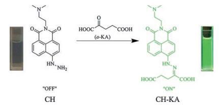
Therefore, to further detect α-KA in actual biological samples, herein, we introduced N, N'-dimethylethylenediamine unit into the meso-position of naphthalimide to enhance its PET efficiency [27-31] and water solubility, and hydrazino group as the recognition site to form probe CH, as shown in Fig. 1. Under optimized measurement conditions, the typical Schiff-base reaction between α-KA and hydrazine group occurred to product CH-KA, which showed a fast selective and sensitive fluorescence enhancement. The probe CH, product CH-KA and related agents were well characterized by NMR and HRMS (Figs. S7–S12 in Supporting information). More significantly, owing to the better properties of water solubility and sensitivity, it was successfully applied to distinguish normal and cancer blood serum via the large different fluorescence response, exhibiting a potential possibility of application in practice, especially in biological fields.
图 1
图 1
Rational design of fluorescent probe CH for α-KA detection. The hydrazino group behaves as the reaction trigger for the specific fluorescence "off-on" response.
Figure 1.
Rational design of fluorescent probe CH for α-KA detection. The hydrazino group behaves as the reaction trigger for the specific fluorescence "off-on" response.
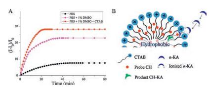
As illustrated in Table S1 (Supporting information), CH (10 μmol/L) shows very weak fluorescence and large stokes shift in all kinds of solvents. The quantum yield (Φ) of CH is only 0.022 in PBS (pH 7.4) with λabs and λem at 440 nm and 531 nm, respectively. Upon addition of 10 equiv. α-KA (100 μmol/L), CH (10 μmol/L) showed a 7.6-fold fluorescence enhancement in more than 40 min (Fig. 2A, black line). However, in some organic solvents such as DMSO, there was a 30-fold fluorescence enhancement within 25 min (Fig. S1 in Supporting information). Hence, a mixture of PBS-DMSO (99:1, v/v) was optimized as the test system solvent (Fig. 2A, pink line). To further accelerate the reaction between CH and α-KA, cetyltrimethyl ammonium bromide (CTAB) was introduced into the detection system. As CH bears a long alkyl chain, it can easily form micelles in the presence of CTAB. Upon formation of micelles, the positive charges on the micelle surface can aid α-KA to gather around (Fig. 2B). Thus the cationic surfactant CTAB can enable or accelerate chemical reactions in aqueous solution by possibly providing a nonpolar solvent-like microenvironment [32], resulting in a 28-fold fluorescence enhancement. In this case, 20 min was found to be the suitable time for the detection of α-KA (Fig. 2A, red line).
图 2
图 2
(A) Fluorescence kinetics of probe CH (10 μmol/L) upon addition of α-KA (100 μmol/L). λex = 440 nm, λem = 552 nm, 37 ℃. (B) Schematic diagram of the micelle-induced microenvironment using CTAB and the process of α-KA detection.
Figure 2.
(A) Fluorescence kinetics of probe CH (10 μmol/L) upon addition of α-KA (100 μmol/L). λex = 440 nm, λem = 552 nm, 37 ℃. (B) Schematic diagram of the micelle-induced microenvironment using CTAB and the process of α-KA detection.
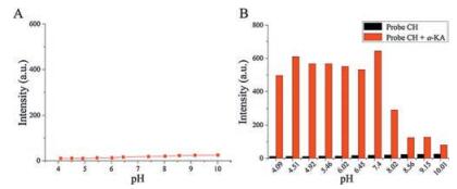
To measure the stability of CH, a series of α-KA detection in different pH buffers were carried out. In the absence of α-KA, there are slightly intensity changes at pH from 4 (acidic) to 10 (alkaline) (Figs. 3 and S2 in Supporting information), indicating that CH is stable in different environments with fluorescence "off" state. Upon the respective addition of α-KA, similar fluorescence enhancements were obtained with short equilibrium time in acidic environments (Figs. 3B and S3 in Supporting information). However, a tiny increase of intensity and long equilibrium time were detected in alkaline environment which means that alkaline environment is not suitable for this detection. Finally, the optimum pH for CH is 6.0, at which the reaction between CH and α-KA is the fastest.
图 3
图 3
(A) Stability of probe CH in different pH. Data are measured in CTAB (1 mmol/L), DMSO-PBS (10 mmol/L, 1:99, v/v) at 37 ℃. (B) Effect of pH on the fluorescence intensities of CH (10μmol/L) in the absence (black) and presence (red) of α-KA (10 equiv.).
Figure 3.
(A) Stability of probe CH in different pH. Data are measured in CTAB (1 mmol/L), DMSO-PBS (10 mmol/L, 1:99, v/v) at 37 ℃. (B) Effect of pH on the fluorescence intensities of CH (10μmol/L) in the absence (black) and presence (red) of α-KA (10 equiv.).
Thus the fluorescence titration of α-KA in probe CH was carried out in 10 mmol/L PBS-DMSO solution (1:99, v/v, pH 6.0, 37 ℃, in the presence of 1 mmol/L CTAB). As shown in Fig. S4 (Supporting information), there is a linear relationship between fluorescence intensity and the concentration of α-KA. The standard deviation s was determined by fluorescence response to be 0.4405. Therefore, the detection limit for α-KA was determined on the basis of signalnoise ratio (S/N = 3) to be 3.13 ×10⁻⁸ mol/L.
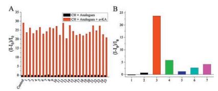
As α-KA exists in serum, the selectivity of probe CH in the presence of various potential interferents, such as amino acids, remains very important. As shown in Fig. 4A, only α-KA led to a 28-fold enhancement among 20 common amino acids or ions (Fig. 4A). This implies that probe CH has good specificity and feasibility towards α-KA, even in the presence of amino acids. In fact, in serum, there are other potential interferents with similar structures, including various dicarbonyl compounds and reactive oxygen species (ROS). We therefore compared the selectivity among some typical species (Fig. 4B), including glyoxal (GO), hydrogen peroxide (H2O2), phenylglyoxal (PGO), methylglyoxal (MGO) and phenylpyruvic acid (PPA). After incubation of various dicarbonyls and ROS with probe CH for 20 min, respectively, only with PPA and PAS showed minor fluorescence enhancement (Fig. 4B). The high selectivity of probe CH in the presence of dicarbonyl compounds with similar structures, such as MGO, could be attributed to the unique keto acid group in α-KA.
图 4
图 4
(A) Fluorescence responses of CH (10 μmol/L) to various analogues (100 μmol/L). Black bars represent CH + analogues, red bars represent CH +α-KA in the presence of other analogues (100 μmol/L). Data was collected at 20 min after addition of each analogue in DMSO-PBS (10 mmol/L, pH 6.0, 1:99, v/v) at 37 ℃. Control: blank, only probe CH; 1. NaF, 2. NaCl, 3. NaBr, 4. NaI, 5. Na2CO3, 6. Na2HPO4, 7. NaH2PO4, 8. Na2SO4, 9. NaAc, 10. KNO3, 11. H2O2, 12. Gly, 13. Leu, 14. Cys, 15. Ala, 16. Gln, 17. Pro, 18. Hcy, 19. His, 20. Thr, 21. Lys, 22. Arg, 23. Asn, 24. Glu, 25. Ser. (B) Fluorescence response of probe CH towards various dicarbonyls and ROS. Probe CH (10 μmol/L) with CTAB (1 mmol/L) was incubated with 100 μmol/L substrate for 20 min in DMSO-PBS (10 mmol/L, pH 6.0, 1:99, v/v). Substrates are glyoxal (GO, 1), hydrogen peroxide (H2O2, 2), α-KA (3), sodium pyruvate (PAS, 4), phenylglyoxal (PGO, 5), methylglyoxal (MGO, 6) and phenylpyruvic acid (PPA, 7).
Figure 4.
(A) Fluorescence responses of CH (10 μmol/L) to various analogues (100 μmol/L). Black bars represent CH + analogues, red bars represent CH +α-KA in the presence of other analogues (100 μmol/L). Data was collected at 20 min after addition of each analogue in DMSO-PBS (10 mmol/L, pH 6.0, 1:99, v/v) at 37 ℃. Control: blank, only probe CH; 1. NaF, 2. NaCl, 3. NaBr, 4. NaI, 5. Na2CO3, 6. Na2HPO4, 7. NaH2PO4, 8. Na2SO4, 9. NaAc, 10. KNO3, 11. H2O2, 12. Gly, 13. Leu, 14. Cys, 15. Ala, 16. Gln, 17. Pro, 18. Hcy, 19. His, 20. Thr, 21. Lys, 22. Arg, 23. Asn, 24. Glu, 25. Ser. (B) Fluorescence response of probe CH towards various dicarbonyls and ROS. Probe CH (10 μmol/L) with CTAB (1 mmol/L) was incubated with 100 μmol/L substrate for 20 min in DMSO-PBS (10 mmol/L, pH 6.0, 1:99, v/v). Substrates are glyoxal (GO, 1), hydrogen peroxide (H2O2, 2), α-KA (3), sodium pyruvate (PAS, 4), phenylglyoxal (PGO, 5), methylglyoxal (MGO, 6) and phenylpyruvic acid (PPA, 7).
To elucidate the mechanism of the fluorescence "off-on" process, CH and CH-KA were optimized, and their frontier molecular orbital (FMO) energies were calculated in water using Gaussian 09 (DFT/TDDFT in B3LYP/6-311G level) [33]. In probe CH, there is PET between naphthalimide and N, N'-dimethylethylenediamine unit, as shown in Fig. S5 (Supporting information). The PET state energy level (-5.567eV) was found to be higher than that of the fluorophore's HOMO(-5.886eV)(Fig.S5, left).Sowhen CHwas excited, there was an electron transition from the HOMO to the LUMO, and simultaneously an electron from the PET state would transfer to the HOMO to initiate the PET process, which quenched the fluorescence of CH. In the presence of α-KA, the binding of the hydrazine moiety to α-KA could force the probe to have an obvious change in electron density (Fig. S5, right). The electron density of HOMO and LUMO orbitals located in naphthalimide unit, and the oscillator strength between fluorophore itself is increasing from 0.002 to 0.6861, which resulted in elimination of the PET fluorescence quenching process. Moreover, the calculated HOMO-LUMO energy gaps of fluorophore in CH-KA (2.913eV), which was lower in comparison to KA (3.286eV). Therefore, the theoretical calculation agrees well with the experimental results and the product CH-KA excited with strong fluorescence.
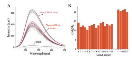
Encouraged by the good performance of CH in aqueous systems, we further carried out its detection of α-KA in human serum. As shown in Fig. 5, the fluorescence responses of CH (2 μmol/L) in addition of 5 samples of normal blood serum (150 μL) and 16 samples of blood cancer serum (150 μL) were detected, respectively, including acute lymphoblastic leukemia (ALL), acute monoblastic and monocytic leukemia (A-MOL), acute myelogenous leukemia (AML), non-hodgkinlymphoma (NHL), T-lymphoblastic lymphoma (T-LBL), acute leukemia (AL), T-acute lymphoblasticleukemia (T-ALL), and chronic myelomonocytic leukemia (CMML). As respected, the fluorescence intensity decreased obviously in blood cancer serums which indicated that the concentration level of α-KA in cancer blood decreased drastically due to the abnormal transformation of α-KA into 2-HG [15-19]. However, as hydrazine has been reported to be very reactive for formaldehyde (FA) [34], the detection result might be affected by FA. Fortunately, when extra 1–5 μmol/L FA was added into the serum, there was little interference (Fig. S6 in Supporting information). These results revealed that the application of CH for α-KA detection was completely feasible in serum.
图 5
图 5
Blood serum tests using probe CH. (A) Fluorescence responses of CH (2 μmol/L) after adding 150 μL blood serum in CTAB (1mmol/L), DMSO-PBS (10mmol/L, pH 6.0, 1:99, v/v) at 37 ℃. (B) (I -I0)/I0 of probe CH after adding serum. Five samples of normal blood serum and sixteen of abnormal blood serum of cancer patients were selected.1. ALL; 2. A-MOL; 3. AML; 4, NHL; 5. T-LBL; 6. AML; 7. AL; 8. AML; 9. T-ALL; 10. CMML; 11. AL; 12. T-ALL; 13. CLL; 14. NHL; 15. B-ALL; 16. AML; 17-21. Normal blood serum.
Figure 5.
Blood serum tests using probe CH. (A) Fluorescence responses of CH (2 μmol/L) after adding 150 μL blood serum in CTAB (1mmol/L), DMSO-PBS (10mmol/L, pH 6.0, 1:99, v/v) at 37 ℃. (B) (I -I0)/I0 of probe CH after adding serum. Five samples of normal blood serum and sixteen of abnormal blood serum of cancer patients were selected.1. ALL; 2. A-MOL; 3. AML; 4, NHL; 5. T-LBL; 6. AML; 7. AL; 8. AML; 9. T-ALL; 10. CMML; 11. AL; 12. T-ALL; 13. CLL; 14. NHL; 15. B-ALL; 16. AML; 17-21. Normal blood serum.
In summary, we designed an "off-on" fluorescent probe CH for the monitoring of α-KA, exhibiting good selectivity towards α-KA among amino acids, dicarbonyls and ROS. With the aid of micelles by inducing CTAB, CH can be applied in aqueous systems with an appropriate response time. It was successfully applied to the detection of α-KA in human serum and could differentiate cancer from normal blood serum. This work provides a potential method which is quick and convenient for sensing the biomarker α-KA in serum as a potential diagnostic tool.
Acknowledgements
This work was financially supported by the National Natural Science Foundation of China (Nos. 21576037, 21422601, 21406028, 21421005), NSFC-Liaoning United Fund (No. U1608222).
Figure 1
Rational design of fluorescent probe CH for α-KA detection. The hydrazino group behaves as the reaction trigger for the specific fluorescence "off-on" response.
Figure 2
(A) Fluorescence kinetics of probe CH (10 μmol/L) upon addition of α-KA (100 μmol/L). λex = 440 nm, λem = 552 nm, 37 ℃. (B) Schematic diagram of the micelle-induced microenvironment using CTAB and the process of α-KA detection.
Figure 3
(A) Stability of probe CH in different pH. Data are measured in CTAB (1 mmol/L), DMSO-PBS (10 mmol/L, 1:99, v/v) at 37 ℃. (B) Effect of pH on the fluorescence intensities of CH (10μmol/L) in the absence (black) and presence (red) of α-KA (10 equiv.).

 下载:
下载:

 下载:
下载:


