图 1
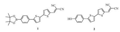
Structures of probe 1 and NIAD-4 (2).
Figure 1.
Structures of probe 1 and NIAD-4 (2).
A pinacol boronate caged NIAD-4 derivative as a near-infrared fluorescent probe for fast and selective detection of hypochlorous acid
Hongjuan Tong , Yajun Zhang , Shengnan Ma , Minghao Zhang , Na Wang , Rui Wang , Kaiyan Lou , Wei Wang

Reactive oxygen species (ROS) play an important role in cell signal transduction and maintaining redox homeostasis. Dysregulation of ROS contributes to many pathological conditions including aging [1], chronic inflammatory disease [1], Alzheimer's disease [2], etc. Among the various ROS, hypochlorous acid (HOCl) is involved in immune defense and inflammation [3]. It is generated in vivo from hydrogen peroxide (H2O2) and chloride ions in the presence of myeloperoxidase (MPO) in activated neutrophils for killing invading microbes [4], while uncontrolled production of HOCl is involved in many disease including inflammatory disease, cardiovascular disease, and neurodegeneration disorders including the Alzheimer's disease [5, 6]. Nevertheless, the exact role of HOCl in these pathological conditions is not fully elucidated, partially due to lack of powerful tools for selective detection of HOCl in the presence of other ROS. Recently, fluorescent methods utilizing specially designed fluorescence probes have become one of the most popular tools to study biomolecules of interest due to their high sensitivity, convenience in operation, and suitability for real time applications [7-9]. Towards this end, a number of fluorescent probes have emerged for detection of HOCl [10, 11]. However, few fluorescent probes were able to detect HOCl with emission in near-infrared (NIR) region between 650–900 nm [12-16], which has additional advantages of low background auto-fluorescence, less light scattering, and deep tissue penetration [17]. Therefore, there is still great need to develop near-infrared fluorescent probe for selective detection of HOCl.
Herein we reported synthesis and characterization of a pinacol boronate caged NIAD-4 derivative (1) as a NIR fluorescent probe for fast and selective detection of hypochlorous acid over other ROS species in both ratiometric and turn-on mode. In our probe design, NIAD-4 (2), a known NIR fluorophore which selectively binds to Aβ aggregates with strong fluorescence enhancement, was chosen as the fluorescence reporting group [18], and pinacol boronate group was selected as the reactive group for hypochlorous acid, as it was known that reaction between boronates and HOCl finishes within second and forms more electron-donating hydroxyl group [19]. Conversion of the boronate group to the hydroxyl group upon exposure to HOCl/ClO− would increase intramolecular charge transfer (ICT) in the excited state, resulting in the red-shift and intensity enhancement of fluorescence emission (Fig. 1).
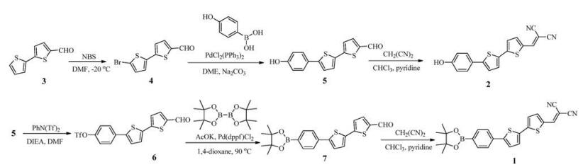
The probe 1 was synthesized from commercial available [2, 2'-bithiophene]-5-carbaldehyde (3) in 5 steps. Bromination of the starting aldehyde with N-bromosuccinimide (NBS) in DMF afforded the bromide 4 in 98% yield [20]. Suzuki coupling with (4-hydroxyphenyl)boronic acid gave the key intermediate 5 in 91% yield. Then, the phenol 5 was transformed to the trifluorometha-nesulfonate 6 and further converted to the pinacol bronate 7 in 95% and 77% yield, respectively. Finally, the aldehyde group, which remained intact in the previous steps, was condensed with malononitrile to afford the target probe 1 in 95% yield. The total yield for probe 1 was 62% (Supporting information). Similarly, condensation of the intermediate 5 with malononitrile gave NIAD-4 in 94% yield (Supporting information). Compared with the reported five-step synthetic route using Stille coupling as the key transformation [18], our synthetic route to NIAD-4 was not only short with just three steps and gave much improved total yield (84% vs. 37%), but also more environmentally friendly without the use of organotin reagents.
All experimental procedures including synthetic procedures, additional UV–vis and fluorescent spectra, NMR and HRMS spectra associated with this article can be found in Supporting information (Scheme 1).
With probe 1 in hand, we set out to study its absorption and fluorescence response to HOCl. In PBS and THF mixed solvent (1:1, v/v), probe 1 exhibits maximum absorption at 463 nm (Fig. S1 in Supporting information) with molar extinction coefficient ε = 36, 500 L mol−1 cm−1 (Fig. S1 in Supporting information). Fluorescence spectra of the probe 1 exhibits maximum emission at 565 nm and maximum excitation at 465 nm (Fig. 2a). Upon addition of NaClO, a significant fluorescence enhancement was observed instantly (Fig. 2b). The maximum fluorescence emission and excitation were red-shifted to 640 nm and 482 nm, respectively (Fig. 2a). Besides, the maximum fluorescence intensity increased about 2-fold (Fig. 2c), suggesting probe 1's potential utility as a ratiometric fluorescence probe for HOCl/ClO−. A concentration dependent fluorescence response (I640/I565) study in the range of 0 to 400 μmol/L NaClO (Fig. S3 in Supporting information) showed that the probe was able to detect NaClO over 50 μmol/L. Meanwhile, the fluorescence intensity at 700 nm of probe 1 increased about 14-fold upon reaction with HOCl/ClO− (Fig. 2c), indicating that probe 1 is also an excellent turn-on fluorescence probe for HOCl/ClO−.
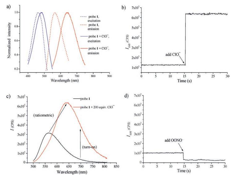
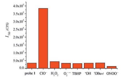
Next, we evaluated the selectivity of probe 1 towards various other reactive oxygen species including H2O2, superoxide anion (·O2−), tert-butyl hydroperoxide (TBHP), hydroxyl radical (·OH), tert-butoxyl radical (t-BuO·), and ONOO−. The fluorescence emission spectra upon addition of 200 μmol/L various other ROS except ONOO− at the incubation time of 30 s were measured and they showed nearly identical fluorescence emission spectra as that of the probe 1 indicating no reaction or slow reactions. For ONOO−, probe 1 also showed instant fluorescence response but turn-off fluorescence response (Fig. 2d). Since the uncaging of boronate by ONOO− was similar to HOCl with a much faster rate [19], this abnormal fluorescence quenching was likely attributed to fluorophore decomposition by ONOO−. This was supported by the blue-shift of λmax from 485 nm to around 300 nm in the absorption spectrum of the probe 1 after reaction with ONOO− (Fig. S4 in Supporting information). For other ROS, the ratio of the fluorescence intensity at 640 nm versus 565 nm was plotted (Fig. S5 in Supporting information). The ratiometric response (I640/I565) of probe 1 to hypochlorite is about 5 times of those of other ROS species tested in 30 s. Probe 1 also showed excellent selectivity at emission wavelength 700 nm (Fig. 2c) for turn-on response (Fig. 3). Further 1H NMR studies (Fig. S12 in Supporting information) verified that the reaction of the probe 1 with hypochlorite forms NIAD-4. We believe the probe's selectivity of HOCl over H2O2 was originated from kinetic difference of the oxidation reaction, as HOCl was known to react with boronate about 1000 times faster than H2O2[19].
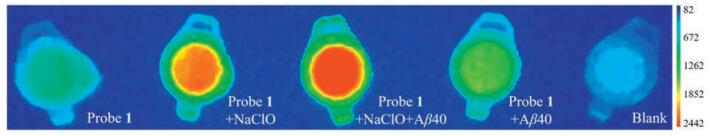
We further investigated the imaging utility of the probe 1 for detection of HOCl/ClO− through phantom imaging condition using mouse brain homogenate as biological relevant media [21] (Supporting information). The brain homogenates from a wild type mouse were employed. The reaction of the probe 1 with NaClO in the presence of or without Aβ40 aggregate were studied through near-infrared imaging (Fig. 4). At the emission wavelength of 700 nm, the probe behaved as a turn-on fluorescence probe for HOCl/ClO− (Fig. 2c). A clear fluorescence increase was seen when NaClO was added. Further fluorescence enhancement upon addition of Aβ40 aggregate was also observed, which agreed well with the reported fluorescence enhancement of NIAD-4 upon interaction with Aβ40 aggregate [18]. Since the level of HOCl in AD brains is estimated in the range of 20–400 μmol/L [22], this imaging studies showed that probe 1 could be used in Aβ-plaque enhanced fluorescence detection of HOCl in AD brains.
In conclusion, we reported here the design and concise synthesis of a new NIAD-4 derived NIR fluorescent probe 1 for selective detection of HOCl/ClO− over other ROS. The probes bears several unique features: (1) It could be used as either a ratiomatic or turn-on fluorescent probe; (2) The boronate group reacts with HOCl/ClO− much faster than with H2O2, which provides the selectivity for detection of HOCl/ClO− over H2O2; (3) The NIAD-4 fluorophore provides additional selectivity for detection of HOCl/ClO− over peroxynitrite. Moreover, the utility of the probe in imaging HOCl/ClO− was demonstrated in in vitro phantom imaging studies using mouse brain homogenate as a biological relevant media.
The work was supported by the China 111 Project (No. B07023, W. Wang), East China University of Science and Technology (start-up funds, W. Wang), the National Natural Science Foundation of China (No. 21577037, K. Lou), the Fundamental Research Funds for the Central Universities (No. ECUST-WY1213013, K. Lou).
Supplementary data associated with this article can be found, in the online version, at http://dx.doi.org/10.1016/j.cclet.2017.07.007.
S.G. Rhee, Science 312(2006) 1882-1883. doi: 10.1126/science.1130481
R. von Bernhardi, J. Eugenin, Antioxid. Redox Signal. 16(2012) 974-1031. doi: 10.1089/ars.2011.4082
D. Roos, C.C. Winterbourn, Science 296(2002) 669-671. doi: 10.1126/science.1071271
D.I. Pattison, M.J. Davies, Biochemistry 45(2006) 8152-8162. doi: 10.1021/bi060348s
Y.W. Yap, M. Whiteman, N.S. Cheung, Cell. Signal. 19(2007) 219-228. doi: 10.1016/j.cellsig.2006.06.013
M.V. Avshalumov, L. Bao, J.C. Patel, M.E. Rice, Antioxid. Redox Signal. 9(2007) 219-231. doi: 10.1089/ars.2007.9.219
A.P. de Silva, H.Q.N. Gunaratne, T. Gunnlaugsson, et al., Chem. Rev. 97(1997) 1515-1566. doi: 10.1021/cr960386p
X. Qian, Y. Xiao, Y. Xu, et al., Chem. Commun. 46(2010) 6418-6436. doi: 10.1039/c0cc00686f
Y. Yang, Q. Zhao, W. Feng, F. Li, Chem. Rev. 113(2013) 192-270. doi: 10.1021/cr2004103
Y. Yue, F. Huo, C. Yin, J.O. Escobedo, R.M. Strongin, Analyst 141(2016) 1859-1873. doi: 10.1039/C6AN00158K
X. Chen, F. Wang, J.Y. Hyun, et al., Chem. Soc. Rev. 45(2016) 2976-3016. doi: 10.1039/C6CS00192K
M. Sun, H. Yu, H. Zhu, et al., Anal. Chem. 86(2014) 671-677. doi: 10.1021/ac403603r
F. Tian, Y. Jia, Y. Zhang, et al., Biosens. Bioelectron. 86(2016) 68-74. doi: 10.1016/j.bios.2016.06.039
L. Yuan, W. Lin, H. Chen, Biomaterials 34(2013) 9566-9571. doi: 10.1016/j.biomaterials.2013.08.081
L. Yuan, W. Lin, Y. Yang, H. Chen, J. Am. Chem. Soc. 134(2012) 1200-1211. doi: 10.1021/ja209292b
G. Cheng, J. Fan, W. Sun, et al., Chem. Commun. 50(2014) 1018-1020. doi: 10.1039/C3CC47864E
J.O. Escobedo, O. Rusin, S. Lim, R.M. Strongin, Curr. Opin. Chem. Biol. 14(2010) 64-70. doi: 10.1016/j.cbpa.2009.10.022
E.E. Nesterov, J. Skoch, B.T. Hyman, et al., Angew. Chem. Int. Ed. 44(2005) 5452-5456. doi: 10.1002/(ISSN)1521-3773
A. Sikora, J. Zielonka, M. Lopez, J. Joseph, B. Kalyanaraman, Free Radic. Biol. Med. 47(2009) 1401-1407. doi: 10.1016/j.freeradbiomed.2009.08.006
S.P.G. Costa, R.M.F. Batista, M.M.M. Raposo, Tetrahedron 64(2008) 9733-9737. doi: 10.1016/j.tet.2008.07.096
J. Yang, J. Yang, S.H. Liang, et al., Sci. Rep. 6(2016) 35613. doi: 10.1038/srep35613
Y.W. Yap, M. Whiteman, B.H. Bay, et al., J. Neurochem. 98(2006) 1597-1609. doi: 10.1111/jnc.2006.98.issue-5

Figure 2 (a) Dashed line: normalized fluorescence excitation (λem = 565 nm) and emission (λex = 465 nm) spectra of probe 1 (1 μmol/L); Full line: normalized fluorescence excitation (λem = 640 nm) and emission spectra (λex = 482 nm) of probe 1 (1 μmol/L) + 200 equiv. of NaClO. (b) Instant fluorescence turn-on response of probe 1 (1 μmol/L) at 640 nm (λex = 482 nm) upon addition of NaClO (200 equiv.) at the time point of 15 s. (c) Fluorescence spectra (λex = 482 nm) of 1 μmol/L probe 1 before and after addition of 200 μmol/L NaClO. (d) Instant fluorescence turn-off response of probe 1 (1 μmol/L) at 640 nm (λex = 482 nm) upon addition of ONOO− (200 equiv.) at the time point of 15 s. All measurements were taken in 10 mmol/L pH 7.4 PBS/THF (1:1, v/v) solution containing 0.1% DMSO at 25 ℃.

Figure 3 Fluorescence intensity of 1 μmol/L probe 1 at 700 nm after incubation with 200 μmol/L various (ROS) for 30 s. All measurements were acquired at 25 ℃ in 10 mmol/L pH 7.4 PBS/THF (1:1, v/v), with λex = 482 nm.

 扫一扫看文章

扫一扫关注我们
 下载:
下载:

 下载:
下载:

