Login In
Patterned surfaces for biological applications: A new platform using two dimensional structures as biomaterials
- Corresponding author: Yang Bai, byangchem@jlu.edu.cn
Figures(15)
Citation:
Liu Wen-Dong, Yang Bai. Patterned surfaces for biological applications: A new platform using two dimensional structures as biomaterials[J]. Chinese Chemical Letters,
;2017, 28(4): 675-690.
doi:
10.1016/j.cclet.2016.09.004

Figures(15)


Bae W.-G., Kim H.N., Kim D.. 25th anniversary article:scalable multiscale patterned structures inspired by nature:the role of hierarchy[J]. Adv. Mater., 2014,26:675-700. doi: 10.1002/adma.201303412
Sun T.L., Qing G.Y., Su B.L., Jiang L.. Functional biointerface materials inspired from nature[J]. Chem.Soc.Rev., 2011,40:2909-2921. doi: 10.1039/c0cs00124d
Feng L., Li S., Li Y.. Super-hydrophobic surfaces:from natural to artificial[J]. Adv.Mater., 2002,14:1857-1860. doi: 10.1002/adma.200290020
Autumn K., Liang Y.A., Hsieh S.T.. Adhesive force of a single gecko foot-hair[J]. Nature, 2000,405:681-685. doi: 10.1038/35015073
Gao X., Yan X., Yao X.. The dry-style antifogging properties of mosquito compound eyes and artificial analogues prepared by soft lithography[J]. Adv. Mater., 2007,19:2213-2217. doi: 10.1002/(ISSN)1521-4095
Becker N., Oroudjev E., Mutz S.. Molecular nanosprings in spider capture-silk threads[J]. Nat.Mater., 2003,2:278-283. doi: 10.1038/nmat858
Zheng Y.M., Bai H., Huang Z.B.. Directional water collection on wetted spider silk[J]. Nature, 2010,463:640-643. doi: 10.1038/nature08729
Liu Y., Shao Z.Z., Vollrath F.. Relationships between supercontraction and mechanical properties of spider silk[J]. Nat.Mater., 2005,4:901-905. doi: 10.1038/nmat1534
Cai Y., Lin L., Xue Z.X.. Filefish-inspired surface design for anisotropic underwater oleophobicity[J]. Adv.Funct.Mater., 2014,24:809-816. doi: 10.1002/adfm.201302034
Chen P.-Y., Lin A.Y.-M., McKittrick J., Meyers M.A.. Structure and mechanical properties of crab exoskeletons[J]. Acta Biomater., 2008,4:587-596. doi: 10.1016/j.actbio.2007.12.010
Rhee H., Horstemeyer M.F., Hwang Y.. A study on the structure and mechanical behavior of the Terrapene carolina carapace:a pathway to design bio-inspired synthetic composites[J]. Mater.Sci.Eng.C, 2009,29:2333-2339. doi: 10.1016/j.msec.2009.06.002
Chen I.H., Kiang J.H., Correa V.. Armadillo armor:mechanical testing and micro-structural evaluation[J]. J.Mech.Behav.Biomed.Mater., 2011,4:713-722. doi: 10.1016/j.jmbbm.2010.12.013
Yang W., Chen I.H., Gludovatz B.. Natural fiexible dermal armor[J]. Adv. Mater., 2013,25:31-48. doi: 10.1002/adma.201202713
Liu K.S., Tian Y., Jiang L.. Bio-inspired superoleophobic and smart materials: design, fabrication, and application[J]. Prog.Mater.Sci., 2013,58:503-564. doi: 10.1016/j.pmatsci.2012.11.001
Wang S.T., Liu K.S., Yao X., Jiang L.. Bioinspired surfaces with superwettability: new insight on theory, design, and applications[J]. Chem.Rev., 2015,115:8230-8293. doi: 10.1021/cr400083y
Li Y.F., Zhang J.H., Yang B.. Antire flective surfaces based on biomimetic nanopillared arrays[J]. Nano Today, 2010,5:117-127. doi: 10.1016/j.nantod.2010.03.001
Nie Z.H., Kumacheva E.. Patterning surfaces with functional polymers[J]. Nat. Mater., 2008,7:277-290. doi: 10.1038/nmat2109
Han H., Huang Z.P., Lee W.. Metal-assisted chemical etching of silicon and nanotechnology applications[J]. Nano Today, 2014,9:271-304. doi: 10.1016/j.nantod.2014.04.013
del Campo A., Arzt E.. Fabrication approaches for generating complex micro-and nanopatterns on polymeric surfaces[J]. Chem.Rev., 2008,108:911-945. doi: 10.1021/cr050018y
Zhang J.H., Li Y.F., Zhang X.M., Yang B.. Colloidal self-assembly meets nanofabrication:from two-dimensional colloidal crystals to nanostructure arrays[J]. Adv.Mater., 2010,22:4249-4269. doi: 10.1002/adma.201000755
Zhang J.H., Yang B.. Patterning colloidal crystals and nanostructure arrays by soft lithography[J]. Adv.Funct.Mater., 2010,20:3411-3424. doi: 10.1002/adfm.v20:20
Ogaki R., Alexander M., Kingshott P.. Chemical patterning in biointerface science[J]. Mater.Today, 2010,13:22-35.
T.Blättler , Huwiler C., Ochsner M.. Nanopatterns with biological functions[J]. J.Nanosci.Nanotechnol., 2006,6:2237-2264. doi: 10.1166/jnn.2006.501
Chow D.C., Johannes M.S., Lee W.-K.. Nanofabrication with biomolecules[J]. Mater.Today, 2005,8:30-39.
Christman K.L., Enriquez-Rios V.D., Maynard H.D.. Nanopatterning proteins and peptides[J]. Soft Matter, 2006,2:928-939. doi: 10.1039/b611000b
Ganesan R., Kratz K., Lendlein A.. Multicomponent protein patterning of material surfaces[J]. J.Mater.Chem., 2010,20:7322-7331. doi: 10.1039/b926690a
Derby B.. Printing and prototyping of tissues and scaffolds[J]. Science, 2012,338:921-926. doi: 10.1126/science.1226340
Srimongkon T., Mandai S., Enomae T.. Application of biomaterials and inkjet printing to develop bacterial culture system[J]. Adv.Mater.Sci.Eng., 2015,2015290790.
Liu W.D., Li Y.F., Yang B.. Fabrication and applications of the protein patterns[J]. Sci.China Chem., 2013,56:1087-1100.
J.El-Ali , Sorger P.K., Jensen K.F.. Cells on chips[J]. Nature, 2006,442:403-411. doi: 10.1038/nature05063
Solak H.H., David C., Gobrecht J.. Sub-50 nm period patterns with EUV interference lithography[J]. Microelectron.Eng.67-, 2003,68:56-62.
Pavli P., Petrou P.S., Douvas A.M.. Protein-resistant cross-linked poly (vinyl alcohol)micropatterns via photolithography using removable polyoxometalate photocatalyst[J]. ACS Appl.Mater.Interfaces, 2014,6:17463-17473. doi: 10.1021/am5053224
Chen Z.J., He S.Q., Butt H.-J., Wu S.. Photon upconversion lithography: patterning of biomaterials using near-infrared light[J]. Adv.Mater., 2015,27:2203-2206. doi: 10.1002/adma.201405933
Waldbaur A., Waterkotte B., Schmitz K., Rapp B.E.. Maskless projection lithography for the fast and flexible generation of grayscale protein patterns[J]. Small, 2012,8:1570-1578. doi: 10.1002/smll.v8.10
Reuther C., Tucker R., Ionov L., Diez S.. Programmable patterning of protein bioactivity by visible light[J]. Nano Lett., 2014,14:4050-4057. doi: 10.1021/nl501521q
Shiu J.-Y., Chen P.L.. Addressable protein patterning via switchable superhydrophobic microarrays[J]. Adv.Funct.Mater., 2007,17:2680-2686. doi: 10.1002/(ISSN)1616-3028
Dubey M., Emoto K., Takahashi H., Castner D.G., Grainger D.W.. A ffinity-based protein surface pattern formation by ligand self-selection from mixed protein solutions[J]. Adv.Funct.Mater., 2009,19:3046-3055. doi: 10.1002/adfm.v19:19
Falconner D., Koenig A., Assi F., Textor M.. A combined photolithographic and molecular-assembly approach to produce functional micropatterns for applications in the biosciences[J]. Adv.Funct.Mater., 2004,14:749-756. doi: 10.1002/(ISSN)1616-3028
Zhang G.M., Surwade S.P., Zhou F., Liu H.T.. DNA nanostructure meets nanofabrication[J]. Chem.Soc.Rev., 2013,42:2488-2496. doi: 10.1039/C2CS35302D
Wang D.H., Ha Y., Gu J.. 2D protein supramolecular nano film with exceptionally large area and emergent functions[J]. Adv.Mater., 2016.
Valsesia A., Colpo P., Meziani T.. Selective immobilization of protein clusters on polymeric nanocraters[J]. Adv.Funct.Mater., 2006,16:1242-1246. doi: 10.1002/(ISSN)1616-3028
T.M.Blättler , Binkert A., Zimmermann M.. From particle self-assembly to functionalized sub-micron protein patterns[J]. Nanotechnology, 2008,19075301. doi: 10.1088/0957-4484/19/7/075301
Wang P.-Y., Bennetsen D.T., Foss M.. Modulation of human mesenchymal stem cell behavior on ordered tantalum nanotopographies fabricated using colloidal lithography and glancing angle deposition[J]. ACS Appl.Mater.Interfaces, 2015,7:4979-4989. doi: 10.1021/acsami.5b00107
Singh G., Griesser H.J., Bremmell K., Kingshott P.. Highly ordered nanometer-scale chemical and protein patterns by binary colloidal crystal lithography combined with plasma polymerization[J]. Adv.Funct.Mater, 2011,21:540-546. doi: 10.1002/adfm.v21.3
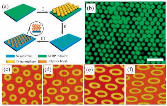
Li Y.F., Zhang J.H., Fang L.P.. Polymer brush nanopatterns with controllable features for protein pattern applications[J]. J.Mater.Chem., 2012,22:25116-25122. doi: 10.1039/c2jm35197h
Liu W.D., Li Y.F., Wang T.Q.. Elliptical polymer brush ring array mediated protein patterning and cell adhesion on patterned protein surfaces[J]. ACS Appl. Mater.Interfaces, 2013,5:12587-12593. doi: 10.1021/am403808s
Liu W.D., Liu X.Y., Ge P.. Hierarchical-multiplex DNA patterns mediated by polymer brush nanocone arrays that possess potential application for specific DNA sensing[J]. ACS Appl.Mater.Interfaces, 2015,7:24760-24771. doi: 10.1021/acsami.5b07577
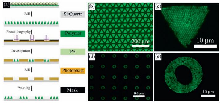
Li Y.F., Zhang J.H., Liu W.D.. Hierarchical polymer brush nanoarrays:a versatile way to prepare multiscale patterns of proteins[J]. ACS Appl.Mater. Interfaces, 2013,5:2126-2132. doi: 10.1021/am3031757
Xia Y.N., Whitesides G.M.. Soft lithography[J]. Annu.Rev.Mater.Sci., 1998,28:153-184. doi: 10.1146/annurev.matsci.28.1.153
Hoff J.D., Cheng L.-J., E.Meyhöfer , Guo L.J., Hunt A.J.. Nanoscale protein patterning by imprint lithography[J]. Nano Lett., 2004,4:853-857. doi: 10.1021/nl049758x
Seo S., Lee J., Kim K.-S.. Anisotropic adhesion of micropillars with spatula pads[J]. ACS Appl.Mater.Interfaces, 2014,6:1345-1350. doi: 10.1021/am4044135
Lebib A., Chen Y., Bourneix J.. Nanoimprint lithography for a large area pattern replication[J]. Microelectron.Eng., 1999,46:319-322. doi: 10.1016/S0167-9317(99)00094-5
Tsioris K., Tao H., Liu M.K.. Rapid transfer-based micropatterning and dry etching of silk microstructures[J]. Adv.Mater., 2011,23:2015-2019. doi: 10.1002/adma.201004771
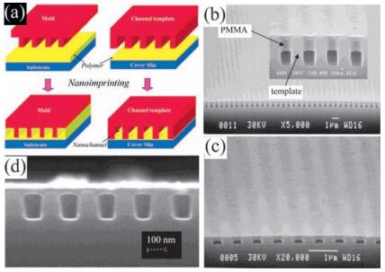
Guo L.J., Cheng X., Chou C.-F.. Fabrication of size-controllable nanofluidic channels by nanoimprinting and its application for DNA stretching[J]. Nano Lett., 2004,4:69-73. doi: 10.1021/nl034877i
Yim E.K.F., Reano R.M., Pang S.W.. Nanopattern-induced changes in morphology and motility of smooth muscle cells[J]. Biomaterials, 2005,26:5405-5413. doi: 10.1016/j.biomaterials.2005.01.058
Kolodziej C.M., Kim S.H., Broyer R.M.. Combination of integrin-binding peptide and growth factor promotes cell adhesion on electron-beam-fabricated patterns[J]. J.Am.Chem.Soc., 2012,134:247-255. doi: 10.1021/ja205524x
Broers A.N., Hoole A.C.F., Ryan J.M.. Electron beam lithography-resolution limits[J]. Microelectron.Eng., 1996,32:131-142. doi: 10.1016/0167-9317(95)00368-1
Kolodziej C.M., Maynard H.D.. Electron-beam lithography for patterning biomolecules at the micron and nanometer scale[J]. Chem.Mater., 2012,24:774-780. doi: 10.1021/cm202669f
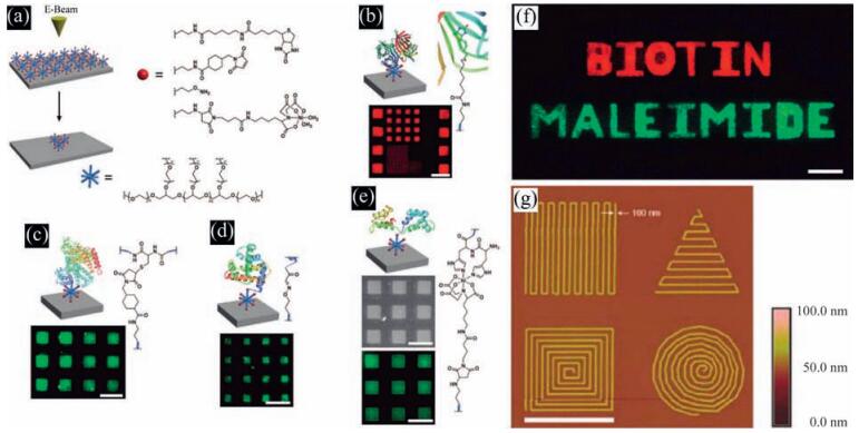
Christman K.L., Schopf E., Broyer R.M.. Positioning multiple proteins at the nanoscale with electron beam cross-linked functional polymers[J]. J.Am. Chem.Soc., 2009,131:521-527. doi: 10.1021/ja804767j
Christman K.L., Vázquez-Dorbatt V., Schopf E.. Nanoscale growth factor patterns by immobilization on a heparin-mimicking polymer[J]. J.Am.Chem. Soc., 2008,130:16585-16591. doi: 10.1021/ja803676r
Schlapak R., Danzberger J., Haselgrübler T.. Painting with biomolecules at the nanoscale:biofunctionalization with tunable surface densities[J]. Nano Lett., 2012,12:1983-1989. doi: 10.1021/nl2045414
Bat E., Lee J., Lau U.Y., Maynard H.D.. Trehalose glycopolymer resists allow direct writing of protein patterns by electron-beam lithography[J]. Nat. Commun., 2015,66654. doi: 10.1038/ncomms7654
Kim S., Marelli B., Brenckle M.A.. All-water-based electron-beam lithography using silk as a resist[J]. Nat.Nanotechnol., 2014,9:306-310. doi: 10.1038/nnano.2014.47
Feng C.L., Embrechts A., Bredebusch I.. Reactive microcontact printing on block copolymer films:exploiting chemistry in microcontacts for sub-micrometer patterning of biomolecules[J]. Adv.Mater., 2007,19:286-290. doi: 10.1002/(ISSN)1521-4095
Strulson M.K., Maurer J.A.. Microcontact printing for creation of patterned lipid bilayers on tetraethylene glycol self-assembled monolayers[J]. Langmuir, 2011,27:12052-12057. doi: 10.1021/la201839w
Feng C.L., Vancso G.J.. H.Schönherr, Fabrication of robust biomolecular patterns by reactive microcontact printing on N-hydroxysuccinimide ester-containing polymer films[J]. Adv.Funct.Mater., 2006,16:1306-1312. doi: 10.1002/(ISSN)1616-3028
Kumar A., Whitesides G.M.. Features of gold having micrometer to centimeter dimensions can be formed through a combination of stamping with an elastomeric stamp and an alkanethiol ink followed by chemical etching[J]. Appl. Phys.Lett., 1993,63:2002-2004. doi: 10.1063/1.110628
Renault J.P., Bernard A., Bietsch A.. Fabricating arrays of single protein molecules on glass using microcontact printing[J]. J.Phys.Chem.B, 2003,107:703-711. doi: 10.1021/jp0263424
Jang M.J., Nam Y.. Aqueous micro-contact printing of cell-adhesive biomolecules for patterning neuronal cell cultures[J]. BioChip J., 2012,6:107-113. doi: 10.1007/s13206-012-6201-9
Bernard A., Renault J.P., Michel B.. Microcontact printing of proteins[J]. Adv.Mater., 2000,12:1067-1070. doi: 10.1002/(ISSN)1521-4095
Piner R.D., Zhu J., Xu F., Hong S.H., Mirkin C.A.. Dip-pen nanolithography[J]. Science, 1999,283:661-663. doi: 10.1126/science.283.5402.661
Wu C.-C., Reinhoudt D.N., Otto C., Subramaniam V., Velders A.H.. Strategies for patterning biomolecules with dip-pen nanolithography[J]. Small, 2011,7:989-1002. doi: 10.1002/smll.201001749
Salazar R.B., Shovsky A., Schänherr H., Vancso G.J.. Dip-pen nanolithography on(bio)reactive monolayer and block-copolymer platforms:deposition of lines of single macromolecules[J]. Small, 2006,2:1274-1282. doi: 10.1002/(ISSN)1613-6829
Garcia R., Knoll A.W., Riedo E.. Advanced scanning probe lithography[J]. Nat. Nanotechnol., 2014,9:577-587. doi: 10.1038/nnano.2014.157
Hong S., Zhu J., Mirkin C.A.. Multiple ink nanolithography:toward a multiple-pen nano-plotter[J]. Science, 1999,286:523-525. doi: 10.1126/science.286.5439.523
Mirkin C.A.. The power of the pen:development of massively parallel dip-pen nanolithography[J]. ACS Nano, 2007,1:79-83. doi: 10.1021/nn700228m
Salaita K., Lee S.W., Wang X.F.. Sub-100 nm centimeter-scale, parallel dip-pen nanolithography[J]. Small, 2005,1:940-945. doi: 10.1002/(ISSN)1613-6829
Salaita K., Wang Y.H., Fragala J.. Massively parallel dip-pen nanolithography with 55000-pen two-dimensional arrays[J]. Angew.Chem. Int.Ed., 2006,45:7220-7223. doi: 10.1002/(ISSN)1521-3773
Lee K.-B., Park S.-J., Mirkin C.A., Smith J.C., Mrksich M.. Protein nanoarrays generated by dip-pen nanolithography[J]. Science, 2002,295:1702-1705. doi: 10.1126/science.1067172
Hong S., Mirkin C.A.. A nanoplotter with both parallel and serial writing capabilities[J]. Science, 2000,288:1808-1811. doi: 10.1126/science.288.5472.1808
Zheng Z.J., Daniel W.L., Giam L.R.. Multiplexed protein arrays enabled by polymer pen lithography:addressing the inking challenge[J]. Angew.Chem.Int. Ed., 2009,121:7762-7765. doi: 10.1002/ange.v121:41
Lenhert S., Sun P., Wang Y.H., Fuchs H., Mirkin C.A.. Massively parallel dip-pen nanolithography of heterogeneous supported phospholipid multilayer patterns[J]. Small, 2007,3:71-75. doi: 10.1002/(ISSN)1613-6829
Lim J.-H., Mirkin C.A.. Electrostatically driven dip-pen nanolithography of conducting polymers[J]. Adv.Mater., 2002,14:1474-1477. doi: 10.1002/1521-4095(20021016)14:20 & lt; 1474::AID-ADMA1474 & gt; 3.0.CO; 2-2
Senesi A.J., Rozkiewicz D.I., Reinhoudt D.N., Mirkin C.A.. Agarose-assisted dip-pen nanolithography of oligonucleotides and proteins[J]. ACS Nano, 2009,3:2394-2402. doi: 10.1021/nn9005945
Fais M., Karamanska R., Russell D.A., Field R.A.. Lectin and carbohydrate microarrays:new high-throughput methods for glycoprotein carbohydrate-binding protein and carbohydrate-active enzyme analysis[J]. J.Cereal Sci., 2009,50:306-311. doi: 10.1016/j.jcs.2009.06.010
Kong H., Liu D., Zhang S.C., Zhang X.R.. Protein sensing and cell discrimination using a sensor array based on nanomaterial-assisted chemiluminescence[J]. Anal.Chem., 2011,83:1867-1870. doi: 10.1021/ac200076c
Chang G., Mori Y., Mori S.. Microarray of human P450 with an integrated oxygen sensing film for high-throughput detection of metabolic activities[J]. Anal.Chem., 2012,84:5292-5297. doi: 10.1021/ac300355w
Baldini L., Wilson A.J., Hong J., Hamilton A.D.. Pattern-based detection of different proteins using an array of fluorescent protein surface receptors[J]. J. Am.Chem.Soc., 2004,126:5656-5657. doi: 10.1021/ja039562j
Liu Y.S., Hu W.H., Lu Z.S., Li C.M. ZnO nanomulberry and its significant nonenzymatic signal enhancement for protein microarray[J]. ACS Appl.Mater. Interfaces, 2014,6:7728-7734. doi: 10.1021/am501015p
Zhao Y.J., Zhao X.W., Pei X.P.. Multiplex detection of tumor markers with photonic suspension array[J]. Anal.Chim.Acta, 2009,633:103-108. doi: 10.1016/j.aca.2008.11.035
Song S.Y., Han Y.D., Hong S.Y.. Chip-based cartilage oligomeric matrix protein detection in serum and synovialfluid for osteoarthritis diagnosis[J]. Anal.Biochem., 2012,420:139-146. doi: 10.1016/j.ab.2011.09.012
Zhu Q.D., Trau D.. Multiplex detection platform for tumor markers and glucose in serum based on a microfluidic microparticle array[J]. Anal.Chim. Acta, 2012,751:146-154. doi: 10.1016/j.aca.2012.09.007
Jagt R.B.C., Gémez-Biagi R.F., Nitz M.. Pattern-based recognition of heparin contaminants by an array of self-assembling fluorescent receptors[J]. Angew. Chem.Int.Ed., 2009,48:1995-1997. doi: 10.1002/anie.v48:11
Wang D.N.. Carbohydrate microarrays[J]. Proteomics, 2003,3:2167-2175. doi: 10.1002/(ISSN)1615-9861
Im H., Bantz K.C., Lee S.H.. Self-assembled plasmonic nanoring cavity arrays for SERS and LSPR biosensing[J]. Adv.Mater., 2013,25:2678-2685. doi: 10.1002/adma.v25.19
Li X.N., Wen F., Creran B.. Colorimetric protein sensing using catalytically amplified sensor arrays[J]. Small, 2012,8:3589-3592. doi: 10.1002/smll.v8.23
Chang M.-J., Pang C.-R., Liu J.. High spatial resolution label-free detection of antigen-antibody binding on patterned surface by imaging ellipsometry[J]. J.Colloid Interface Sci., 2011,360:826-833. doi: 10.1016/j.jcis.2011.04.107
Wu Z.F., Yang P.. Simple multipurpose surface functionalization by phase transited protein adhesion[J]. Adv.Mater.Interfaces, 2015,21400401. doi: 10.1002/admi.201400401
Linman M.J., Yu H., Chen X., Cheng Q.. Fabrication and characterization of a sialoside-based carbohydrate microarray biointerface for protein binding analysis with surface plasmon resonance imaging[J]. ACS Appl.Mater. Interfaces, 2009,1:1755-1762. doi: 10.1021/am900290g
Son K.J., Kim S., Kim J.-H.. Dendrimer porphyrin-terminated polyelectrolyte multilayer micropatterns for a protein microarray with enhanced sensitivity[J]. J.Mater.Chem., 2010,20:6531-6538. doi: 10.1039/c0jm00498g
Nam K., Eom K., Yang J.. Aptamer-functionalized nano-pattern based on carbon nanotube for sensitive, selective protein detection[J]. J.Mater.Chem., 2012,22:23348-23356. doi: 10.1039/c2jm33688j
Wang H., Xu Q.H., Shang L.R.. Boronate affinity molecularly imprinted inverse opal particles for multiple label-free bioassays[J]. Chem.Commun., 2016,52:3296-3299. doi: 10.1039/C5CC09371F
Walt D.R.. Protein measurements in microwells[J]. Lab Chip, 2014,14:3195-3200. doi: 10.1039/C4LC00277F
Lu W.B., Fu C., Chen Y.. Multiplex detection of B-type natriuretic peptide, cardiac troponin I and C-reactive protein with photonic suspension array[J]. PLoS One, 2012,7e41448. doi: 10.1371/journal.pone.0041448
Zhou H.C., Baldini L., Hong J., Wilson A.J., Hamilton A.D.. Pattern recognition of proteins based on an array of functionalized porphyrins[J]. J.Am.Chem.Soc., 2006,128:2421-2425. doi: 10.1021/ja056833c
Morales-Narváez E., Guix M., Medina-Sánchez M., Mayorga-Martinez C.C., Merkoći A.. Micromotor enhanced microarray technology for protein detection[J]. Small, 2014,10:2542-2548. doi: 10.1002/smll.v10.13
Mu Z.D., Zhao X.W., Huang Y., Lu M., Gu Z.Z.. Photonic crystal hydrogel enhanced plasmonic staining for multiplexed protein analysis[J]. Small, 2015,11:6036-6043. doi: 10.1002/smll.201501829
Yuan J.J., Zhao X.W., Wang X.X., Gu Z.Z.. Image decoding of photonic crystal beads array in the microfluidic chip for multiplex assays[J]. Sci.Rep., 2014,46755.
Gaster R.S., Hall D.A., Wang S.X. Autoassembly protein arrays for analyzing antibody cross-reactivity[J]. Nano Lett., 2011,11:2579-2583. doi: 10.1021/nl1026056
Zhang X.M., Li Z.B., Ye S.S.. Elevated Ag nanohole arrays for high performance plasmonic sensors based on extraordinary optical transmission[J]. J.Mater.Chem., 2012,22:8903-8910. doi: 10.1039/c2jm30525a
Ye S.S., Zhang X.M., Chang L.X.. High-performance plasmonic sensors based on two-dimensional Ag nanowell crystals[J]. Adv.Opt.Mater., 2014,2:779-787. doi: 10.1002/adom.v2.8
Lau U.Y., Saxer S.S., Lee J., Bat E., Maynard H.D.. Direct write protein patterns for multiplexed cytokine detection from live cells using electron beam lithography[J]. ACS Nano, 2016,10:723-729. doi: 10.1021/acsnano.5b05781
Hu W.H., Liu Y.S., Lu Z.S., Li C.M.. Poly[J]. Adv.Funct.Mater., 2010,20:3497-3503. doi: 10.1002/adfm.v20:20
Duan R.X., Zuo X.L., Wang S.T.. Lab in a tube:ultrasensitive detection of microRNAs at the single-cell level and in breast cancer patients using quadratic isothermal amplification[J]. J.Am.Chem.Soc., 2013,135:4604-4607. doi: 10.1021/ja311313b
Schlapak R., Danzberger J., Armitage D.. Nanoscale DNA tetrahedra improve biomolecular recognition on patterned surfaces[J]. Small, 2012,8:89-97. doi: 10.1002/smll.201101576
Zhao Y.J., Zhao X.W., Tang B.C.. Quantum-dot-tagged bioresponsive hydrogel suspension array for multiplex label-free DNA detection[J]. Adv.Funct. Mater., 2010,20:976-982. doi: 10.1002/adfm.200901812
Bajaj A., Miranda O.R., Phillips R.. Array-based sensing of normal cancerous, and metastatic cells using conjugated fluorescent polymers[J]. J.Am. Chem.Soc., 2010,132:1018-1022. doi: 10.1021/ja9061272
Siltanen C., Shin D.-S., Sutcliffe J.. Revzin A.Micropatterned photodegradable hydrogels for the sorting of microbeads and cells[J]. Angew.Chem.Int.Ed., 2013,52:9224-9228. doi: 10.1002/anie.201303965
Toccafondi C., Thorat S., R.La Rocca. Multifunctional substrates of thin porous alumina for cell biosensors[J]. J.Mater.Sci.:Mater.Med., 2014,25:2411-2420.
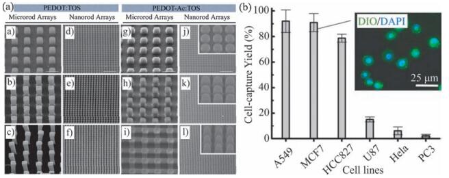
Wang S.T., Wang H., Jiao J.J.. Three-dimensional nanostructured substrates toward efficient capture of circulating tumor cells[J]. Angew.Chem. Int.Ed., 2009,121:9132-9135. doi: 10.1002/ange.v121:47
Lin M., Chen J.-F., Lu Y.-T.. Nanostructure embedded microchips for detection isolation, and characterization of circulating tumor cells[J]. Acc. Chem.Res., 2014,47:2941-2950. doi: 10.1021/ar5001617
Amin Y.Y.I., Runager K., Simoes F.. Combinatorial biomolecular nanopatterning for high-throughput screening of stem-cell behavior[J]. Adv. Mater., 2016,28:1472-1476. doi: 10.1002/adma.v28.7
Zhao S., Zhao H., Zhang X.Y., Li Y.Q., Du Y.. Off-the-shelf microsponge arrays for facile and efficient construction of miniaturized 3D cellular microenvironments for versatile cell-based assays[J]. Lab Chip, 2013,13:2350-2358. doi: 10.1039/c3lc50183c
Zhang B., Cai Y.L., Shang L.R.. A photonic crystal hydrogel suspension array for the capture of blood cells from whole blood[J]. Nanoscale, 2016,8:3841-3847. doi: 10.1039/C5NR06368J
Jin J., Xing Y., Xi Y.. A triggered DNA hydrogel cover to envelop and release single cells[J]. Adv.Mater., 2013,25:4714-4717. doi: 10.1002/adma.v25.34
Kumashiro Y., Ishihara J., Umemoto T.. Stripe-patterned thermo-responsive cell culture dish for cell separation without cell labeling[J]. Small, 2015,11:681-687. doi: 10.1002/smll.201400787
Chen L., Liu X.L., Su B.. Aptamer-mediated efficient capture and release of T lymphocytes on nanostructured surfaces[J]. Adv.Mater., 2011,23:4376-4380. doi: 10.1002/adma.201102435
Zhang F.L., Jiang Y., Liu X.L.. Hierarchical nanowire arrays as three-dimensional fractal nanobiointerfaces for high efficient capture of cancer cells[J]. Nano Lett., 2016,16:766-772. doi: 10.1021/acs.nanolett.5b04731
Hsiao Y.-S., Luo S.-C., Hou S.. 3D bioelectronic interface:capturing circulating tumor cells onto conducting polymer-based micro/nanorod arrays with chemical and topographical control[J]. Small, 2014,10:3012-3017. doi: 10.1002/smll.v10.15
Custódio C.A., San Miguel-Arranz V., Gropeanu R.A.. Photopatterned antibodies for selective cell attachment[J]. Langmuir, 2014,30:10066-10071. doi: 10.1021/la502688h
Meng J.X., Zhang P.C., Zhang F.L.. A self-cleaning TiO2 nanosisal-like coating toward disposing nanobiochips of cancer detection[J]. ACS Nano, 2015,9:9284-9291. doi: 10.1021/acsnano.5b04230
Liu X.L., Wang S.T.. Three-dimensional nano-biointerface as a new platform for guiding cell fate[J]. Chem.Soc.Rev., 2014,43:2385-2401. doi: 10.1039/c3cs60419e
Ye X.Z., Qi L.M.. Two-dimensionally patterned nanostructures based on monolayer colloidal crystals:controllable fabrication, assembly, and applications[J]. Nano Today, 2011,6:608-631. doi: 10.1016/j.nantod.2011.10.002
Shao Y., Sang J.M., Fu J.P.. On human pluripotent stem cell control:the rise of 3D bioengineering and mechanobiology[J]. Biomaterials, 2015,52:26-43. doi: 10.1016/j.biomaterials.2015.01.078
Khan F., Tanaka M., Ahmad S.R.. Fabrication of polymeric biomaterials:a strategy for tissue engineering and medical devices[J]. J.Mater.Chem.B, 2015,3:8224-8249. doi: 10.1039/C5TB01370D
Phong H.Q., Wang S.-L., Wang M.-J.. Cell behaviors on micro-patterned porous thin films[J]. Mater.Sci.Eng.B, 2010,169:94-100. doi: 10.1016/j.mseb.2010.01.009
Skorb E.V., Andreeva D.V.. Surface nanoarchitecture for bio-applications: self-regulating intelligent interfaces[J]. Adv.Funct.Mater., 2013,23:4483-4506. doi: 10.1002/adfm.v23.36
Zhang Y., Gordon A., Qian W.Y., Chen W.Q. Engineering nanoscale stem cell niche:direct stem cell behavior at cell-matrix interface[J]. Adv.Healthc.Mater., 2015,4:1900-1914. doi: 10.1002/adhm.201500351
Kim H.N., Jiao A., Hwang N.S.. Nanotopography-guided tissue engineering and regenerative medicine[J]. Adv.Drug Deliv.Rev., 2013,65:536-558. doi: 10.1016/j.addr.2012.07.014
Schwarz U.S., Nelson C.M., Silberzan P.. Proteins, cells, and tissues in patterned environments[J]. Soft Matter, 2014,10:2337-2340. doi: 10.1039/c4sm90028f
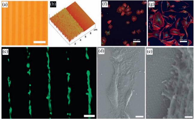
Chen S.S., Lu X.M., Hu Y., Lu Q.H.. Biomimetic honeycomb-patterned surface as the tunable cell adhesion scaffold[J]. Biomater.Sci., 2015,3:85-93. doi: 10.1039/C4BM00233D
Premnath P., Tavangar A., Tan B., Venkatakrishnan K.. Tuning cell adhesion by direct nanostructuring silicon into cell repulsive/adhesive patterns[J]. Exp.Cell Res., 2015,337:44-52. doi: 10.1016/j.yexcr.2015.07.028
Meng F.W., Hlady V., Tresco P.A. Inducing alignment in astrocyte tissue constructs by surface ligands patterned on biomaterials[J]. Biomaterials, 2012,33:1323-1335. doi: 10.1016/j.biomaterials.2011.10.034
Bae W.-G., Kim J., Choung Y.-H.. Bio-inspired configurable multiscale extracellular matrix-like structures for functional alignment and guided orientation of cells[J]. Biomaterials, 2015,69:158-164. doi: 10.1016/j.biomaterials.2015.08.006
Subramani C., Saha K., Creran B.. Cell alignment using patterned biocompatible gold nanoparticle templates[J]. Small, 2012,8:1209-1213. doi: 10.1002/smll.201102405
Zhou X.T., Shi J., Zhang F.. Reversed cell imprinting, AFM imaging and adhesion analyses of cells on patterned surfaces[J]. Lab Chip, 2010,10:1182-1188. doi: 10.1039/b926325j
van Hoorn H., Harkes R., Spiesz E.M.. The nanoscale architecture of force-bearing focal adhesions[J]. Nano Lett., 2014,14:4257-4262. doi: 10.1021/nl5008773
Gautrot J.E., Trappmann B., Oceguera-Yanez F.. Exploiting the superior protein resistance of polymer brushes to control single cell adhesion and polarisation at the micron scale[J]. Biomaterials, 2010,31:5030-5041. doi: 10.1016/j.biomaterials.2010.02.066
Théry M., Racine V., Piel M.. Anisotropy of cell adhesive microenvironment governs cell internal organization and orientation of polarity[J]. Proc.Natl.Acad.Sci.U.S.A., 2006,103:19771-19776. doi: 10.1073/pnas.0609267103
Zhang J.-T., Nie J.Q., Mühlstädt M.. Stable extracellular matrix protein patterns guide the orientation of osteoblast-like cells[J]. Adv.Funct.Mater., 21,2011:4079-4087.
Jiang X.Y., Bruzewicz D.A., Wong A.P., Piel M., Whitesides G.M.. Directing cell migration with asymmetric micropatterns[J]. Proc.Natl.Acad.Sci.U.S.A., 2005,102:975-978. doi: 10.1073/pnas.0408954102
Kim D.-H., Seo C.-H., Han K.. Guided cell migration on microtextured substrates with variable local density and anisotropy[J]. Adv.Funct.Mater., 2009,19:1579-1586. doi: 10.1002/adfm.v19:10
Kim D.-H., Han K., Gupta K.. Mechanosensitivity of fibroblast cell shape and movement to anisotropic substratum topography gradients[J]. Biomaterials, 2009,30:5433-5444. doi: 10.1016/j.biomaterials.2009.06.042
Tseng P., Carlo D.D.. Substrates with patterned extracellular matrix and subcellular stiffness gradients reveal local biomechanical responses[J]. Adv. Mater., 2014,26:1242-1247. doi: 10.1002/adma.v26.8
Li Y., Jiang X.Q., Zhong H.X.. Hierarchical patterning of cells with a microeraser and electrospun nanofibers[J]. Small, 2016,12:1230-1239. doi: 10.1002/smll.v12.9
Benson K., Prasetyanto E.A., Galla H.-J., Kehr N.S.. Self-assembled monolayers of bifunctional periodic mesoporous organosilicas for cell adhesion and cellular patterning[J]. Soft Matter, 2012,8:10845-10852. doi: 10.1039/c2sm26563j
Jang J.-W., Collins J.M., Nettikadan S.. User-friendly universal and durable subcellular-scaled template for protein binding:application to single-cell patterning[J]. Adv.Funct.Mater., 2013,23:5840-5845. doi: 10.1002/adfm.v23.47
You J., Yoshida A., Heo J.S.. Protein coverage on polymer nanolayers leading to mesenchymal stem cell patterning[J]. Phys.Chem.Chem.Phys., 2011,13:17625-17632. doi: 10.1039/c1cp21732a
Matsui T., Arima Y., Takemoto N., Iwata H.. Cell patterning on polylactic acid through surface-tethered oligonucleotides[J]. Acta Biomater., 2015,13:32-41. doi: 10.1016/j.actbio.2014.11.011
Seo H., Choi I., Lee J.. Facile method for development of ligand-patterned substrates induced by a chemical reaction[J]. Chem.Eur.J., 2011,17:5804-5807. doi: 10.1002/chem.v17.21
Li B.Q., Wang L., Xu F.. UV-crosslinkable and injectable chitosan for patterned cell-laden microgel and rapid transdermal curing hydrogel in vivo[J]. Acta Biomater., 2015,22:59-69. doi: 10.1016/j.actbio.2015.04.026
Fisher O.Z., Khademhosseini A., Langer R., Peppas N.A.. Bioinspired materials for controlling stem cell fate[J]. Acc.Chem.Res., 2010,43:419-428. doi: 10.1021/ar900226q
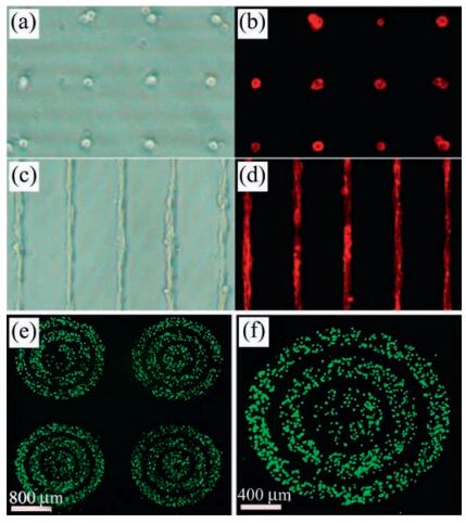
Xin H.B., Li Y.C., Li B.J. Controllable patterning of different cells via optical assembly of 1D periodic cell structures[J]. Adv.Funct.Mater., 2015,25:2816-2823. doi: 10.1002/adfm.201500287
Tuft B.W., Zhang L.C., Xu L.J.. Material stiffness effects on neurite alignment to photopolymerized micropatterns[J]. Biomacromolecules, 2014,15:3717-3727. doi: 10.1021/bm501019s
Yang K., Jung H., Lee H.-R.. Multiscale, hierarchically patterned topography for directing human neural stem cells into functional neurons[J]. ACS Nano, 2014,8:7809-7822. doi: 10.1021/nn501182f
Swarup V.P., Hsiao T.W., Zhang J.X.. Exploiting differential surface display of chondroitin sulfate variants for directing neuronal outgrowth[J]. J. Am.Chem.Soc., 2013,135:13488-13494. doi: 10.1021/ja4056728
Tuft B.W., Xu L.J., White S.P.. Neural pathfinding on uni-and multidirectional photopolymerized micropatterns[J]. ACS Appl.Mater. Interfaces, 2014,6:11265-11276. doi: 10.1021/am501622a
Jia C., Yu D., Lamarre M.. Patterned electrospun nanofiber matrices via localized dissolution:potential for guided tissue formation[J]. Adv.Mater., 2014,26:8192-8197. doi: 10.1002/adma.201403509
Wittenbrink I., Hausmann A., Schickle K.. Low-aspect ratio nanopatterns on bioinert alumina influence the response and morphology of osteoblast-like cells[J]. Biomaterials, 2015,62:58-65. doi: 10.1016/j.biomaterials.2015.05.026
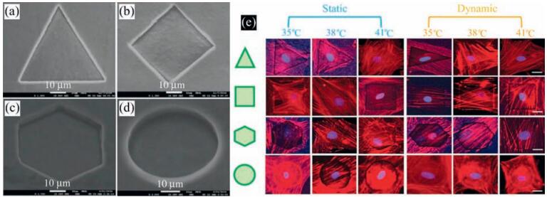
Gong T., Lu L.X., Liu D.. Dynamically tunable polymer microwells for directing mesenchymal stem cell differentiation into osteogenesis[J]. J.Mater. Chem.B, 2015,3:9011-9022.
Zhao C.C., Xia L.G., Zhai D.. Designing ordered micropatterned hydroxyapatite bioceramics to promote the growth and osteogenic differentiation of bone marrow stromal cells[J]. J.Mater.Chem.B, 2015,3:968-976.
Tien L.W., Gil E.S., Park S.-H., Mandal B.B., Kaplan D.L.. Patterned silk film scaffolds for aligned lamellar bone tissue engineering[J]. Macromol.Biosci., 2012,12:1671-1679. doi: 10.1002/mabi.201200193
Kim J., Bae W.-G., Lim K.-T.. Density of nanopatterned surfaces for designing bone tissue engineering scaffolds[J]. Mater.Lett., 2014,130:227-231. doi: 10.1016/j.matlet.2014.05.107
Marino A., Ciofani G., Filippeschi C.. Two-photon polymerization of sub-micrometric patterned surfaces:investigation of cell-substrate interactions and improved differentiation of neuron-like cells[J]. ACS Appl.Mater.Interfaces, 2013,5:13012-13021. doi: 10.1021/am403895k
Limongi T., Cesca F., Gentile F.. Nanostructured superhydrophobic substrates trigger the development of 3D neuronal networks[J]. Small, 2013,9:402-412. doi: 10.1002/smll.201201377
Dalby M.J., Gadegaard N., Tare R.. The control of human mesenchymal cell differentiation using nanoscale symmetry and disorder[J]. Nat.Mater., 2007,6:997-1003. doi: 10.1038/nmat2013
Tserepi A., Gogolides E., Bourkoula A.. Plasma nanotextured polymeric surfaces for controlling cell attachment and proliferation:a short review[J]. Plasma Chem.Plasma Process., 2016,36:107-120. doi: 10.1007/s11090-015-9674-1
Bettinger C.J., Langer R., Borenstein J.T.. Engineering substrate topography at the micro-and nanoscale to control cell function[J]. Angew.Chem.Int.Ed., 2009,48:5406-5415. doi: 10.1002/anie.v48:30
Tay C.Y., Irvine S.A., Boey F.Y.C., Tan L.P., Venkatraman S.. Micro-/nano-engineered cellular responses for soft tissue engineering and biomedical applications[J]. Small, 2011,7:1361-1378. doi: 10.1002/smll.201100046
Zheng T., Peelen D., Smith L.M.. Lectin arrays for profiling cell surface carbohydrate expression[J]. J.Am.Chem.Soc., 2005,127:9982-9983. doi: 10.1021/ja0505550
Chen C.S., Mrksich M., Huang S., Whitesides G.M., Ingber D.E.. Geometric control of cell life and death[J]. Science, 1997,276:1425-1428. doi: 10.1126/science.276.5317.1425
Théry M., Racine V., Pépin A.. The extracellular matrix guides the orientation of the cell division axis[J]. Nat.Cell Biol., 2005,7:947-953. doi: 10.1038/ncb1307
Zhang P.C., Wang S.T.. Designing fractal nanostructured biointerfaces for biomedical applications[J]. ChemPhysChem, 2014,15:1550-1561. doi: 10.1002/cphc.201301230
Yan L., Yang Y., Zhang W.J., Chen X.F.. Advanced materials and nanotechnology for drug delivery[J]. Adv.Mater., 2014,26:5533-5540. doi: 10.1002/adma.201305683
Cahill D.J.. Protein and antibody arrays and their medical applications[J]. J. Immunol.Methods, 2001,250:81-91. doi: 10.1016/S0022-1759(01)00325-8
Chen X.F., Corbett H.J., Yukiko S.R.. Site-selectively coated, densely-packed microprojection array patches for targeted delivery of vaccines to skin[J]. Adv.Funct.Mater., 2011,21:464-473. doi: 10.1002/adfm.v21.3
Chen X.F., Zhu G.Y., Yang Y.. A diamond nanoneedle array for potential high-throughput intracellular delivery[J]. Adv.Healthc.Mater., 2013,2:1103-1107. doi: 10.1002/adhm.v2.8
DeMuth P.C., Min Y., Irvine D.J., Hammond P.T.. Implantable silk composite microneedles for programmable vaccine release kinetics and enhanced immunogenicity in transcutaneous immunization[J]. Adv.Healthc.Mater., 2014,3:47-58. doi: 10.1002/adhm.v3.1
Rasekh M., Ahmad Z., Day R., Wickham A., Edirisinghe M.. Direct writing of polycaprolactone polymer for potential biomedical engineering applications[J]. Adv.Eng.Mater., 2011,13:B296-B305. doi: 10.1002/adem.201080126
Liu W.D., Liu X.Y., Fangteng J.. Bioinspired polyethylene terephthalate nanocone arrays with underwater superoleophobicity and anti-bioadhesion properties[J]. Nanoscale, 2014,6:13845-13853. doi: 10.1039/C4NR04471A
Luz G.M., Boesel L., del Campo A., Mano J.F.. Micropatterning of bioactive glass nanoparticles on chitosan membranes for spatial controlled biomineralization[J]. Langmuir, 2012,28:6970-6977. doi: 10.1021/la300667g
You J., Shin D.-S., Patel D., Gao Y.D.. Revzin A.Multilayered heparin hydrogel microwells for cultivation of primary hepatocytes[J]. Adv.Healthc.Mater., 2014,3:126-132. doi: 10.1002/adhm.v3.1
Sakimoto K.K., Liu C., Lim J., Yang P.D.. Salt-induced self-assembly of bacteria on nanowire arrays[J]. Nano Lett., 2014,14:5471-5476. doi: 10.1021/nl502946j
Deshuai Zhen , Chunlin Liu , Qiuhui Deng , Shaoqi Zhang , Ningman Yuan , Le Li , Yu Liu . A review of covalent organic frameworks for metal ion fluorescence sensing. Chinese Chemical Letters, 2024, 35(8): 109249-. doi: 10.1016/j.cclet.2023.109249
Ya-Ping Liu , Zhi-Rong Gui , Zhen-Wen Zhang , Sai-Kang Wang , Wei Lang , Yanzhu Liu , Qian-Yong Cao . A phenylphenthiazide anchored Tb(Ⅲ)-cyclen complex for fluorescent turn-on sensing of ClO−. Chinese Chemical Letters, 2025, 36(2): 109769-. doi: 10.1016/j.cclet.2024.109769
Chao Liu , Chao Jia , Shi-Xian Gan , Qiao-Yan Qi , Guo-Fang Jiang , Xin Zhao . A luminescent one-dimensional covalent organic framework for organic arsenic sensing in water. Chinese Chemical Letters, 2024, 35(11): 109750-. doi: 10.1016/j.cclet.2024.109750
Meihui Liu , Xinyuan Zhou , Xiao Li , Zhenjie Xue , Tie Wang . Pushing the frontiers: Chip-based detection based on micro- and nano-structures. Chinese Chemical Letters, 2024, 35(4): 108875-. doi: 10.1016/j.cclet.2023.108875
Zian Fang , Qianqian Wen , Yidi Wang , Hongxia Ouyang , Qi Wang , Qiuping Li . The Test Paper for Metal Ion: A Popular Science Experiment Based on Color Aesthetics. University Chemistry, 2024, 39(5): 108-115. doi: 10.3866/PKU.DXHX202310032
Yiming Yang , Lichao Sun , Qingfeng Zhang . Plasmonic nanocrystals with intrinsic chirality: Biomolecule-directed synthesis and applications. Chinese Journal of Structural Chemistry, 2025, 44(1): 100467-100467. doi: 10.1016/j.cjsc.2024.100467
Binhan Zhao , Zheng Li , Lan Zheng , Zhichao Ye , Yuyang Yuan , Shanshan Zhang , Bo Liang , Tianyu Li . Recent progress in the biomedical application of PEDOT:PSS hydrogels. Chinese Chemical Letters, 2024, 35(10): 109810-. doi: 10.1016/j.cclet.2024.109810
Rui Wang , Yuan Tian , Xuefeng Gao , Lei Jiang . Design and fabrication of triangle-pattern superwettability hybrid surface with high-efficiency condensation heat transfer performance. Chinese Chemical Letters, 2025, 36(3): 110395-. doi: 10.1016/j.cclet.2024.110395
Bingyang Lu , Dehui Wang , Junchang Guo , Yang Shen , Qian Feng , Jinlong Yang , Xiao Han , Huali Yu , Luohuizi Li , Jiaxin Liu , Jing Luo , Huan Liu , Zhongwei Zhang , Xu Deng . High-efficiency exudates drainage of anti-adhesion dressings for chronic wound. Chinese Chemical Letters, 2025, 36(4): 110601-. doi: 10.1016/j.cclet.2024.110601
Manoj Kumar Sarangi , L․D Patel , Goutam Rath , Sitansu Sekhar Nanda , Dong Kee Yi . Metal organic framework modulated nanozymes tailored with their biomedical approaches. Chinese Chemical Letters, 2024, 35(11): 109381-. doi: 10.1016/j.cclet.2023.109381
Yating Zheng , Yulan Huang , Jing Luo , Xuqi Peng , Xiran Gui , Gang Liu , Yang Zhang . Supercritical fluid technology: A game-changer for biomacromolecular nanomedicine preparation and biomedical application. Chinese Chemical Letters, 2024, 35(7): 109169-. doi: 10.1016/j.cclet.2023.109169
Shuaiwen Li , Zihui Chen , Feng Yang , Wanqing Yue . The age of vanadium-based nanozymes: Synthesis, catalytic mechanisms, regulation and biomedical applications. Chinese Chemical Letters, 2024, 35(4): 108793-. doi: 10.1016/j.cclet.2023.108793
Bharathi Natarajan , Palanisamy Kannan , Longhua Guo . Metallic nanoparticles for visual sensing: Design, mechanism, and application. Chinese Journal of Structural Chemistry, 2024, 43(9): 100349-100349. doi: 10.1016/j.cjsc.2024.100349
Quan Zhang , Shunjie Xing , Jingqian Han , Li Feng , Jianchun Li , Zhaosheng Qian , Jin Zhou . Organic pollutant sensing for human health based on carbon dots. Chinese Chemical Letters, 2025, 36(1): 110117-. doi: 10.1016/j.cclet.2024.110117
Liyong DU , Yi LIU , Guoli YANG . Preparation and triethylamine sensing performance of ZnSnO3/NiO heterostructur. Chinese Journal of Inorganic Chemistry, 2025, 41(4): 729-740. doi: 10.11862/CJIC.20240404
Ruikui YAN , Xiaoli CHEN , Miao CAI , Jing REN , Huali CUI , Hua YANG , Jijiang WANG . Design, synthesis, and fluorescence sensing performance of highly sensitive and multi-response lanthanide metal-organic frameworks. Chinese Journal of Inorganic Chemistry, 2024, 40(4): 834-848. doi: 10.11862/CJIC.20230301
Yu Deng , Yan Liu , Yonghui Deng , Jinsheng Cheng , Yidong Zou , Wei Luo . In situ sulfur-doped mesoporous tungsten oxides for gas sensing toward benzene series. Chinese Chemical Letters, 2024, 35(7): 108898-. doi: 10.1016/j.cclet.2023.108898
Ting WANG , Peipei ZHANG , Shuqin LIU , Ruihong WANG , Jianjun ZHANG . A Bi-CP-based solid-state thin-film sensor: Preparation and luminescence sensing for bioamine vapors. Chinese Journal of Inorganic Chemistry, 2024, 40(8): 1615-1621. doi: 10.11862/CJIC.20240134
Xiaxia Xing , Xiaoyu Chen , Zhenxu Li , Xinhua Zhao , Yingying Tian , Xiaoyan Lang , Dachi Yang . Polyethylene imine functionalized porous carbon framework for selective nitrogen dioxide sensing with smartphone communication. Chinese Chemical Letters, 2024, 35(9): 109230-. doi: 10.1016/j.cclet.2023.109230
Liangji Chen , Zhen Yuan , Fudong Feng , Xin Zhou , Zhile Xiong , Wuji Wei , Hao Zhang , Banglin Chen , Shengchang Xiang , Zhangjing Zhang . A hydrogen-bonded organic framework containing fluorescent carbazole and responsive pyridyl units for sensing organic acids. Chinese Chemical Letters, 2024, 35(9): 109344-. doi: 10.1016/j.cclet.2023.109344
 DownLoad: