图 1
The sensing mechanism of MNP toward thiols.
Figure 1.
The sensing mechanism of MNP toward thiols.
Anaphthalimide-based fluorescent probe for mercaptocontaining compounds
Lun Song , Xu-Dong Sun , Yu Ge , Yu-Hua Yao , Jie Shen , Wei-Bing Zhang , Jun-Hong Qian

Biothiols, including cysteine (Cys), homocysteine (Hcy), and glutathione (GSH), play pivotal roles in physiological and pathological events including redox homeostasis, biocatalysis, detoxification of xenobiotics, metal biding, signal transduction, etc. [1-3]. Abnormal levels of biothiols are thought to be implicated in various diseases, such as liver damage, cancer, hematopoiesis decrease, cardiovascular, Alzheimer's disease, and HIV [4-6]. Therefore, it is of intense interest to develop sensitive and selective methods for the detection of biothiols. Fluorescence technique is a frequently used method due to its advantages of the convenience, real time monitoring, in vivo and in vitro bioimaging. Many fluorescent probes have been developed for biothiols in view of their strong nucleophilicity, high binding affinity toward metal ions [7-9].
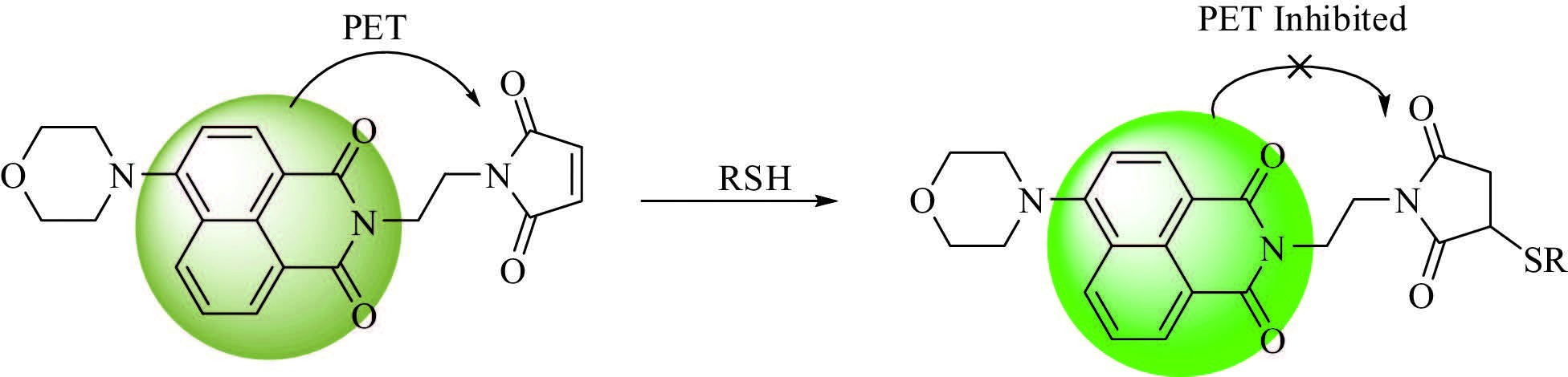
In the past few decades, maleimide is widely used as a receptor for thiols because of its merits of highly selective reaction with thiols, very fast response to mercapto-containing compounds and ease of being appended to different fluorophores. We present here a rationally designed fluorescent probe MNP for thiols. The probe consists of the following three units: 1) maleimide, a selective thiol receptor; 2) naphthalimide fluorophore with absorption and emission in visible region; 3) a morpholine molecule was incorporated into the 4-position of naphthalimide to improve the water solubility. The photoinduced electron transfer (PET) takes place from the fluorophore to the electron deficient maleimide receptor; therefore, MNP is expected to be weakly fluorescent. 1, 4-Michael addition of a mercapto group to the C=C in maleimide blocks the PET process, which leads to a significant fluorescence enhancement (Scheme 1). We envisioned that small weighted thiols and mercapto-containing proteins could be detected by MNP due to the high activity of maleimide.
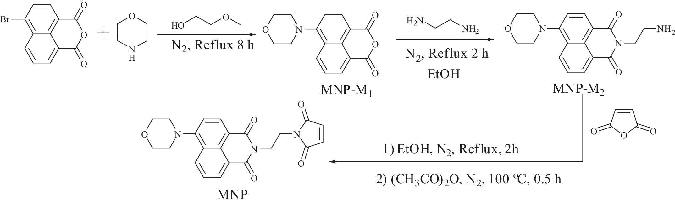
MNP was synthesized according to the following procedures (Scheme 2).
MNP-M1: 4-Bromine-1, 8-naphthalic anhydride (1.1 g, 4 mmol) and morpholine (1.4 g, 16 mmol) were added to 20 mL of ethylene glycol monomethyl ether. The mixture was refluxed for 4 h under a nitrogen atmosphere. The solvent was evaporated under vacuum. The remaining solid was purified through column chromatography (PE:DCM = 1:4, v/v) to give MNP-M1 as yellow powder, yield 71.2%. 1H NMR (CDCl3): d 8.60 (d, 1H, J = 7.2 Hz), 8.54 (d, 1H, J = 8.1 Hz), 8.48 (d, 1H, J = 8.5 Hz), 7.76 (t, 1H, J = 7.5 Hz), 7.26 (d, 1H, J = 8.0 Hz), 4.44 (t, 4H, J = 4.5 Hz), 4.04 (t, 1H, J = 4.1 Hz) (Fig. S1 in Supporting information).
MNP-M2: Ethanediamine (120 mg, 2 mmol) was dissolved in 5 mL EtOH, MNP-M1 (566 mg, 2 mmol) was dissolved in 10 mL EtOH. MNP-M1 solution was added dropwise into the ethanediamine solution. The mixture was refluxed under nitrogen atmosphere for 2 h. The solvent was evaporated under vacuum. The remaining solid was purified through column chromatography (DCM:MeOH:Et3N = 500:10:1, v/v/v) to give MNP-M2 as yellow powder, yield 60.9%. 1H NMR (CDCl3): d 8.60 (d, 1H, J = 7.2 Hz), 8.54 (d, 1H, J = 8.1 Hz), 8.43 (d, 1H, J = 8.4 Hz), 7.73 (t, 1H, J = 7.5 Hz), 7.24 (d, 1H, J = 8.0 Hz), 4.33 (t, 2H, J = 6.3 Hz), 4.03 (t, 4H, J = 3.8 Hz), 3.28 (t, 4H, J = 4.0 Hz), 3.14 (t, 2H, J = 6.3 Hz) (Fig. S1).
MNP: MNP-M2 (325.1 mg, 1 mmol) was dissolved in 8 mL anhydrous EtOH, maleic anhydride (98 mg, 1 mmol) was dissolved in 2 mL anhydrous EtOH. MNP-M2 solution was added dropwise into the maleic anhydride solution. The mixture was refluxed under nitrogen atmosphere for 2 h. The solvent was evaporated under vacuum. The remaining solid was dissolved in 5 mL acetic anhydride, and then sodium acetate (820 mg, 10 mmol) was added into the solution. The mixture was stirred at 100℃ for 0.5 h. After evaporating the solvent, the remaining solid was purified through column chromatography (DCM:MeOH:HOAc = 500:10:2, v/v/v) to give MNP as yellowy powder, yield 53.2%. 1H NMR (CDCl3): d 8.55 (d, 1H, J = 7.3 Hz), 8.49 (d, 1H, J = 8.0 Hz), 8.42 (d, 1H, J = 8.4 Hz), 7.70 (t, 1H, J = 7.8 Hz), 7.21 (d, 1H, J = 8.1 Hz), 6.62 (s, 1H), 4.42 (t, 2H, J = 4.6 Hz), 4.03 (t, 4H, J = 4.1 Hz), 4.00 (t, 2H, J = 5.1 Hz), 3.27 (t, 2H, J = 4.1 Hz) (Fig. S2 in Supporting information). 13C NMR (DMSO-d6): d 163.42, 163.24, 149.08, 148.94, 146.85, 145.40, 143.80, 139.31, 133.44, 132.70, 132.58, 131.75, 131.56, 130.45, 130.39, 129.88, 129.33, 129.18, 125.25, 123.40, 123.18, 44.88. ESI: calcd. for [M + H]+: 406.1403, found: 406.1400.
With probe MNP in hand, we first measured its spectral responses toward small weighted thiols.
Fig. S3a-S3b in Supporting information shows the absorption and emission spectra of MNP and MNP mixed with Cys. MNP is weakly fluorescent and absorbs at 405 nm. The addition of Cys led to about eight-fold fluorescence enhancement at 545 nm, however, the absorption spectrum almost kept the same. The fluorescence intensity of Cys-MNP system had no obvious change after 1 min (Fig. S3c), demonstrating that the reaction betweenMNPand Cys can be finished within 1 min. Similar spectral changes of MNP were observed when Hcy and GSH were added instead of Cys.
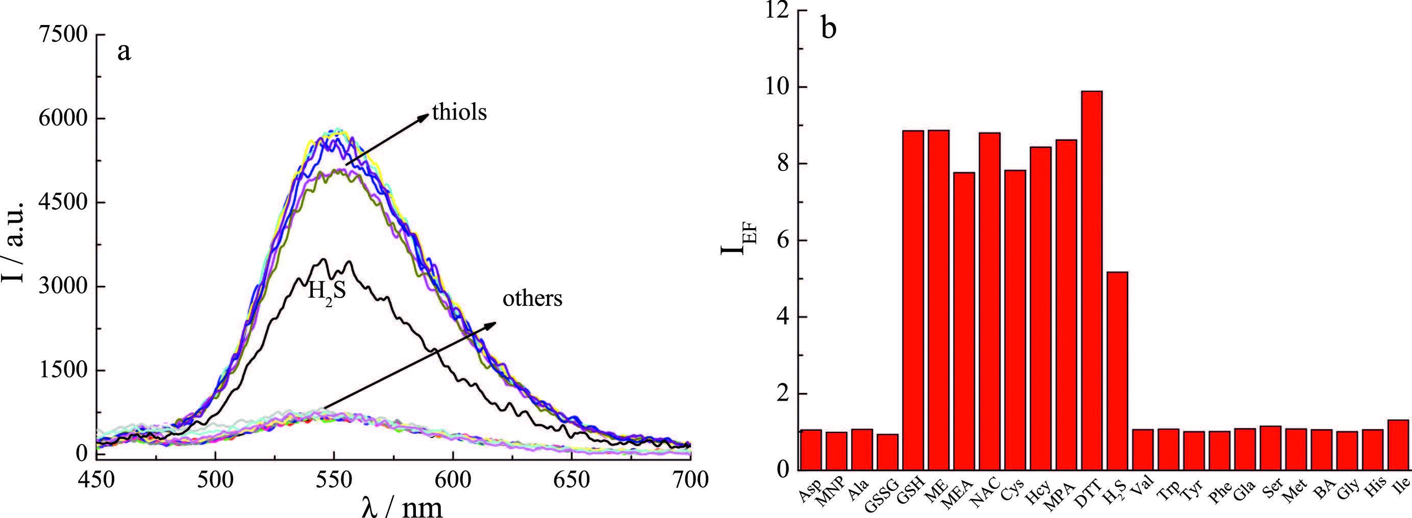
Biological samples are composed of many kinds of components; other coexisted species may interfere with the detection of thiols. Consequently, the high selectivity is very important for the probe applied in biosamples. To evaluate the selectivity and competition of MNP toward thiols over other biologically relevant species, a series of amino acids and anions were examined. As shown in Fig. 1 and Fig. S4 in Supporting information, other additives except for H2S did not trigger noticeable spectral changes of MNP. H2S induced about five-fold fluorescence enhancement of MNP, because Michael addition reaction between HS- (the pKa1 of H2S is about 6.89) and maleimide took place in physiological pH [10-12]. The above results reveal that MNP is highly selective toward thiols; other species do not interfere with the detection of thiols (Fig. S4).
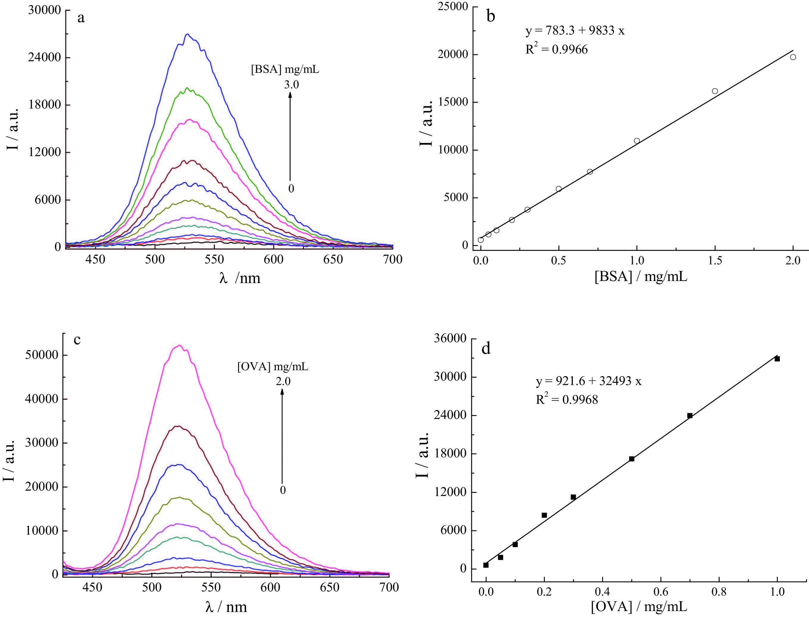
Mercapto-containing proteins, such as bovine serum albumin (BSA) and ovalbumin (OVA), play critical roles in various physiological and biological processes [13-16]. In view of the high reactivity ofMNPtoward thiols, wefurther explored the potential of MNPin the detection of thiol-bearing proteins. Upon the addition of BSA/OVA, the fluorescence intensity of MNP at 540 nm (I540) increased steadily companied with clear blue-shifts (Fig. S5 in Supporting information). After 30 min, the I540 leveled off, indicating that the reaction between MNP and BSA/OVA was finished within 30 min.The proteins without free sulfhydryl group (mucin and pepsin) [17] did not exertmuchinfluenceon the emission signal of MNP(Fig. 2), which suggests thatMNPcould be a potential probe for thiol-bearing proteins. It is noting that compared to small weighted thiols, mercapto-containing proteins initiated much higher fluorescence enhancements (>30-fold for OVA/BSA vs. eight-fold for thiols) companied with about 20 nm blue-shifts in the emission maximum (Fig. 2), which could be ascribed to the hydrophobic microenvironment provided by proteins [18, 19].
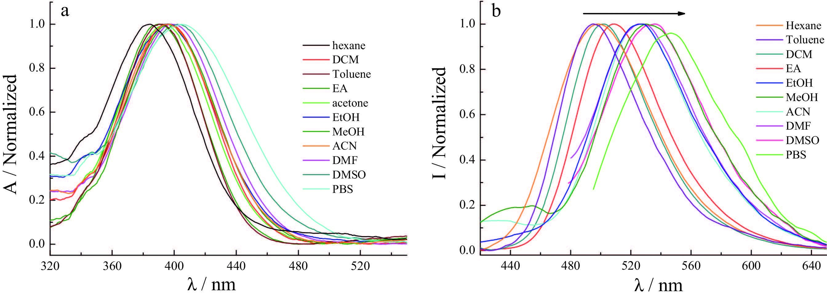
MNP is a push-pull electron system with 4-amino as the electron donor and 1, 2-imide as the acceptor. Its absorption and emission spectra are sensitive to the solvent polarity [20], namely, clear blue shifts in the absorption and emission maxima and fluorescence enhancement are expected to occur with decreasing solvent polarity. The absorption and emission spectra of MNP in different solvents were then tested to understand the polarity effect (Fig. 3). It is known from Fig. 3 that MNP displays solvatochromic UV-vis and fluorescence spectra: the absorption maximum changes from 384 nm in apolar hexane to 406 nm in PBS, while the emission peak shifts from 495 to 545 nmin the same series of solvents. The fluorescence quantum yield of MNP increases with decreasing solvent polarity (Table S1 in supporting information). About four-fold fluorescence enhancement with 20 nm blue-shift induced by non-mercapto protein casein [21, 22] (Fig. 2) could be attributed to the possibility that the probe may exist inside the hydrophobic cavity of casein.
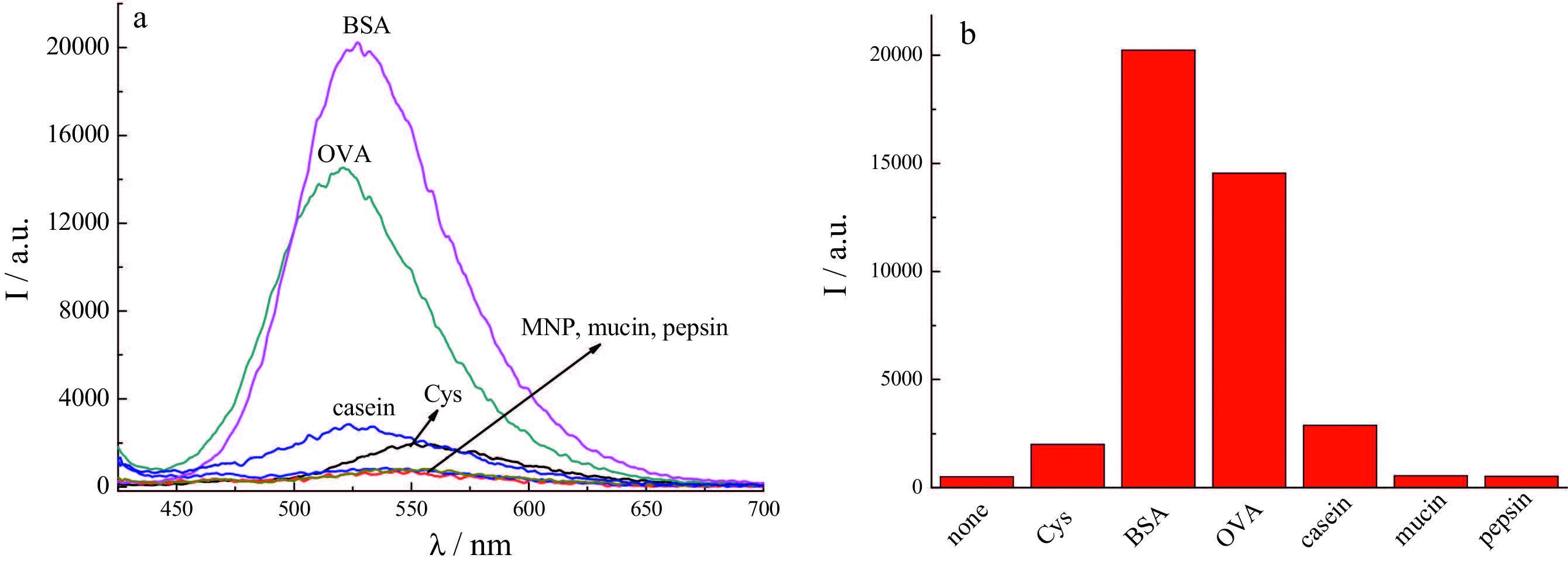
Both OVA and BSA are proteins bearing sulfhydryl groups [23, 24]. When they are mixed with MNP, Michael addition reaction between the mercapto groups in the proteins and maleimide group in the probe takes place, leading to a remarkable fluorescence enhancement. In addition, proteins can provide hydrophobic cavities [25-28] for organic molecules, which increase the emission intensity on the other hand. Therefore, the maximum fluorescence increment caused by BSA/OVA (more than 30-fold) is much higher than that triggered by small weighted thiols (eightfold). The selectivity of MNP was evaluated by measuring its emission spectrum in the presence of different proteins. Only BSA and OVA induced significant fluorescence enhancements, other species including small weighted thiols initiated much smaller spectral changes (Fig. 2). The results reveal that MNP is highly selective toward mercapto-containing proteins over other species.
To study the possibility of precise quantitation of BSA/OVA, MNP was treated with different concentrations of BSA/OVA in PBS. Upon the addition of BSA, the fluorescence intensity increased significantly (Fig. 4). Good linear relationship between the fluorescence intensity at 530 nm and BSA concentration was observed in the range of 0-2.0 mg/mL (0-1.0 mg/mL for OVA, Fig. 4). The detection limits of BSA and OVA were calculated to be 6.8 and 2.1 mg/mL, respectively. The main protein in fetal bovine serum (FBS) is BSA [20], therefore, MNP was applied in the detection of BSA in FBS. From Table S2 in Supporting information, the BSA concentration in FBS is estimated to be 36.36 mg/mL with good recovery, which is consist with the normal BSA concentration in blood plasma (35-55 mg/mL) [29, 30].
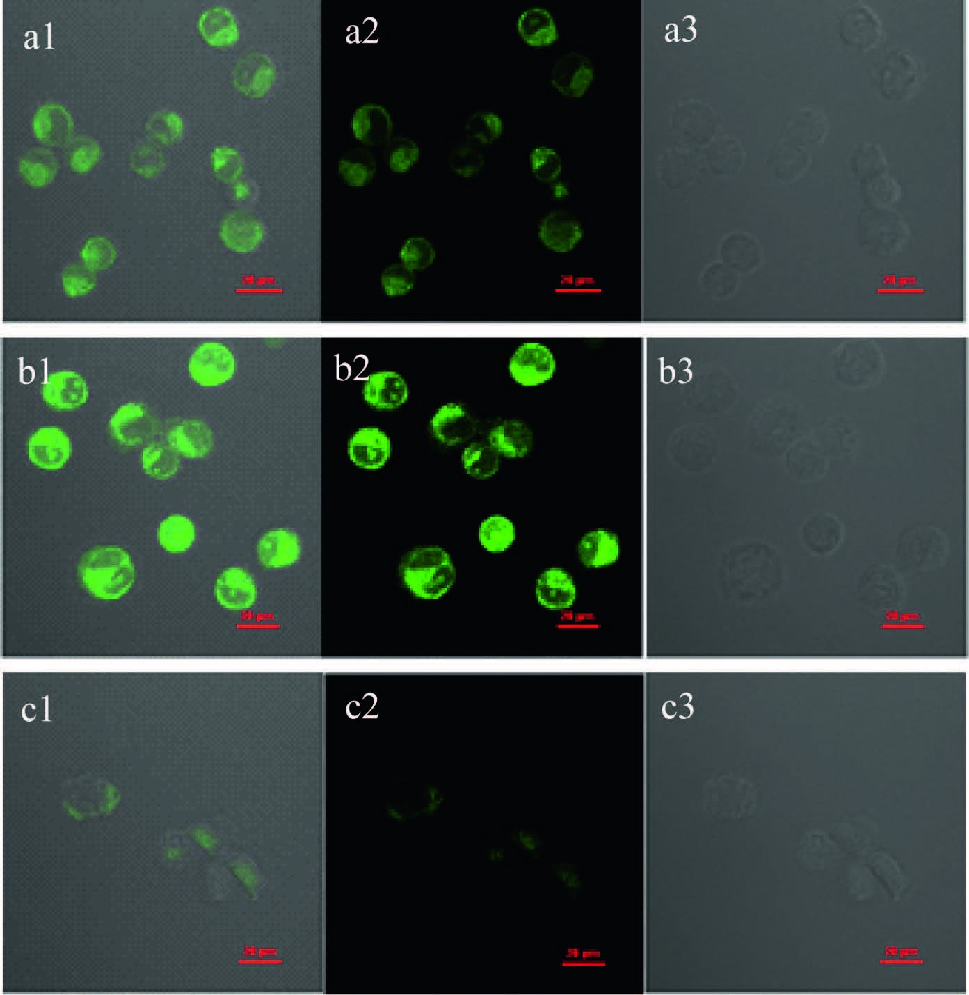
To demonstrate the capability ofMNP for fluorescent imaging of thiols in living cells, Hela cells were incubated with MNP for 15 min, and strong green fluorescence was observed (Fig. 5a). A marked increase in the green emission was seen when Hela cells pretreated with 0.5 mmol/L GSH for 15 min, followed by incubated with MNP for another 15 min (Fig. 5b). By contrast, remarkable green emission decrease was observed when Hela cells were treated with 0.5 mmol/L N-ethylmaleimide (NME, a thiol-reactive reagent) for 30 min followed by further incubated with MNP for 15 min (Fig. 5c). These results imply that MNP is a potential candidate for fluorescent imaging of thiols in living cells.
In conclusion, a fluorescent probe MNP for thiols with maleimide as the receptor and naphthalimide as the fluorophore was designed and synthesized. MNP can be used to identify thiols based on the Michael addition reaction between -SH and maleimide. Mercapto-bearing proteins induced more obvious spectral changes of MNP due to their hydrophobic pockets. MNP has potential application in fluorescent imaging of intracellular thiols as well as in the detection of BSA in biological samples.
K.G. Reddie, K.S. Carroll. Expanding the functional diversity of proteins through cysteine oxidation[J]. Curr. Opin. Chem. Biol., 2008, 12: 746-754. doi: 10.1016/j.cbpa.2008.07.028
G. Bulaj, T. Kortemme, D.P. Goldenberg. Ionization-reactivity relationships for cysteine thiols in polypeptides[J]. Biochemistry, 1998, 37: 8965-8972. doi: 10.1021/bi973101r
E. Weerapana, C. Wang, G.M. Simon. Quantitative reactivity profiling predicts functional cysteines in proteomes[J]. Nature, 2010, 468: 790-795. doi: 10.1038/nature09472
S. Shahrokhian. Lead phthalocyanine as a selective carrier for preparation of a cysteine-selective electrode[J]. Anal. Chem., 2001, 73: 5972-5978. doi: 10.1021/ac010541m
A. Meister, M.E. Anderson. Glutathione[J]. Ann. Rev. Biochem., 1983, 52: 711-760. doi: 10.1146/annurev.bi.52.070183.003431
L.M. Hyman, K.J. Franz. Probing oxidative stress:small molecule fluorescent sensors of metal ions, reactive oxygen species, and thiols[J]. Coord. Chem. Rev., 2012, 256: 2333-2356. doi: 10.1016/j.ccr.2012.03.009
Y. Zhou, X.J. Peng, J. Yoon. Fluorescent and colorimetric probes for detection of thiols[J]. Chem. Soc. Rev., 2010, 39: 2120-2135. doi: 10.1039/b925092a
H.S. Jung, X.Q. Chen, J.S. Kim, J. Yoon. Recent progress in luminescent and colorimetric chemosensors for detection of thiols[J]. Chem. Soc. Rev., 2013, 42: 6019-6031. doi: 10.1039/c3cs60024f
K. Chen, Q.H. Shu, M. Schmittel. Design strategies for lab-on-a-molecule probes and orthogonal sensing[J]. Chem. Soc. Rev., 2015, 44: 136-160. doi: 10.1039/C4CS00263F
S. Girouard, M.H. Houle, A. Grandbois, J.W. Keillor, S.W. Michnick. Synthesis and characterization of dimaleimide fluorogens designed for specific labeling of proteins[J]. J. Am. Chem. Soc., 2005, 127: 559-566. doi: 10.1021/ja045742x
Q.N. Lin, C.Y. Bao, S.Y. Cheng. Target-activated coumarin phototriggers specifically switch on fluorescence and photocleavage upon bonding to thiolbearing protein[J]. J. Am. Chem. Soc., 2012, 134: 5052-5055. doi: 10.1021/ja300475k
G.Q. Chen, Y.Q. Hua, C.W. Ou. Nanostructure formation-induced fluorescence turn-on for selectively detecting protein thiols in solutions, bacteria and live cells[J]. Chem. Commun., 2015, 51: 10758-10761. doi: 10.1039/C5CC01349F
K. Nishimura, M. Goto, T. Higasa, S.I. Kawase, Y. Matsumura. Aggregation behaviour of bovine serum albumin as a cause of sauce liquid separation by heating[J]. J. Sci. Food Agric., 2001, 81: 76-81. doi: 10.1002/(ISSN)1097-0010
D. Bulone, V. Martorana, P.L.S. Biagio. Effects of intermediates on aggregation of native bovine serum albumin[J]. Biophys. Chem., 2001, 91: 61-69. doi: 10.1016/S0301-4622(01)00155-7
S.M. Zhao, W. Xu, W.Q. Jiang. Regulation of cellular metabolism by protein lysine acetylation[J]. Science, 2010, 237: 1000-1004.
Y.L. Yang, S.Y. Fang, J.P. Jensen, A.M. Weissman, J.D. Ashwell. Ubiquitin protein ligase activity of IAPs and their degradation in proteasomes in response to apoptotic stimuli[J]. Science, 2000, 288: 874-877. doi: 10.1126/science.288.5467.874
L. Song, T. Jia, W.J. Lu. Multi-channel colorimetric and fluorescent probes for differentiating between cysteine and glutathione/homocysteine[J]. Org. Biomol. Chem., 2014, 12: 8422-8427. doi: 10.1039/C4OB01219D
O.E. Sponton, A.A. Perez, C.R. Carrara, L.G. Santiago. Impact of environment conditions on physicochemical characteristics of ovalbumin heat-induced nanoparticles and on their ability to bind PUFAs[J]. Food Hydrocoll., 2015, 48: 165-173. doi: 10.1016/j.foodhyd.2015.02.011
A. Antoniou, G. Herlem, C. André, Y. Guillaume, T. Gharbi. Simple method for detection of extremely diluted anti beta-casein antibodies from glass bead based receptors[J]. Talanta, 2011, 84: 632-637. doi: 10.1016/j.talanta.2011.01.038
S. Singha, D. Kim, B. Roy. A structural remedy toward bright dipolar fluorophores in aqueous media[J]. Chem. Sci., 2015, 6: 4335-4342. doi: 10.1039/C5SC01076D
L.K. Rasmussen, P. Højrup, T.E. Petersen. The multimeric structure and disulfidebonding pattern of bovine k-casein[J]. Eur. J. Biochem., 1992, 207: 215-222. doi: 10.1111/ejb.1992.207.issue-1
J.W. Holland, H.C. Deeth, P.F. Alewood. Analysis of disulphide linkages in bovine k-casein oligomers using two-dimensional electrophoresis[J]. Electrophoresis, 2008, 29: 2402-2410. doi: 10.1002/(ISSN)1522-2683
F. Tani, N. Shirai, F. Venelle. Temperature control for kinetic refolding of heat-denatured ovalbumin[J]. Protein Sci., 1997, 6: 1491-2150. doi: 10.1002/pro.v6:7
R. Wang, S.Y. Sun, E.J. Bekos, F.V. Bright. Dynamics surrounding Cys-34 in native, chemically denatured, and silica-adsorbed bovine serum albumin[J]. Anal. Chem., 1995, 67: 149-159. doi: 10.1021/ac00097a024
H.Y. Bai, J.H. Qian, H.Y. Tian. Fluorescent polarity probes for identifying bovine serum albumin:amplification effect of para-substituted benzene[J]. Dyes Pigm., 2014, 103: 1-8. doi: 10.1016/j.dyepig.2013.11.018
Y.D. Zhuang, P.Y. Chiang, C.W. Wang, K.T. Tan. Environment-sensitive fluorescent turn-on probes targeting hydrophobic ligand-binding domains for selective protein detection[J]. Angew. Chem. Int. Ed., 2013, 52: 8124-8128. doi: 10.1002/anie.v52.31
J.H. Qian, Y.F. Xu, X.H. Qian. Logically sensing aggregate process and discriminating SDS from other surfactants with the assistance of BSA[J]. Chin. J. Chem., 2012, 30: 1283-1288. doi: 10.1002/cjoc.201200434
G. Signore, R. Nifosì, L. Albertazzi, B. Storti, R. Bizzarri. Polarity-sensitive coumarins tailored to live cell imaging[J]. J. Am.Chem. Soc., 2010, 132: 1276-1288. doi: 10.1021/ja9050444
D.H. Li, H.H. Yang, H. Zhen. Fluorimetric determination of albumin and globulin in human serum using tetra-substituted sulphonated aluminum phthalocyanine[J]. Anal. Chim. Acta, 1999, 401: 185-189. doi: 10.1016/S0003-2670(99)00483-3
Y.C. Ma, L. Zuo, J.H. Chen. Modified glomerular filtration rate estimating equation for Chinese patients with chronic kidney disease[J]. J. Am. Soc. Nephrol., 2006, 17: 2937-2944. doi: 10.1681/ASN.2006040368

Figure 1 The emission spectra (a) and the fluorescence intensity at 550 nm (b) of MNP in the presence of different additives. [MNP] = 20 mmol/L, [analyte] = 400 mmol/L, recorded 1 min after the addition of the reagent, PBS (20 mmol/L, pH 7.4) containing 2% MeCN, 25℃, λex= 405 nm.

Figure 2 The emission spectra of MNPin the presence of different proteins (a) and their corresponding fluorescence intensities at 540 nm(b). PBS (20 mmol/L, pH 7.4) containing 2% MeCN, recorded 30 min after each addition, λex = 405 nm, 25℃.

Figure 3 Normalized absorption and emission spectra of MNP in different solvents, [MNP] = 10 mmol/L.

Figure 4 The effect of BSA/OVA concentration on the emission spectra of MNP (a, c, lex = 405 nm); the plot of fluorescence intensity at 540 nm vs BSA/OVA concentration (b, d). [MNP] = 20 mmol/L, PBS (20 mmol/L, pH 7.4) containing 2% MeCN, the samples was equilibrated at 25℃ for 30 min for each addition.

Figure 5 Fluorescence images of Hela cells incubated with 20 mmol/L MNP at 37℃. 1) the merged images of 2 and 3; 2) fluorescence images at the green channel; 3) the bright field image. a) Hela cells incubated with MNP for 15 min; b) Hela cells pretreated with 0.5 mmol/L GSH for 30 min followed by incubated with MNP for another 15 min; c) Hela cells pretreated with 0.5 mmol/L NME for 30 min, then incubated with MNP for another 15 min, excited with a 488 nm laser.

 扫一扫看文章

扫一扫关注我们
 下载:
下载:

 下载:
下载:
