图 1
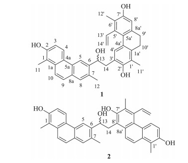
Structures of compounds 1 and 2.
Figure 1.
Structures of compounds 1 and 2.
Two new phenanthrenoid dimers from Juncus effusus
Fang Xiao , Qiong Li , Chun-Ping Tang , Chang-Qiang Ke , Yang Ye , Sheng Yao

Juncus effusus L.(Juncaceae) is a perennial herbaceous plant distributed in the wetlands and coastal marshes of South China.Its stem pith has long been used as a sedative, anxiolytic, antipyretic and diuretic agent for the treatment of fidgetiness, insomnia and edema in traditional Chinese medicine [1, 2].Previous phytochemical investigations on this genus have reported the existence of secondary metabolites such as phenanthrenoids [1-17], dihydro-dibenzoxepins [13, 18], benzocoumarins [19], phenylpropane glycerides [6, 20, 21], flavonoids [7], triterpenes [22], and phenolic acid derivatives [1].Phenanthrenoids are characteristic constituents of the genus Juncus, exhibiting a wide range of biological activities like anti-algal [3, 8-10, 12, 13, 15], cytotoxic [1, 2], antitumor [17], anti-inflammatory [2, 4], anxiolytic [5, 14], and antioxidant [7] activities.Phenanthrenoid dimers were previously reported from this genus, however, their number is still few [2, 7-9, 12].In our efforts on searching for bioactive natural products from Chinese medicinal plants, an investigation of the whole plant of J.effusus was carried out, which led to the isolation of two phenanthrenoid dimers named dijuncuenins A and B (1, 2, Fig. 1).These two compounds possessed linkages of C-14-C-3' and C14-C-8', respectively, between two phenanthrenoid halves, both of which have not been reported before.This paper describes the isolation and structure elucidation of compounds 1 and 2, and their plausible biosynthetic pathways as well.
Optical rotations were measured on a Rudulph Autopol Ⅵ Automatic polarimeter.IR and UV data were recorded on a Nicolet Magna FTIR-750 and a Shimadzu UV-2550 spectrophotometer, respectively.NMR spectra were recorded on a Bruker AM-400 (Bruker, Ettlingen, Germany) and a Bruker Avance Ⅲ (Bruker, Ettlingen, Germany) 500 NMR spectrometer with TMS as internal standard.HRESIMS were measured on a Waters Xevo QTof mass detector.Analytical HPLC was applied on a Waters 2695 instrument coupled with a 2998 PDA, a Waters Acquity ELSD, and a Waters 3100 SQDMS detector.Chiral HPLC analysis was detected on a JASCO 2000 instrument by using Daicel columns with 2-propanol/hexane as the eluents.TLC was performed on pre-coated silica gel GF254 plates (Qingdao Marine Chemical Industrials).MCI gel CHP20P (75-150 μm, Mitsubishi Chemical Industries, Japan), Silica gel (Qingdao Marine Chemical Industrials), and Sephadex LH-20 (Pharmacia Biotech AB, Uppsala, Sweden) were used for column chromatography (CC).Preparative HPLC was performed on a Varian PrepStar system with an Alltech 3300 ELSD using a Waters Sunfire RP C18, 5 μm, 30 × 150 mm column.All solvents used for CC and HPLC were of analytical grade (Shanghai Chemical Reagents Co., Ltd.) and gradient grade (Merck KGaA, Germany), respectively.
The whole plant of J.effusus was collected in Jinxiu County, Guangxi Province, China, and identified by Professor Jing-Gui Shen of Shanghai Institute of Materia Medica. A voucher specimen (No.20120618) was deposited at the herbarium of Shanghai Institute of Materia Medica, Chinese Academy of Sciences.
The air-died and powdered whole plant of J.effusus (10 kg) was extracted with 95% EtOH (4 × 30 L) at room temperature (each 72 h).The combined extracts were concentrated to yield a dark brownish residue (605 g).The residue was suspended in water and then partitioned with petroleum ether (PE) and CH2Cl2 successively to yield a PE (85 g) and a CH2Cl2 extract (150 g).The CH2Cl2 extract was applied to a MCI gel column eluted with EtOH-H2O in gradient (50:50 to 95:5) to obtain six fractions (Fr.1-Fr.6).Fr.5 was separated over silica gel CC (200-300 mesh) eluted gradiently with PE-acetone (15:1 to 0:1) to afford nine subfractions (Fr.5A-Fr.5I).Fr.5I was purified by CC over Sephadex LH-20 gel and then preparative HPLC (MeCN-H2O, 50-60%, 0-100 min, 25.0 mL/min) to yield 1 (20 mg) and 2 (3 mg).
Dijuncuenin A (1): Yellow powder; 
 下载:
导出CSV
下载:
导出CSV

|
Dijuncuenin B (2): Yellow powder;
Compound 1 had the molecular formula of C36H34O4 as determined by HRESIMS (m/z, 529.2355, [M-H]-) with 20 degrees of unsaturation. The IR absorption bands at 3513 and 3378 cm-1 indicated the presence of hydroxyl groups. The 1H NMR spectrum (Table 1) displayed signals attributable to four ortho-coupled aromatic protons (δH 8.45, 7.84, 7.72, 7.28), four singlet aromatic protons (δH 8.76, 7.70, 7.30, 6.67), a vinyl group (δH 6.53, 5.25, 5.09), an oxygenated aliphatic methine (δH 5.23, dd, J=7.8, 1.9 Hz), and four methyls (δH 2.52, 2.48, 2.21, 2.11). The 13C NMR spectrum (Table 1) exhibited 36 resonances including 18 sp2 quaternary (three oxygenated), nine sp2 methine, one oxygenated sp3 methine, four methylene (three sp3 and one sp2), and four methyl carbons. These data suggested the existence of a phenanthrene unit and a 9, 10-dihydrophenanthrene unit in the molecule. These two units accounted for all the 20 degrees of unsaturation. Therefore, a C-C linkage was assigned between the two units. In the 1H-1H COSY spectrum (Fig. 2), the proton at δH 5.23 (H-13) was observed to correlate with the methylene at δH 2.83 and 2.99 (H2-14), indicating that they might be derived from the vinyl substituent of one unit. The HMBC correlations from H2-14 to C-6 (δC 142.8), C-20 (δC 151.8), and C-4'(δC 129.7), and from H-13 to C-5 (δC 118.4) (Fig. 2) revealed the connections between C-14 and C-3' and between C-13 and C-6, respectively. In addition, the ROESY correlations of H3-11↔H-10, H-3↔H-4↔H-5, H-8↔H3-12, H3-11'↔H2-10', H2-14↔H-4'↔H-13'↔H3-12', and H-8'↔H2-9' (Fig. 2), with the aid of the HMBC correlation from H-9 to C-8 (δC 129.0), determined the substitution pattern of 1: two methyls at C-1 and C-7, and a hydroxyl group at C-2 in one phenanthrene unit; two methyls at C-1' and C-6', two hydroxyl groups at C-2' and C-7', and a vinyl group at C-5' in the other 9, 10-dihydrophenanthrene unit. The planar structure of 1 was thus constructed while the stereochemistry of C-13 still remained uncertain. The specific optical rotation of 1 was recorded as zero. When compound 1 was run on HPLC with optically active stationary phase, two peaks with a ratio of 1:1 were observed, suggesting that 1 was racemic. Accordingly, the structure of 1 was established as the first phenanthrenoid dimer with a 14-3' linkage and named dijuncuenin A.
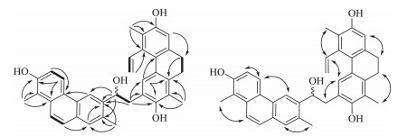
Compound 2 was obtained as yellow powder and had a molecular formula of C36H32O4 as determined by HRESIMS (m/z 527.2231, [M-H]-). The IR absorption band at 3404 cm-1 indicated the presence of hydroxyl groups. The 1D NMR data analysis (Table 1) of 2 indicated that it consisted of two phenanthrene units. In comparison to 1, a detailed 2D NMR analysis revealed that 2 possessed the same phenanthrene unit of 1 while the other unit and the connection site between two units were different. In the 1H NMR spectrum, the signals observed for a vinyl group (δH 7.35, dd, J=18.0, 11.3 Hz; 5.64, dd, J=11.3, 2.0 Hz; 5.20, dd, J=18.0, 2.0 Hz), and an oxygenated aliphatic methine (δH 5.60, dd, J=9.0, 1.9 Hz) suggested that 2 might possess a similar linkage to that of 1. In the HMBC spectrum, the key correlations from H2-14 (δH 3.63, m) to C-7' (δC 153.5, oxygenated), C-8' (δC 119.8), and C-8a0 (δC 130.1), as well as correlations from H-9' (δH 7.98, d, J=9.6 Hz) and H-100 (dH 7.86, d, J=9.6 Hz) to C-8a', confirmed a linkage between C-14 and C-8'. The correlations from H2-140 (δH 5.65, 5.25) to C-5', and from H3-120 (δH 2.51, s) to C-5' (δC 136.1), C-6' (δC 126.6), and C-7', assigned a vinyl, a methyl and a hydroxyl group at C-5', C-6', and C-7', respectively. In addition, the ROESY correlations of H3-11↔H-10, H-3↔H-4↔H-5, H3-12↔H-8, H-13↔H3-12, H3-11'↔H-10', H-3'↔H-4', and H-9'↔H2-14 further confirmed the substitution pattern of 2. Similarly, compound 2 was proved to be racemic due to the optical rotation value and the HPLC behavior on a chiral column. Therefore, the structure of 2 was established as the first 14-80 linked phenanthrenoid dimer and named dijuncuenin B.
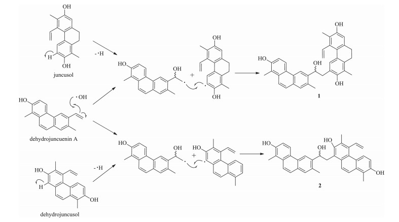
The plausible biosynthetic pathways of 1 and 2 are proposed from three known compounds (Scheme 1). Compound 1 could be derived from a coupling reaction between dehydrojuncuenin A and juncusol, both of which are metabolites and also isolated in this study. The mechanism might involve the attack of a hydroxyl radical at C-13 of dehydrojuncuenin A first, and the loss of the hydrogen free radical from juncusol at C-3 at the same time, and consequently the linking between C-14 of dehydrojuncuenin A and C-3 of juncusol. Compound 2 was presumably to have a similar mechanism to that of 1, but with a different linkage formed between C-14 of dehydrojuncuenin A and C-8 of dehydrojuncusol.
Compounds 1 and 2 were evaluated for their neuroprotective activity against Aβ25-35-induced neurotoxicity in cultured SHSY5Y cells by MTT method. Unfortunately, they did not show any positive effect at concentrations of 1 μmol/L and 10 μmol/L.
Phenanthrenoid dimers have been reported rarely from the plants of the genus Juncus so far. The dimerization pattern between two monomeric halves could be classified into two types. One features a cage-like carbon framework with complicated stereochemistry, and the other is characteristic by a single C-C' linkage between two halves, such as the reported 3-3', 7-7', 8-3', 8-8', and 8-11' linkages. Our investigation of two new phenanthrenoid dimers, dijuncuenins A (1) and B (2) with novel 14-3' and 14-8' linkages, respectively, enriches the dimerization patterns of phenanthrenoids of the genus Juncus. Our findings suggest that additional novel dimeric phenanthrenoids are to be expected from the Juncus plants since there are a variety of possible linkage patterns between two halves.
We are thankfulfor thefinancial support ofthe NationalScience & Technology Major Project "Key New Drug Creation and Manufacturing Program" (No. 2012ZX09301001-001). Our thanks are also given to the National Natural Science Foundation of China (Nos. 81302657, 81473112, 81573305), Youth Innovation Promotion Association CAS and for grants from the Chinese Academy of Sciences (No. KSZD-EW-Z-004-01), and the State Key Laboratory of Drug Research (No. SIMM1501ZZ-03).
Supplementary data associated with this article can be found, in the online version, at http://dx.doi.org/10.1016/j.cclet.2016.04.008.
Su X.H., Yuan Z.P., Li C.Y.. Phenanthrenes from Juncus effusus[J]. Planta Med., 2013, 79: 1447-1452. doi: 10.1055/s-00000058
Ma W., Liu F., Ding Y.Y.. Fournewphenanthrenoid dimers from Juncus effusus L. with cytotoxic and anti-inflammatory activities[J]. Fitoterapia, 2015, 105: 83-88. doi: 10.1016/j.fitote.2015.06.006
DellaGreca M., Fiorentino A., Isidori M.. Phenanthrenoids from the wetland Juncus acutus[J]. Phytochemistry, 2002, 60: 633-638. doi: 10.1016/S0031-9422(02)00152-8
Behery F.A.A., Naeem Z.E.M., Maatooq G.T.. Phenanthrenoids from Juncus acutus L. new natural lipopolysaccharide-inducible nitric oxide synthase inhibitors[J]. Chem. Pharm. Bull., 2007, 55: 1264-1266. doi: 10.1248/cpb.55.1264
Wang Y.G., Wang Y.L., Zhai H.F.. Phenanthrenes from Juncus effusus with anxiolytic and sedative activities[J]. Nat. Prod. Res., 2012, 26: 1234-1239. doi: 10.1080/14786419.2011.561491
Shima K., Toyota M., Asakawa Y.. Phenanthrene derivatives from the medullae of Juncus effusus[J]. Phytochemistry, 1991, 30: 3149-3151. doi: 10.1016/S0031-9422(00)98276-1
Behery F.A.A., Naeem Z.E.M., Maatooq G.T.. A novel antioxidant phenanthrenoid dimer from Juncus acutus L[J]. Nat. Prod. Res., 2013, 27: 155-163. doi: 10.1080/14786419.2012.662759
DellaGreca M., Fiorentino A., Monaco P.. New dimeric phenanthrenoids from the rhizomes of Juncus acutus. Structure determination and antialgal activity[J]. Tetrahedron, 2003, 59: 2317-2324. doi: 10.1016/S0040-4020(03)00237-0
DellaGreca M., Fiorentino A., Monaco P.. A new dimeric 9, 10-dihydrophenanthrenoid from the rhizome of Juncus acutus[J]. Tetrahedron Lett., 2002, 43: 2573-2575. doi: 10.1016/S0040-4039(02)00308-8
DellaGreca M., Monaco P., Previtera L.. Minor bioactive dihydrophenanthrenes from Juncus effusus[J]. J. Nat. Prod., 1997, 60: 1265-1268. doi: 10.1021/np970268c
DellaGreca M., Fiorentino A., Monaco P.. Effusides I-V: 9, 10-dihydrophenanthrene glucosides from Juncus effusus[J]. Phytochemistry, 1995, 40: 533-535. doi: 10.1016/0031-9422(95)00287-H
DellaGreca M., Previtera L., Zarrelli A.. Dimeric phenanthrenoids from Juncus acutus[J]. Nat. Prod. Res., 2005, 19: 69-74. doi: 10.1080/1478641042000196115
DellaGreca M., Isidori M., Monaco M.P.. Bioactivity of phenanthrenes from Juncus acutus on Selenastrum capricornutum[J]. J. Chem. Ecol., 2004, 30: 867-879. doi: 10.1023/B:JOEC.0000028437.96654.2c
Liao Y.J., Zhai H.F., Zhang B.. Anxiolytic and sedative effects of dehydroeffusol from Juncus effusus in mice[J]. Planta Med., 2011, 77: 416-420. doi: 10.1055/s-0030-1250517
DellaGreca M., Fiorentino A., Monaco P.. Action of antialgal compounds from Juncus effusus L. on Selenastrum capricornutum[J]. J. Chem. Ecol., 1996, 22: 587-603. doi: 10.1007/BF02033657
Wang X.Y., Ke C.Q., Tang C.P.. 9, 10-Dihydrophenanthrenes and phenanthrenes from Juncus setchuensis[J]. J. Nat. Prod., 2009, 72: 1209-1212. doi: 10.1021/np9000834
DellaGreca M., Fiorentino A., Mangoni L.. 9, 10-Dihydrophenanthrene metabolites from Juncus effusus L[J]. Tetrahedron Lett., 1992, 33: 5257-5260. doi: 10.1016/S0040-4039(00)79148-9
DellaGreca M., Fiorentino A., Molinaro A.. A bioactive dihydrodibenzoxepin from Juncus effusus[J]. Phytochemistry, 1993, 34: 1182-1184. doi: 10.1016/S0031-9422(00)90742-8
DellaGreca M., Fiorentino A., Isidori M.. Benzocoumarins from the rhizomes of Juncus acutus[J]. Tetrahedron, 2003, 59: 4821-4825. doi: 10.1016/S0040-4020(03)00698-7
Jin D.Z., Min Z.D., Chiou G.C.Y.. Two p-coumaroyl glycerides from Juncus effusus[J]. Phytochemistry, 1996, 41: 545-547. doi: 10.1016/0031-9422(95)00648-6
DellaGreca M., Fiorentino A., Monaco P.. Antialgal phenylpropane glycerides from Juncus effusus[J]. Nat. Prod. Lett., 1998, 12: 263-270. doi: 10.1080/10575639808048300
DellaGreca M., Fiorentino A., Monaco P.. Cycloartane triterpenes from Juncus effusus[J]. Phytochemistry, 1994, 35: 1017-1022. doi: 10.1016/S0031-9422(00)90659-9

 扫一扫看文章

扫一扫关注我们

 下载:
下载:

