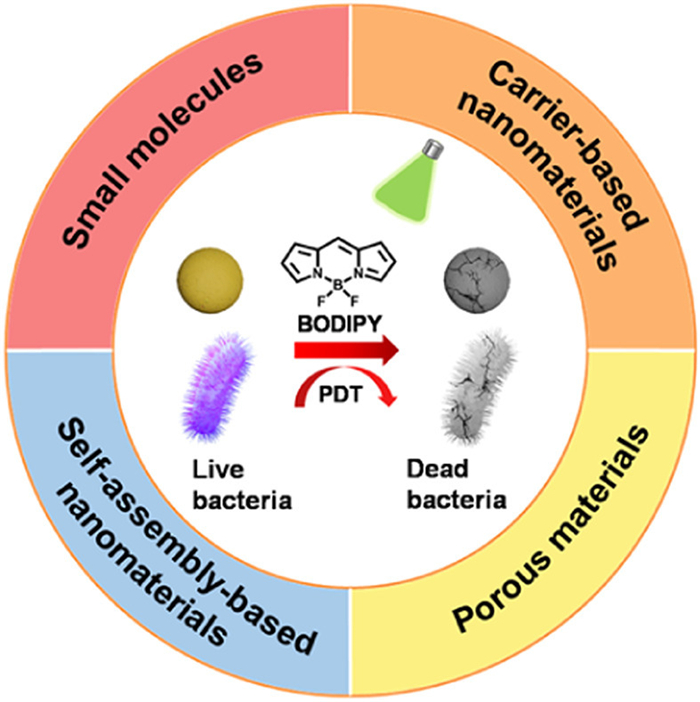
Scheme 1.
Schematic illustration of BODIPY-based materials prepared with different strategies for aPDT.
BODIPY photosensitizers for antibacterial photodynamic therapy
Yuyao Guan , Baoting Yu , Jun Ding , Tingting Sun , Zhigang Xie

Pathogenic bacterial infections, characterized by high incidence and mortality rates, have evoked millions of incidences and death per year worldwide, which seriously threaten the health of human life [1–3]. Especially in clinical work, bacterial infections severely affect the prognosis of postoperative patients [4–8]. Although the development of antibiotics has solved this problem well in the past few decades, the emergence of multi-drug-resistant (MDR) bacteria as a result of the overuse of antibiotics still poses a new threat [9–11]. Therefore, the development of novel antibacterial drugs or methods is imperative [12].
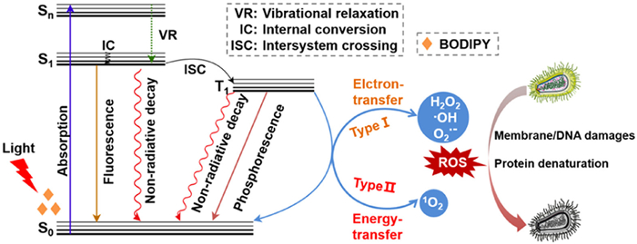
Due to the benefits of high selectivity, low invasiveness, minor side effects, wide application range, and repeatable treatment, photodynamic therapy (PDT) has attracted much attention and is considered a potential alternative method to treat pathogenic microbial infections [13–18]. Under the irradiation of visible or near-infrared (NIR) light, photosensitizers (PSs) are excited by the absorbed light energy and transfer energy or electrons to the surrounding receptors or molecular oxygen (3O2) to produce cytotoxic reactive oxygen species (ROS) [19,20]. The reaction generating superoxide anions (O2•−) or hydroxyl radicals (HO•) belongs to the type Ⅰ mechanism [21–23], and the process producing singlet oxygen (1O2) pertains to the type Ⅱ pathway [24,25]. ROS reacts with biomacromolecules in antibacterial therapy, leading to the death of pathogenic bacteria without developing resistance [26–29].
The selection of PSs is a hinge component of PDT. Recently, researchers have paid much attention on various organic dyes as efficient PSs for PDT, such as 4,4-difluoro-4-bora-3a,4a-diaza-s-indacene (BODIPY) [30], porphyrin [31,32], phthalocyanine [33], phenothiazinium [34], cyanine [35], and xanthene [36]. Benefiting from the excellent physical and chemical properties, photostability and adjustable functions, BODIPY scaffolds have been universally used as fluorescent markers/sensors for bioimaging and PSs for PDT in the biomedical field [37–49]. Recent reports have shown that BODIPY-based PSs have adequate outcomes in the PDT of bacterial infections [50–54].
Up to now, there have been several reviews about BODIPY-based PSs for antitumoral and antimicrobial PDT [55–57]. For example, the review by Wang et al. highlights the BODIPY-assembled supramolecular PSs for PDT [55]. Tomé et al. reviewed the applications of BODIPYs in antibacterial and antitumoral therapies from a structural perspective about five years ago [57]. In this review, we will provide a summary of the newly developed materials and methods based on BODIPYs for antibacterial PDT (aPDT) in recent years (Scheme 1 and Table 1 [58–68]). The design concept is well elaborated. It is of great significance to tease out the material design and antibacterial mechanisms (Scheme 2), which could guide the clinical transformation of aPDT. Finally, we will analyze the potential challenges and promising perspectives of BODIPY PSs in aPDT.
 DownLoad:
CSV
DownLoad:
CSV
| Type | Material | Illumination | MIC | Bacteria | Ref. |
| Small molecules | LIBDP | Green LED light, 5 min, 12 mW/cm2 | 0.3 µg/mL | S. aureus | [58] |
| IBDPPy-Ph/IBDPPy-PBA | Green light, 10 min, 18 mW/cm2 | 0.9 or 0.3 µmol/L 2 or 3 µmol/L |
S. aureus
E. coli |
[59] | |
| 3c | >450 nm, 15 min, 90 mW/cm2 | 0.63 µmol/L 1.25 µmol/L 0.63 µmol/L 0.63 µmol/L |
S. aureus
E. coli C. Albicans MRSA |
[60] | |
| Carrier-based nanomaterials | p-BDP | Green LED light, 5 min, 18 mW/cm2 | 128 ng/mL | S. aureus | [61] |
| IBPAAs | 550 nm, 5 min/10 min, 0.2 W/cm2 | - | S. aureus
E. coli |
[62] | |
| N-BDPI | Green light, 5 min, 18 mW/cm2 | 50 nmol/L | S. aureus | [63] | |
| Porous materials | BODIPY-ZIF-8 | LED, 1.5 h, 7 W | 1.25 µg/mL | S. aureus
E. coli |
[64] |
| Tph-BDP-COF | Visible light, 30 min, 50 mW/cm2 | - | E. coli | [65] | |
| Self-assembly-based nanomaterials | BDPI-TPP | Green light, 10 min, 12 mW/cm2 | 0.3 µg/mL | S. aureus | [66] |
| 1a | 660 nm, 10 min, 50 mW/cm2 | 1 µmol/L | MRSA | [67] | |
| BDP-LAsp | Green LED light, 10 min, 12 mW/cm2 | - | S. aureus | [68] |
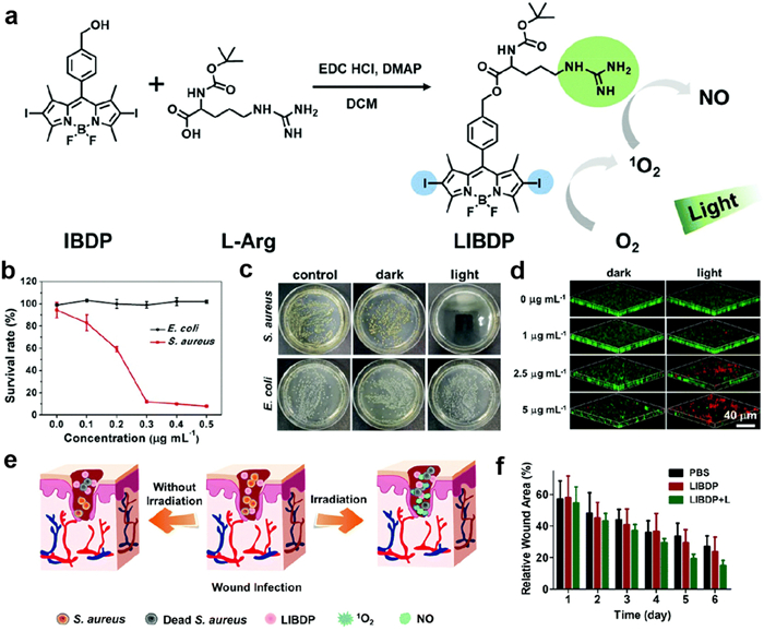
Small molecule-based drugs may transition more seamlessly from the laboratory to clinical applications [69–73]. In recent years, BODIPY-based small-molecule PSs for aPDT applications have garnered significant attention. Our group designed and synthesized a guanidine group-containing BODIPY PS (LIBDP) for antibacterial infection without drug resistance (Fig. 1a) [58]. Under green light emitting diode (LED) light irradiation, LIBDP can generate ROS to induce potent antibacterial activity and disrupt the preformed biofilms (Figs. 1b–d). Additionally, the generated ROS can also oxidize the positively charged guanidine group in LIBDP to nitric oxide (NO). As the earliest gasotransmitter to be identified, NO has already been a potent therapeutic agent used to address a variety of maladies, including bacterial infections [74–77], cardiovascular diseases [78–81], inflammatory conditions [82,83], cancer [84–86], and other rehabilitation treatments [87]. NO exerts a modulatory effect on cell proliferation and collagen deposition processes that are vital for wound healing [88,89]. Consequently, the synergistic application of NO and PDT potentiates the antibacterial impact while also accelerating wound healing (Figs. 1e and f).
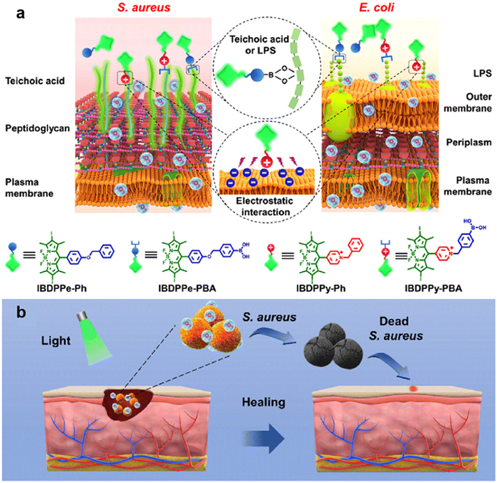
By forming borate esters, the phenylboronic acid (PBA) group is known to target teichoic acid and lipopolysaccharide (LPS), which are essential components of the cell walls in Gram-positive (G+) and Gram-negative (G−) bacteria, respectively [90–92]. On the other hand, the positively charged pyridinium (Py) cations can adhere to bacterial surfaces through strong electrostatic interactions with the negatively charged bacterial membranes [93–96]. To evaluate the comparative antibacterial efficacy of two distinct function modalities, we have developed four BODIPY conjugates with bacteria-targeting PBA or positively charged Py cations to study their photodynamic antibacterial activity (Fig. 2a) [59]. Under light irradiation, the BODIPY conjugated with PBA group (IBDPPe-PBA) demonstrated potent inhibitory activity against S. aureus, while the BODIPY with Py cation (IBDPPy-Ph) or that with both PBA group and Py cation (IBDPPy-PBA) could markedly inhibit the growth of S. aureus and E. coli. In particular, BODIPY with Py cation could adhere to both S. aureus and E. coli via electrostatic interaction, which could not only effectively remove the established biofilms in vitro, but also enhance the healing of the infected wounds (Fig. 2b).
Quaternary ammonium compounds are a kind of cationic surfactant with excellent performance. The introduction of quaternary ammonium salt can enhance the water solubility and provide positive charge for BODIPY dyes. Yan's group successfully synthesized a BODIPY quaternary ammonium with a C2 bridge at the 8-position [97], and it demonstrated a robust ability to generate 1O2 upon light activation, which was potent enough to completely eradicate the G+ Bacillus subtilis. This study underscores the critical role of achieving a equilibrium between the stability and the efficiency of 1O2 production in the design of effective PSs.
To elucidate the correlation between the BODIPY structure and the activity of aPDT, the antibacterial attributes of BODIPYs incorporating varying numbers of heavy atoms and electron-withdrawing/electron-donating substituents at diverse positions were studied [98]. It was observed that the incorporation of electron-withdrawing groups into the BODIPY moiety may diminish the light-induced electron transfer capacity, leading to a reduction in the generation efficiency of 1O2. Moreover, the photosensitization efficiency was found to inversely correlate with the electron-withdrawing power of the substituents. In addition, Orlandi and colleagues synthesized a series of BODIPY dyes, comprising 12 non-ionic and 3 cationic variants [99]. Consistent with previous reports, the synthesized cationic BODIPYs exhibited the strongest binding affinity to microbial cell walls, irrespective of their structural compositions. Unexpectedly, a neutral compound demonstrated the best photoinactivation effect against Pseudomonas aeruginosa (P. aeruginosa). Both neutral and monocationic BODIPYs were capable of inducing photoinactivation in Candida albicans (C. albicans). Anionic BODIPYs, on the other hand, showed efficient inhibitory effects on the formation of biofilms across all tested microorganisms. This study probed the impact of varying substituents at a single position on the properties of BODIPYs and highlighted the potential of neutral BODIPYs as broad-spectrum antibacterial PSs.
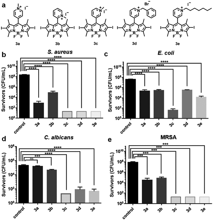
Further, Zhao et al. reported a series of PSs modified at the meso position of a BODIPY scaffold with various pyridinium moieties and investigated the effects of substituent cationization and the position of the cations on the antimicrobial activity of the PSs (Fig. 3a) [60]. All the synthesized cationic BODIPY derivatives (3a–3e) exhibited potent antimicrobial activity in PDT against S. aureus, E. coli, C. albicans, and methicillin-resistant S. aureus (MRSA) (Figs. 3b–e). Notably, even against G− bacteria, the methyl meso-(meta-pyridinium) BODIPY PS (3c) demonstrated exceptional antibacterial efficacy, which could be attributed to its less aggregation behavior, higher 1O2 quantum yield, and enhanced affinity with microbial cells.
Similarly, Okutan's team developed Carbazole (CB)-BODIPY dyads and investigated the correlation of the position and quantity of substituents with the antibacterial attributes of the BODIPY derivatives [100]. The presence of the CB moiety on the BODIPY scaffold influenced the antibacterial activity. The most potent compound featured two CB units. They also designed three naphthalimide-BODIPY (NI-BODIPY) dyads [101]. The most active NI-BODIPY dyads contained three NI units and demonstrated robust inhibition against the growth of both E. coli and S. aureus. These experimental data indicate that the type, location, and quantity of the substituents on the BODIPY scaffold significantly influence the photophysical properties and the photodynamic inactivation efficacy of the resulting PSs.
Small-molecule BODIPY PSs have shown significant potential in aPDT due to their excellent photophysical and photochemical properties. However, they still face limitations in terms of solubility and multifunctionality. To overcome these limitations, researchers began to explore the integration of BODIPY small molecules into nanomaterials. The application of nanotechnology can not only enhance the solubility of BODIPY molecules but also improve the stability and availability within the biological system, further enhancing their aPDT efficacy.
In terms of antimicrobial action, nanoparticles as carriers may promote the accumulation of BODIPYs within the biofilms. The formation of biofilms can protect bacteria from general cleaning techniques such as disinfection and facilitate further colonization. Therefore, inhibiting the formation of biofilms or eliminating existing mature ones is a persistent goal to combat bacteria. Kromer et al. reported an efficient diiodinated BODIPY derivative embedded within polystyrene nanoparticles (BODIPY-NPs), which can effectively eradicate both biofilms and planktonic bacteria [102]. BODIPY-NPs are easily dispersed in aqueous media because of their hydrophilic surface functionalization. BODIPY-NPs could achieve 100% eradication of Streptococcus mutans and S. aureus biofilms. Additionally, the NPs exhibited a remarkable clearing effect on E. coli biofilms.
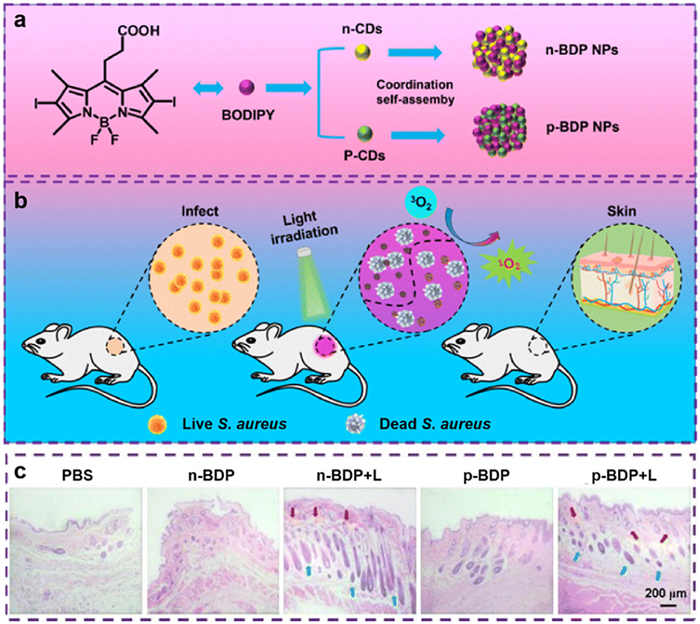
In addition, previous studies have highlighted the relationship between the cationic charge of PSs and their antibacterial effect [103–105]. Based on this, our group conjugated BODIPY with cationic and anionic CDs (p-CDs and n-CDs) to fabricate positively and negatively charged BODIPY@CD nanocomposites named p-BDP NPs and n-BDP NPs, respectively (Fig. 4a) [61]. Under laser irradiation, p-BDP NPs and n-BDP NPs can resist bacterial infection and accelerate wound healing (Fig. 4b). However, due to the electrostatic interaction between the anionic components of the bacterial cell walls and the cationic nanoparticles, p-BDP NPs exhibited superior antibacterial activity compared to n-BDP NPs. The minimum inhibitory concentration (MIC) values were determined to be 128 ng/mL for p-BDP NPs and 256 ng/mL for n-BDP NPs. Moreover, p-BDP NPs can more effectively promote the healing and tissue regeneration of S. aureus-infected wounds (Fig. 4c).
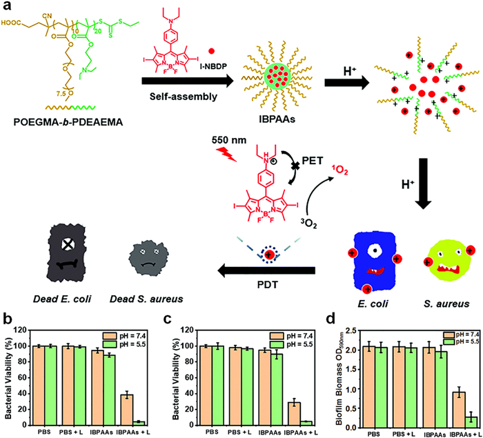
Through co-assembly of acid-responsive BODIPY (I-NBDP) and amphiphilic block copolymer polyoligo(ethylene glycol)methyl ether methyl methacrylate-b-polydiethylaminoethyl methacrylate (POEGMA-b-PDEAEMA), Zhang et al. designed an acid-triggered photodynamic antibacterial nanoplatform (IBPAAs) (Fig. 5a) [62]. On one hand, IBPAAs releases I-NBDP through the acid-triggered process, and I-NBDP is further protonated under acidic microenvironment of bacterial infections to weaken the photoelectron transfer effect under neutral conditions, resulting in increased 1O2 production. On the other side, the protonation of IBPAAs facilitates their adsorption on the negatively charged bacterial surfaces to improve the aPDT performance. Antibacterial assays demonstrated that the IBPAAs exhibit a potent aPDT effect (Figs. 5b and c) and an effective capacity to dismantle biofilms (Fig. 5d), which could effectively accelerate the healing of wounds in vivo. Therefore, this acid-triggered PDT strategy holds promise in offering innovative insights for augmenting the therapeutic potential of aPDT.
The intrinsic limitation of PDT is that the hypoxic microenvironment of the infected sites constrains the production of ROS. Zhang's group proposed that self-oxygenated nanotherapeutics can alleviate the hypoxic microenvironment to improve the photodynamic efficacy and interfere with bacterial colonization [106]. In their work, perfluoropolyether was used as an oxygen carrier, and biocompatible glycomimetics comprising galactose with fucose were introduced. Amphiphilic copolymers were formed by polymerization with iodinated BODIPY. Ultimately, an oxygen-self-supplying nanotherapeutic agent was formed using ultrasonic emulsification method, which had a strong affinity for P. aeruginosa, leading to effective bacterial aggregation and elimination. This self-oxygenating nanotherapy represents a possible approach to treat drug-resistant microbial keratitis.
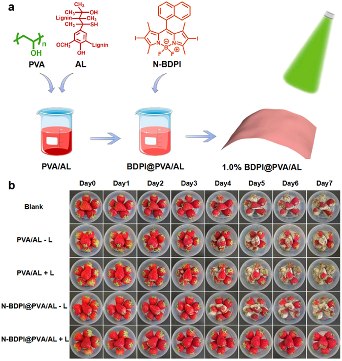
PDT is not only widely employed in the biomedical field, but also plays a crucial role in the food industry. Previous studies have shown that a small amount of ROS can be used as a signal transduction molecule to control cell growth, thereby enhancing the innate resistance of fruits and vegetables to pathogens during development [107–109]. Zheng's group synthesized N-BDPI with naphthyl group on the 8-position of BODIPY (Fig. 6a), which exhibited a potent ROS generation capacity and could effectively combat S. aureus [63]. A 1.0% BDPI@PVA/AL composite film was prepared by doping N-BDPI into polyvinyl alcohol (PVA) and alkaline lignin (AL). This film possesses a dense structure and uniform N-BDPI dispersion. Strawberry preservation tests revealed that the 1.0% BDPI@PVA/AL film could efficiently inhibit mold growth and lower gas permeability as well as water loss, significantly extending the shelf life of strawberries (Fig. 6b). This work demonstrates the broad prospects of BODIPY-based aPDT in convenient food packaging.
Furthermore, studies have shown that aza-BODIPY derivatives can cause a significant redshift in the absorption band of BODIPY [110–116], which positions aza-BODIPY as a promising candidate for NIR dual-mode PSs [117,118]. Yu and colleagues prepared an asymmetrical aza-BODIPY probe and encapsulated it with cholesterol-modified polyethylene glycol polymer to construct PDT/photothermal therapy (PTT) dual-mode antibacterial nanomedicine [119]. Dong et al. have also synthesized a donor-acceptor-donor structure NIR aza-BODIPY and prepared corresponding NPs by co-precipitation method, which can also synergistically kill drug-resistant bacteria through PDT/PTT [120].
By ingeniously introducing various carriers, researchers have been able to regulate the physical and chemical properties of BODIPY molecules, thereby significantly improving their aPDT effect. Although these advances are encouraging, the introduction of carriers has also brought some complexity, which has prompted scientists to continue to explore simpler and more efficient aPDT strategies.
The inherent pores of porous materials allow for the facilitation of oxygen and ROS diffusion, while the expanded π-conjugation within their networks provides them with a wide light absorption spectrum [121]. It has been reported in the literatures that incorporating PSs into porous materials, including nanofibers and porous silica, can effectively inhibit the agglomeration and improve the PDT efficiency [122–124].
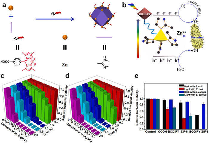
Capitalizing on the benefits of the BODIPY PSs and zeolitic imidazolate framework-8 (ZIF-8), Qu and colleagues synthesized a novel visible-light-induced nano-antibacterial composite BODIPY-ZIF-8 by incorporating BODIPY into the ZIF-8 framework via a coordination-driven self-assembly reaction (Fig. 7a) [64]. The plausible antibacterial mechanism is shown in Fig. 7b. The introduction of BODIPY did not alter the original metal-organic framework (MOF), and BODIPY-ZIF-8 exhibited a broad and strong absorption across the visible light spectrum. The antibacterial efficacy of BODIPY-ZIF-8 exhibited a time-concentration dependency (Figs. 7c and d) a superior antibacterial potency than ZIF-8 (Fig. 7e). BODIPY serves as a structure-directing agent, governing the morphology of BODIPY-ZIF-8, expanding its light absorption range, diminishing the band gap energy, and efficiently reducing the recombination rate of photoinduced electron-hole pairs, which is conducive to generating more ROS and releasing more Zn2+, thereby enhancing the photodynamic antibacterial activity. Similarly, they synthesized another photoactive nano-antibacterial agent BODIPY-ZIF-90 by introducing BODIPY into the ZIF-90 framework through aldehyde-amine condensation reaction [125]. With the uniform rhombic dodecahedral structure, BODIPY-ZIF-90 exhibits potent synergistic visible light-induced antibacterial properties.
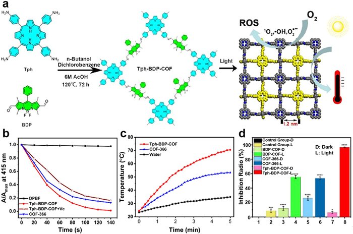
Researchers have discovered that the combination of PDT and PTT in antibacterial treatment could sterilize pathogens at lower temperatures effectively. When ROS appears, protective heat shock proteins are suppressed by biochemical reactions at high temperatures [126,127]. Covalent organic frameworks (COFs) possess permanent porous structures, excellent thermal stability, and extended π-conjugated systems, making them ideal choices for the structural design and integration of PDT and PTT [128]. In accordance with Kasha's exciton model, due to the overlap and interaction of the monomer wave functions, molecular orbitals can form band-like structures in COFs. The long-range ordered arrangement of the aggregates could reduce the energy gap between the singlet (S1) and triplet (T1) states, which is beneficial for PDT applications. Additionally, this arrangement can modulate the energy relaxation pathways, enhancing the efficacy of PTT [129]. Cao et al. proposed an “all in one” strategy for constructing a porphyrin and BODIPY-based COF, named Tph-BDP-COF (Fig. 8a) [65]. The Tph-BDP-COF possesses a narrower optical bandgap (1.33 eV), a longer carrier lifetime (1084.3 ps), and efficient energy and electron transfer, which enhance the intersystem crossing (ISC) process (Fig. 8b) and photothermal conversion (Fig. 8c), further achieving better synergistic antibacterial effects, reaching an antibacterial rate of 97% against E. coli (Fig. 8d). Moreover, the photoinduced energy and electron transfer from the COF to O2 molecules also facilitates both Type I and Type II PDT, enabling antibacterial therapy under both hypoxic and normoxic conditions.
Wang et al. designed an “all in one” light-driven NO-releasing nanoporous organic polymer (POP) composite (BG-SNP) for PDT of S. aureus and E. coli infections, demonstrating a bacteriostatic efficacy exceeding 99% [130]. BG-SNP is a composite material that incorporates guanidine, BODIPY, and sodium nitroprusside (SNP) motifs, enabling a multifaceted approach to antibacterial therapy that combines cation, PDT, PTT and NO gas-based mechanisms. The temperature rise induced by PTT not only triggers the release of NO but also markedly enhances the permeability of the ROS and NO for PDT and gas therapy. This synergistic bactericidal effect not only rapidly eliminates bacteria but also activates the immune response, facilitating the secretion of cytokines, chemokines, and other mediators during wound healing, thereby aiding in the regeneration of wound tissue. Consequently, the BG-SNP composite material is a safe and reliable multi-mode synergistic antibacterial material that not only achieves sterilization and prevents wound infection but also actively promotes wound healing.
These BODIPY-based porous materials are capable of not only integrating various functional units into a unified structure but also manipulating multiple energy dissipation pathways to realize diverse and optimal phototherapeutic outcomes, including both PDT and PTT. They exhibit robust broad-spectrum antibacterial properties and emerge as a promising category of antibacterial agents. However, the biosafety and biodegradability of many carriers and porous materials might still pose a threat to the clinical application of these BODIPY-based nanomaterials. Moreover, the introduction of carriers or porous materials will reduce the loading contents of BODIPY PSs in these nanomaterials. Therefore, preparation of nanomaterials that can reduce or even avoid the use of carriers and increase the loading contents of BODIPY PSs will be more advantageous for their practical applications.
The self-assembly strategy refers to the formation of nanoscale materials with specific structures and functions from molecules with the same structure through non-covalent interactions, such as hydrophobic interaction, π-π interaction, and hydrogen bonding. This self-assembly approach eliminates the potential toxicity associated with carriers, while also offering a high drug loading efficiency, precise molecular structure, and robust experimental reproducibility [131]. Generally speaking, it is uncommon for BODIPYs with simple structures to form NPs without any surfactants or additional additives [132]. Our group has successfully prepared pure BODIPY nanoparticles by self-assembly strategy [133]. These NPs exhibit quasi-spherical morphologies and possess potent ROS generation capabilities, effectively eliminating S. aureus and inhibiting biofilm formation.
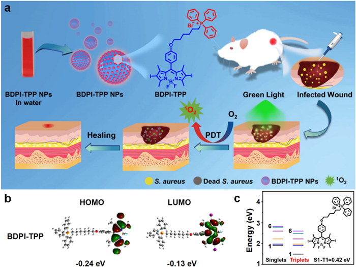
Additionally, we have developed trimethylamine and triphenylphosphine-modified cationic BODIPY derivatives (BODIPY-TMA and BDPI-TPP) for antibacterial therapy [66,134]. This design is based on the principle that positively charged NPs have an enhanced affinity for the negatively charged membranes of bacteria, potentially improving their therapeutic efficacy. BDPI-TPP NPs were formed by self-assembly of BDPI-TPP in water via a nanoprecipitation method. They can effectively destroy the preformed biofilms under green light irradiation, leading to efficient eradication of S. aureus and promoting wound healing in mice (Fig. 9a). We utilize density functional theory (DFT) to calculate the energy band gaps of BDPI-TPP. Fig. 9b displays the electronic cloud distributions of the highest occupied molecular orbital (HOMO) and the lowest unoccupied molecular orbital (LUMO). There is a small energy gap between the S1 and T1 states of BDPI-TPP (Fig. 9c). It is well-documented that a smaller energy difference facilitates the ISC of a PS, thereby enhancing the generation of ROS [135–139].
Previous researches have demonstrated that the fluorination of drugs can significantly enhance their cellular uptake, improve serum stability, and ultimately augment their therapeutic efficacy [140–142]. Moreover, the hydrophobic and oleophobic properties of fluorinated compounds can enhance their capability for self-assembly into nanostructured materials [143]. In light of this, our group prepared fluoroalkylated BODIPY (named B-F) and its counterpart alkylated BODIPY (named B-H), respectively [144]. In the absence of any carriers or auxiliaries, B-F and B-H can form NPs via self-assembly. In comparison to the alkylated B-H NPs, B-F NPs exhibit a more tightly packed structure and possess enhanced cellular internalization. Most significantly, B-F NPs possess a superior capability for generating ROS and exhibit dual efficacy in eliminating G+ bacteria and cancerous cells.
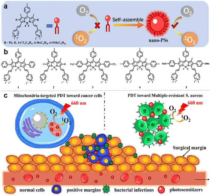
In addition, other self-assembled PSs based on BODIPY have been reported to achieve both antibacterial and antitumor effects [67,68]. For example, a BODIPY-based PS incorporating two pyridinium groups was synthesized, and nano-PSs were prepared using a supramolecular self-assembly strategy (Fig. 10a) [67]. To elucidate the structure-activity relationship, they functionalized the β-position of the BODIPY core with diverse groups, resulting in the synthesis of compounds 1–5 (Fig. 10b), and the corresponding self-assembled nanoparticles were named 1a-5a. These compounds demonstrated a striking enhancement in the generation of 1O2 in water, exhibiting a 24-124 fold increase in efficiency compared to their performance in organic solvents, with 1a showing the most pronounced 1O2 production efficiency in an aqueous milieu. 1a selectively localized to the mitochondria of HeLa cells, induced apoptosis by inflicting oxidative damage to the mitochondria under irradiation, and exhibited potent bactericidal activity against MRSA (Fig. 10c). The exceptional antitumor and antibacterial properties position this nano-PS as a promising candidate for eradicating positive surgical margins and preventing infections at surgical sites, thereby reducing the risk of tumor recurrence and postoperative infections.
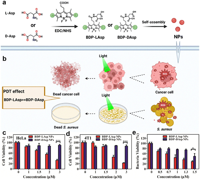
To explore the impact of the chirality of functional groups on molecular properties, L-aspartic acid and D-aspartic acid were selected to modify BODIPY, and BDP-LAsp and BDP-DAsp were obtained (Fig. 11a). Owing to the hydrophilic nature of aspartic acid, the resulting BODIPY compounds were capable of self-assembly into uniform NPs in aqueous solutions. It is worth noting that these chiral amino acid-modified BODIPY PSs also exhibited outstanding PDT effects against cancer cells and bacteria (Fig. 11b) [68]. Experimental data revealed that BDP-LAsp NPs exhibited superior photodynamic activity toward both cancer cells and S. aureus (Figs. 11c–e).
These self-assembled nanomaterials feature a simple preparation process and effective antimicrobial properties, offering new perspectives and directions for the advancement of BODIPY-based PSs in aPDT. Without the assistance of complex carriers, self-assembled nanomaterials with precise compositions are easier for repeated and large-scale preparation, thus possessing more promise for clinical translation.
aPDT with BODIPY PSs has yielded promising outcomes. To overcome the limitations of BODIPY PSs such as poor water solubility and low membrane permeability to enhance their photodynamic activity, many exceptional materials have been developed. In this review, the aPDT materials based on BODIPYs reported in recent years are summarized, also their synthesis, performance optimization strategies, and antibacterial efficacy are reviewed. Firstly, cationic PSs can enhance the interactions with bacteria, since bacterial surfaces are negatively charged. Secondly, the location and number of substituents on the BODIPY core can strongly influence the photophysical properties, and consequently, the photodynamic activity of the PSs. Thirdly, the preparation of nanocomposite materials, for example, combining BODIPY with porous materials to form light-induced nano-antibacterial composite materials, enhances their photodynamic antibacterial effects. In addition, multi-mode synergistic antibacteria, such as combining two or more of PDT, PTT, NO gas therapy, metal ions, and antibiotics, can enhance the overall antibacterial efficacy.
Photodynamic antimicrobial materials based on BODIPYs are currently limited to the treatment of skin surface infections, with inevitable challenges in treating deep tissue infections. Moreover, the existing studies are mainly focused on the theoretical exploration, but how to transition to clinical applications is the direction for future development. Additionally, the application fields of antimicrobial materials are continuously expanded. Beyond the biomedical field and hospital hygiene, there are also broad prospects in areas like food storage and water purification. We believe that BODIPY PSs will broaden their applications in the photodynamic antimicrobial field.
The authors declare that they have no known competing financial interests or personal relationships that could have appeared to influence the work reported in this paper.
Yuyao Guan: Writing – original draft. Baoting Yu: Writing – original draft. Jun Ding: Writing – review & editing, Supervision. Tingting Sun: Writing – review & editing, Supervision. Zhigang Xie: Writing – review & editing, Conceptualization.
Y.H. Yan, Y.Z. Li, Z.W. Zhang, et al., Colloids Surf. B: Biointerfaces 202 (2021) 111682.
Y.W. Ren, H.P. Liu, X.M. Liu, et al., Cell Rep. Phys. Sci. 1 (2020) 100245.
Y.C. Tian, R. Zhang, B.B. Guan, et al., Pharmaceutics 13 (2021) 1399.
M. Blondel, C. Machet, B. Wildemann, Y. Abidine, P. Swider, J. Orthop. Res. 42 (2024) 1861–1869. doi: 10.1002/jor.25822
D. Nicolotti, S. Grossi, V. Palermo, et al., Crit. Care 28 (2024) 44.
R. Martin-Mateos, L. Martínez-Arenas, Á. Carvalho-Gomes, et al., J. Hepatol. 80 (2024) 904–912.
G.H. Liu, R.C. Ma, P. Liu, K. Wang, K.Y. Cai, J. Colloid Interface Sci. 655 (2024) 809–821.
X.Y. Li, D.D. Xing, Y.J. Bai, et al., Biomed. Mater. 19 (2024) 025028.
P. Xue, R. Sang, N. Li, et al., Front. Cell. Infect. Microbiol. 13 (2023) 1119037.
O.M. Abdallah, H.R. Shebl, E. Abdelsalam, S.I. Mehrez, World J. Microbiol. Biotechnol. 40 (2024) 72.
W.T. Wang, Y.Z. Chen, Y.T. Chen, et al., Microbiol. Spectrum 12 (2024) e0127923.
L.H. Zhou, Y.Y. Wu, X.Q. Meng, et al., Small 14 (2018) e1801008.
V.N. Nguyen, Z. Zhao, B.Z. Tang, J. Yoon, Chem. Soc. Rev. 50 (2022) 3324–3340. doi: 10.1039/d1cs00647a
Y. Liu, Y.H. Wang, S.Y. Song, H.J. Zhang, Natl. Sci. Rev. 9 (2022) nwab139.
Q.Y. Zheng, X.M. Liu, Y.F. Zheng, et al., Chem. Soc. Rev. 50 (2021) 5086–5125. doi: 10.1039/d1cs00056j
G.N. Zhang, Z.Z. Wu, Y.Q. Yang, et al., Chem. Eng. J. 428 (2022) 131155.
J.P. Shi, J. Li, Y. Wang, C.Y. Zhang, Chem. Eng. J. 431 (2022) 133714.
A. Maleki, J. He, S. Bochani, et al., ACS Nano 15 (2021) 18895–18930. doi: 10.1021/acsnano.1c08334
C.Y. Mao, Y.M. Xiang, X.M. Liu, et al., ACS Appl. Mater. Interfaces 11 (2019) 17902–17914. doi: 10.1021/acsami.9b05787
A. Alabugin, Photochem. Photobiol. 95 (2019) 722–732. doi: 10.1111/php.13068
D.P. Chen, Q. Yu, X. Huang, et al., Small 16 (2020) 2001059.
D.P. Chen, Q. Xu, W.J. Wang, et al., Small 17 (2021) e2006742.
J.Y. Liu, Y. Tian, L. Dong, RSC Adv. 14 (2024) 8735–8739. doi: 10.1039/d4ra00041b
R.Z. Zhang, K.K. Niu, Y.S. Bi, et al., Green Chem. 26 (2024) 2241–2247. doi: 10.1039/d3gc04412b
R.Z. Dong, X.H. Shi, X.D. Wang, et al., Mol. Catal. 553 (2024) 113749.
X.J. Cai, J. Tian, J.W. Zhu, et al., Chem. Eng. J. 426 (2021) 131919.
H. Chen, S.L. Li, M. Wu, et al., Angew. Chem. Int. Ed. 59 (2020) 632–636. doi: 10.1002/anie.201907343
X.L. Sun, J. Sun, Y. Sun, et al., Adv. Funct. Mater. 31 (2021) 2101040.
L.G. Ding, S. Wang, B.J. Yao, et al., Adv. Healthc. Mater. 10 (2021) 2001821.
A. Turksoy, D. Yildiz, E.U. Akkaya, Coord. Chem. Rev. 379 (2019) 47–64.
J. Tian, B.X. Huang, M.H. Nawaz, W.A. Zhang, Coord. Chem. Rev. 420 (2020) 213410.
A.K. Pujari, R. Kaur, Y.N. Reddy, et al., J. Med. Chem. 67 (2024) 2004–2018. doi: 10.1021/acs.jmedchem.3c01841
X. Li, B.D. Zheng, X.H. Peng, et al., Coord. Chem. Rev. 379 (2019) 147–160.
M. Wainwright, A. McLean, Dyes Pigm. 136 (2017) 590–600.
K. Bilici, S. Cetin, E. Aydındogan, H.Y. Acar, S. Kolemen, Front. Chem. 9 (2021) 707876.
M. Maia, D.I.S.P. Resende, F. Durães, M.M.M. Pinto, E. Sousa, Eur. J. Med. Chem. 210 (2021) 113085.
J. Wang, C.J. Yu, E.H. Hao, L.J. Jiao, Coord. Chem. Rev. 470 (2022) 214709.
N.A. Bumagina, E.V. Antina, A.A. Ksenofontov, et al., Coord. Chem. Rev. 469 (2022) 214684.
Z.Y. Wang, X. Guo, Z.X. Kang, et al., Org. Lett. 25 (2023) 744–749.
F. Lv, X. Guo, Z.Y. Li, et al., Dyes Pigm. 210 (2023) 111030.
F. Lv, H. Li, Q.H. Wu, et al., Chem. Commun. 58 (2022) 3937–3940. doi: 10.1039/d2cc00297c
R.B. Liu, Y. Qian, Spectrochim. Acta. A Mol. Biomol. Spectrosc. 304 (2024) 123387.
C. Kim, D.K. Mai, W.J. Kim, et al., Biomater. Sci. 12 (2024) 1536–1548. doi: 10.1039/d3bm01520c
X. Chen, B.B. Mendes, Y.H. Zhuang, et al., J. Am. Chem. Soc. 146 (2024) 1644–1656. doi: 10.1021/jacs.3c12416
X. Guo, J.M. Yang, M. Li, et al., Angew. Chem. Int. Ed. 61 (2022) e202211081.
Z.Q. Mao, J.H. Kim, J. Lee, et al., Coord. Chem. Rev. 476 (2023) 1–34.
B.W. Lu, X. Lu, M.M. Mu, et al., Heliyon 10 (2024) e26907.
T. Tao, X. Hu, D. Sun, et al., Dyes Pigm. 224 (2024) 111996.
M. Hu, X.C. Dong, W.L. Zhao, Bioorg. Med. Chem. 99 (2024) 117583.
P. Sen, A. Sindelo, N. Nnaji, J. Mack, T. Nyokong, Photochem. Photobiol. 99 (2022) 947–956.
Q.H. Wu, H. Wen, W.H. Lin, T.T. Sun, Z.G. Xie, Chin. Chem. Lett. 35 (2024) 109692.
O. Santoro, M.C. Malacarne, F. Sarcone, et al., Int. J. Mol. Sci. 24 (2023) 4340. doi: 10.3390/ijms24054340
D. Navarro-Barreda, R. de Llanos, J.F. Miravet, et al., J. Photochem. Photobiol. B 235 (2022) 112543.
S. Kirar, Y.N. Reddy, U. Chand Banerjee, J. Bhaumik, ChemPhotoChem 7 (2022) e202200172.
J. Wang, Q.B. Gong, L.J. Jiao, E.H. Hao, Coord. Chem. Rev. 496 (2023) 215367.
A.M. Durantini, D.A. Heredia, J.E. Durantini, E.N. Durantini, Eur. J. Med. Chem. 144 (2018) 651–661.
M.L. Agazzi, M.B. Ballatore, A.M. Durantini, E.N. Durantini, A.C. Tomé, J. Photochem. Photobiol. C 40 (2019) 21–48.
C.N. Li, Y.T. Li, Q.H. Wu, T.T. Sun, Z.G. Xie, Biomater. Sci. 9 (2021) 7648–7654. doi: 10.1039/d1bm01384j
H. Wen, Q.H. Wu, L.Q. Liu, et al., Biomater. Sci. 11 (2023) 2870–2876. doi: 10.1039/d3bm00073g
G.Y. Lin, M. Hu, R. Zhang, et al., J. Med. Chem. 64 (2021) 18143–18157. doi: 10.1021/acs.jmedchem.1c01643
C.J. Mou, X.Y. Wang, Y.C. Liu, Z.G. Xie, M. Zheng, J. Mater. Chem. B 10 (2022) 8094–8099. doi: 10.1039/d2tb01539k
X.N. Liang, L. Xia, Y.C. Zhu, et al., Biomater. Sci. 10 (2022) 4235–4242. doi: 10.1039/d2bm00780k
Y.C. Liu, M. Zheng, Food Chem. 427 (2023) 136691.
Z.Q. Shen, L.L. Qu, X.L. Kan, et al., Colloids Surf. A 644 (2022) 128835.
S.S. Zhao, X.P. Guo, X.H. Pan, Y.B. Huang, R. Cao, Chem. Eng. J. 457 (2023) 141017.
H. Wen, Q.H. Wu, C.N. Li, T.T. Sun, Z.G. Xie, ACS Appl. Nano Mater. 5 (2022) 1500–1507. doi: 10.1021/acsanm.1c04143
B.K. Liu, J. Zheng, H. Wang, L.Y. Niu, Q.Z. Yang, Mater. Chem. Front. 7 (2023) 5879–5890. doi: 10.1039/d3qm00753g
W.T. Lei, Q.H. Wu, H. Wen, et al., J. Mater. Chem. B 11 (2023) 6853–6858. doi: 10.1039/d3tb00684k
J.K. Gao, H.F. Jiang, P.W. Chen, R.S. Zhang, N. Liu, Bioorg. Chem. 136 (2023) 106554.
A.G. Robertson, L.M. Rendina, Chem. Soc. Rev. 50 (2021) 4231–4244. doi: 10.1039/d0cs01075h
P. Bhutani, G. Joshi, N. Raja, et al., J. Med. Chem. 64 (2021) 2339–2381. doi: 10.1021/acs.jmedchem.0c01786
Y.P. Wu, S.M. Li, Y.C. Chen, W.J. He, Z.J. Guo, Chem. Sci. 13 (2022) 5085–5106. doi: 10.1039/d1sc05478c
N.M. Amal, M. Shiddiq, B. Armynah, D. Tahir, Luminescence 37 (2022) 2006–2017. doi: 10.1002/bio.4388
M. Garren, P. Maffe, A. Melvin, et al., ACS Appl. Mater. Interfaces 13 (2021) 56931–56943. doi: 10.1021/acsami.1c17248
R. Devine, M. Douglass, M. Ashcraft, N. Tayag, H. Handa, ACS Appl. Mater. Interfaces 13 (2021) 19613–19624. doi: 10.1021/acsami.1c01330
H.J. Wang, Y. Wang, W.H. Xu, et al., ACS Appl. Mater. Interfaces 15 (2023) 54266–54279. doi: 10.1021/acsami.3c10862
C.Y. Qi, J. Chen, Y. Zhuang, et al., Int. J. Pharm. 640 (2023) 123014.
C. Farah, L.Y.M. Michel, J.L. Balligand, Nat. Rev. Cardiol. 15 (2018) 292–316. doi: 10.1038/nrcardio.2017.224
Y.F. Zhao, C. Li, S. Zhang, et al., Front. Microbiol. 14 (2023) 1277552.
M.Y. He, D.P. Wang, Y.M. Xu, et al., Pharmaceutics 14 (2022) 1345.
A. Olah, B.A. Barta, M. Ruppert, et al., Eur. Heart J. 44 (2023) 1965 ehad655-.
J.K. Jiang, J.L. Xie, L. Zhou, et al., Chem. Eng. J. 480 (2024) 147850.
S.Y. Tao, J. Cheng, G. Su, et al., Angew. Chem. Int. Ed. 59 (2020) 21864–21869. doi: 10.1002/anie.202010009
R. Ren, D.H. Bremner, W.L. Chen, et al., Int. J. Biol. Macromol. 238 (2023) 124087.
D. Gao, S. Asghar, R.F. Hu, et al., Acta Pharm. Sin. B 13 (2023) 1498–1521.
H.N. Tian, J.Y. Lin, F.K. Zhu, et al., Chin. Chem. Lett. 34 (2023) 107577.
V.A. Zaborova, A.V. Butenko, A.B. Shekhter, et al., Med. Gas Res. 13 (2023) 128–132. doi: 10.4103/2045-9912.344983
Y.T. Duan, K.W. He, G.Y. Zhang, J.M. Hu, Biomacromolecules 22 (2021) 2160–2170. doi: 10.1021/acs.biomac.1c00251
M.M. Wan, H. Chen, Q. Wang, et al., Nat. Commun. 10 (2019) 966.
L. Wang, S.X. Li, J.X. Yin, et al., Nano Lett. 20 (2020) 5036–5042. doi: 10.1021/acs.nanolett.0c01196
A. Galstyan, R. Schiller, U. Dobrindt, Angew. Chem. Int. Ed. 56 (2017) 10362–10366. doi: 10.1002/anie.201703398
S.S. Liu, B.N. Wang, Y.W. Yu, et al., ACS Nano 16 (2022) 9130–9141. doi: 10.1021/acsnano.2c01206
L. Liu, X.Y. Wang, S.X. Zhu, et al., Chem. Mater. 32 (2019) 438–447.
H.L. Zhou, D.S. Tang, X.X. Kang, et al., Adv. Sci. 9 (2022) 2200732.
C. Zhao, X. Wang, L. Yu, et al., Acta Biomater. 138 (2022) 528–544.
S.M. Wu, C. Xu, Y.W. Zhu, et al., Adv. Funct. Mater. 31 (2021) 2103591.
D.K. Joshi, F. Betancourt, A. McAdorey, et al., J. Photochem. Photobiol. A 434 (2023) 114213.
Q.X. Shi, C.J. Mou, Z.G. Xie, M. Zheng, Photodiagn. Photodyn. Ther. 39 (2022) 102901.
V.T. Orlandi, E. Martegani, F. Bolognese, E. Caruso, Photochem. Photobiol. Sci. 21 (2022) 1233–1248. doi: 10.1007/s43630-022-00212-4
H. Eserci Gürbüz, E. Öztürk Gündüz, M. Bat-Ozmatara, A. Şenocak, E. Okutan, J. Photochem. Photobiol. A 443 (2023) 114890.
H. Eserci, M. Çetin, F. Aydınoğlu, E.T. Eçik, E. Okutan, J. Mol. Struct. 1265 (2022) 133440.
C. Kromer, K. Schwibbert, S. Radunz, et al., Front. Microbiol. 14 (2023) 1274715.
B.L. Carpenter, F. Scholle, H. Sadeghifar, et al., Biomacromolecules 16 (2015) 2482–2492. doi: 10.1021/acs.biomac.5b00758
J. Piskorz, W. Porolnik, M. Kucinska, et al., ChemMedChem 16 (2021) 399–411. doi: 10.1002/cmdc.202000529
Y.B. Palacios, S.C. Santamarina, J. Durantini, et al., J. Photochem. Photobiol. B 212 (2020) 112049.
Y.Y. Bai, Y.Q. Hu, Y.C. Gao, et al., ACS Appl. Mater. Interfaces 13 (2021) 33790–33801. doi: 10.1021/acsami.1c04996
S. Mansoor, O. Ali Wani, J.K. Lone, et al., Antioxidants 11 (2022) 225. doi: 10.3390/antiox11020225
R. Singh, S. Singh, P. Parihar, et al., Front. Plant Sci. 7 (2016) 1299.
M.S. Katarzyna, W. Natalia, N. Klaudia, et al., Antioxidants 9 (2020) 199.
J.J. Zhang, Y.J. Li, M.L. Jiang, et al., ACS Biomater. Sci. Eng. 9 (2023) 821–830. doi: 10.1021/acsbiomaterials.2c01539
S.M. Wu, W.Z. Zhang, C.R. Li, et al., Chem. Sci. 15 (2024) 5973–5979. doi: 10.1039/d3sc06976a
Y.M. Zhang, S.C. Li, J. Wang, et al., J. Mater. Chem. B 12 (2024) 1372–1378. doi: 10.1039/d3tb02623j
C. Liu, X. Ji, Z.l. Yu, et al., J. Med. Chem. 66 (2023) 7205–7220. doi: 10.1021/acs.jmedchem.2c01653
Y.N. Sun, C.S. Dong, D.X. Zhang, et al., Dyes Pigm. 228 (2024) 112213.
L.L. Cao, Y.K. Li, D.X. Zhang, et al., Chin. Chem. Lett. 35 (2024) 109735.
Z.Z. Yong, B.X. Wen, Q.Q. Dou, et al., Dyes Pigm. 219 (2023) 111614.
X.H. Ruan, M. Wei, X.Y. He, et al., Colloids Surf. B 231 (2023) 113547.
D.P. Chen, Z.H. Zhong, Q.L. Ma, et al., ACS Appl. Mater. Interfaces 12 (2020) 26914–26925. doi: 10.1021/acsami.0c05021
S. Song, G.C. Xu, N. Yang, et al., J. Mater. Sci. 57 (2022) 21206–21218. doi: 10.1007/s10853-022-07924-z
Q. Yu, X. Huang, T. Zhang, et al., Chem. Res. Chin. Univ. 37 (2021) 951–959.
X.Y. Wang, L.J. Chen, S.Y. Chong, et al., Nat. Chem. 10 (2018) 1180–1189. doi: 10.1038/s41557-018-0141-5
D. Thakur, Q.T.H. Ta, J.S. Noh, ACS Appl. Nano Mater. 5 (2022) 1533–1541. doi: 10.1021/acsanm.1c04254
G.F. Su, S.F. Qiu, J.Q. Lin, et al., Colloids Surf. A 640 (2022) 128414.
G. Gupta, Y. Sun, A. Das, P.J. Stang, C.Y. Lee, Coord. Chem. Rev. 452 (2022) 214308.
D.M. Chen, G.Q. He, Q.Y. Chen, et al., New J. Chem. 47 (2023) 8152–8160. doi: 10.1039/d3nj00397c
X.L. Hu, L.Y. Chu, X.J. Dong, et al., Adv. Funct. Mater. 29 (2019) 1806981.
Y.Q. Wang, Y.Y. Jin, W. Chen, et al., Chem. Eng. J. 358 (2019) 74–90. doi: 10.1364/dp.2019.74
Q. Guan, L.L. Zhou, Y.A. Li, et al., ACS Nano 13 (2019) 13304–13316. doi: 10.1021/acsnano.9b06467
X. Chang, M.B. Qarai, F.C. Spano, J. Chem. Phys. 157 (2022) 209901.
J. Wang, X.M. Zhang, N. Wang, et al., ACS Appl. Nano Mater. 6 (2023) 16716–16729. doi: 10.1021/acsanm.3c02922
T. Zhang, C. Ma, T.T. Sun, Z.G. Xie, Coord. Chem. Rev. 390 (2019) 76–85.
F.F. An, J.Q. Xin, C.T. Deng, et al., J. Mater. Chem. B 9 (2021) 9308–9315. doi: 10.1039/d1tb01883c
Q.X. Shi, X.Y. Wang, H.X. Liu, Z.G. Xie, M. Zheng, J. Photochem. Photobiol. B 241 (2023) 112674.
H.Y. Wang, C.N. Li, Q.H. Wu, et al., J. Mater. Chem. B 10 (2022) 4967–4973. doi: 10.1039/d2tb00778a
C.N. Li, W.H. Lin, S. Liu, T.T. Sun, Z.G. Xie, Mater. Chem. Front. 5 (2021) 284–292. doi: 10.1039/d0qm00624f
S.W. Wang, W.B. Wu, P. Manghnani, et al., ACS Nano 13 (2019) 3095–3105. doi: 10.1021/acsnano.8b08398
D.S. Zhang, L.Y. Peng, K.X. Teng, et al., ACS Mater. Lett. 5 (2023) 180–188.
Y.F. Kang, W.K. Chen, K.X. Teng, et al., CCS Chem. 4 (2022) 3516–3528. doi: 10.31635/ccschem.021.202101600
J.X. Zhang, S. Mukamel, J. Jiang, J. Phys. Chem. B. 124 (2020) 2238–2244. doi: 10.1021/acs.jpcb.0c00654
X. Wang, B. Yang, L.X. Li, et al., Theranostics 11 (2021) 7896–7910. doi: 10.7150/thno.61337
J. Lv, H. Wang, G.Y. Rong, Y.Y. Cheng, Acc. Chem. Res. 55 (2022) 722–733. doi: 10.1021/acs.accounts.1c00766
B.W. Jiang, D.Y. Hao, C.N. Li, et al., J. Mater. Chem. B 9 (2021) 9971–9979. doi: 10.1039/d1tb02165f
C. Zhang, T.Q. Liu, W.Q. Wang, et al., ACS Nano 14 (2020) 7425–7434. doi: 10.1021/acsnano.0c02954
X.Y. Wang, B.W. Jiang, Z.G. Xie, M. Zheng, Colloids Surf. B 220 (2022) 112966.

Scheme 1 Schematic illustration of BODIPY-based materials prepared with different strategies for aPDT.

Scheme 2 Schematic illustration of the photodynamic antimicrobial mechanism of BODIPY-based PSs.

Figure 1 (a) Synthetic step of LIBDP. (b) Survival rates and (c) photographs of Staphylococcus aureus (S. aureus) and Escherichia coli (E. coli) treated with LIBDP under irradiation. (d) 3D confocal laser scanning microscope (CLSM) images of biofilms under different conditions. (e) Schematic illustration of LIBDP in promoting wound healing. (f) Relative wound surface areas with time. Reproduced with permission [58]. Copyright 2021, Royal Society of Chemistry.

Figure 2 (a) Interactions between the BODIPYs functionalized with PBA or Py cation with bacterial membranes. (b) Schematic diagram of aPDT application in promoting wound healing. Reproduced with permission [59]. Copyright 2023, Royal Society of Chemistry.

Figure 3 (a) Structural formulas of BODIPY derivatives. Survivors of (b) S. aureus, (c) E. coli, (d) C. albicans, and (e) MRSA treated with 3a–3e after irradiation. ***P < 0.001; ****P < 0.0001; ns, P > 0.05. Reproduced with permission [60]. Copyright 2021, American Chemical Society.

Figure 4 (a) Preparation of p-BDP NPs and n-BDP NPs. (b) Application of p-BDP NPs and n-BDP NPs in accelerating wound healing. (c) Hematoxylin and eosin (H&E) staining of wound tissues after treatment (blood vessels, red arrows; hair follicles, blue arrows). Reproduced with permission [61]. Copyright 2022, Royal Society of Chemistry.

Figure 5 (a) Preparation IBPAAs for aPDT. Viability of (b) S. aureus and (c) E. coli under different conditions. (d) Optical density (OD) values of the residual biofilms. Reproduced with permission [62]. Copyright 2022, Royal Society of Chemistry.

Figure 6 (a) Preparation of 1.0% BDPI@PVA/AL food packing film. (b) Photos of strawberries with different treatments. Reproduced with permission [63]. Copyright 2023, Elsevier.

Figure 7 Schematic illustration of (a) the synthesis of BODIPY-ZIF-8 and (b) the plausible mechanism of antibacterial activity under LED irradiation. Viability of (c) E. coli and (d) S. aureus at different concentrations of BODIPYD-ZIF-8 and incubation times under LED irradiation. (e) Viability of E. coli and S. aureus treated by COOH-BODIPY, ZIF-8, and BODIPY-ZIF-8 with/without irradiation. Reproduced with permission [64]. Copyright 2022, Elsevier.

Figure 8 (a) Schematic illustration of the synthesis of Tph-BDP-COF and photodynamic and photothermal activity under irradiation. (b) ROS generation efficiency under different treatments. (c) Temperature increasing under different treatments. (d) Inhibition ratios of E. coli under different treatments. Reproduced with permission [65]. Copyright 2023, Elsevier.

Figure 9 (a) Preparation and antimicrobial application of BDPI-TPP NPs. (b) HOMO and LUMO of of BDPI-TPP. (c) Excited singlet and triplet levels of BDPI-TPP. Reproduced with permission [66]. Copyright 2022, American Chemical Society.

Figure 10 (a) Assembly-enhanced 1O2 generation of BODIPY molecules. (b) Structural formulas of molecules 1–5. (c) Nano-PSs for simultaneous photodynamic antitumoral and antibacterial therapy. Reproduced with permission [67]. Copyright 2023, Royal Society of Chemistry.

Figure 11 (a) Synthesis of BDP-LAsp and BDP-DAsp for (b) photodynamic antitumoral and antibacterial therapy. Viability of (c) HeLa cells, (d) 4T1 cells, and (e) S. aureus after treatments with different concentrations of the NPs with irradiation. Reproduced with permission [68]. Copyright 2023, Royal Society of Chemistry.
Table 1. Typical examples of the latest studies on the use of BODIPY-based PSs for PDT against bacterial infections.
| Type | Material | Illumination | MIC | Bacteria | Ref. |
| Small molecules | LIBDP | Green LED light, 5 min, 12 mW/cm2 | 0.3 µg/mL | S. aureus | [58] |
| IBDPPy-Ph/IBDPPy-PBA | Green light, 10 min, 18 mW/cm2 | 0.9 or 0.3 µmol/L 2 or 3 µmol/L |
S. aureus
E. coli |
[59] | |
| 3c | >450 nm, 15 min, 90 mW/cm2 | 0.63 µmol/L 1.25 µmol/L 0.63 µmol/L 0.63 µmol/L |
S. aureus
E. coli C. Albicans MRSA |
[60] | |
| Carrier-based nanomaterials | p-BDP | Green LED light, 5 min, 18 mW/cm2 | 128 ng/mL | S. aureus | [61] |
| IBPAAs | 550 nm, 5 min/10 min, 0.2 W/cm2 | - | S. aureus
E. coli |
[62] | |
| N-BDPI | Green light, 5 min, 18 mW/cm2 | 50 nmol/L | S. aureus | [63] | |
| Porous materials | BODIPY-ZIF-8 | LED, 1.5 h, 7 W | 1.25 µg/mL | S. aureus
E. coli |
[64] |
| Tph-BDP-COF | Visible light, 30 min, 50 mW/cm2 | - | E. coli | [65] | |
| Self-assembly-based nanomaterials | BDPI-TPP | Green light, 10 min, 12 mW/cm2 | 0.3 µg/mL | S. aureus | [66] |
| 1a | 660 nm, 10 min, 50 mW/cm2 | 1 µmol/L | MRSA | [67] | |
| BDP-LAsp | Green LED light, 10 min, 12 mW/cm2 | - | S. aureus | [68] |
 下载: 导出CSV

 扫一扫看文章

扫一扫关注我们