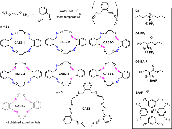
Scheme 1.
The synthesis of crown aldoxime ethers and structures of guests.
Crown aldoxime ethers: Their synthesis, structure, acid-catalyzed/photo-induced isomerization and adjustable guest binding
Yulin Mao , Jingyu Ma , Jiecheng Ji , Yuliang Wang , Wanhua Wu , Cheng Yang

Macrocyclic molecules constitute the majority of molecular hosts by leveraging the cooperative effects of the well-prearranged functional groups [1-6]. Crown ethers, being among the foremost and extensively studied macrocycles, have been applied across various research domains [7-11]. Besides classic crown ethers, exploration has extended to include other heteroatoms to tailor cavity size [12,13] and the binding behavior [14,15]. Moreover, chemists have ventured into adjustable molecular macrocycles to manipulate molecular complexation in vivo by incorporating isomerizable function groups, such as ethene [16], imine [17] and azobenzene [18-21], into a macrocycle. However, in most cases, the switchable functional groups serve solely as regulators within the macrocycle rather than directly participating in guest molecule complexation. This not only complicates synthesis but also often alters the intrinsic binding ability of protogenic macrocyclic hosts. Constructing an adjustable macrocycle that is readily available and capable of manipulating binding properties poses a significant challenge. It occurred to us that macrocycles based on aldoxime ethers may exhibit novel binding behaviors distinct from traditional crown ethers [22], and isomerization of aldoximes occur directly at the binding atoms [23,24], offering an opportunity to significantly modify macrocycle binding. In this work, we designed and synthesized a new series of novel crown aldoxime ethers and demonstrated their ability to complex with ammonium salts, resulting in chiroptical induction. By combining acid-catalyzed and photo-induced isomerization, the crown aldoxime ethers demonstrated a regulation of complexation through isomerization at binding heteroatoms.
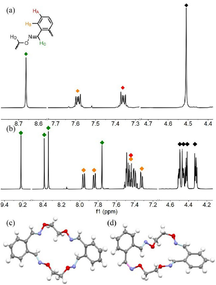
Synthesis of crown aldoxime ethers was conducted by the condensation of a bis-hydroxylamine (Scheme 1) and o-phthalaldehyde in the presence of a catalytic amount of hydrochloric acid. HPLC tracking (Fig. S1 in Supporting information) suggested that the reaction yielded several products, with two predominant ones. Silica gel column chromatography separated the crude products into two major components, identified as CAE2–1 in 26.7% yield and CAE2–2 in 7.0% yield (Fig. 1). Their HR-MS spectra revealed molecular ion peaks at 381.1555 and 381.1563, respectively, confirming both as [2 + 2] macrocycles (Figs. S34 and S37 in Supporting information). Their chemical structures were assigned based on NMR spectra and X-ray single crystal diffraction (Fig. 1, Figs. S2-S5 and S32-S37 in Supporting information). The 1H NMR spectrum of CAE2–1 showed a single peak of HC at 8.66 ppm, indicating a symmetrical structure with four HC atoms in identical environments (Fig. 1a). In contrast, CAE2–2 exhibited four different types HC signals at 9.23, 8.40, 8.36 and 7.77 ppm, respectively, suggesting varied chemical environments for each HC atom (Fig. 1b). From single-crystal analyses (Figs. 1c and d), the averaged CN bond lengths measured 1.27 Å, supporting their double bond nature. For CAE2–1 (CCDC 2335210), the torsion angle of H—C=N—O was −0.86° to 4.32° For CAE2–2 (CCDC 2335211), one particular torsion angle was measured as 176.65° and attributed to cis configuration. These results revealed that CAE2–1 and CAE2–2 were [2 + 2] isomers having all trans C=N, and one cis, three trans C=N double bonds, respectively.
In addition to CAE2–1 and CAE2–2, the reaction also yielded other isomers in trace amounts as observed in HPLC analyses. These isomers displayed molecular ion peaks characteristic of [2 + 2] macrocycles (Figs. S40, S43, S46 and S49 in Supporting information) and were assigned as CAE2–3, CAE2–4, CAE2–5, and CAE2–6 (Fig. S1). However, direct purification of these [2 + 2] macrocycles proved challenging due to their small quantities and their co-elution with other reaction products during column chromatography, including the [3 + 3] macrocycle CAE3 and its isomers. CAE2–3, −4, −5 and −6 were obtained via photoisomerization of CAE2–1 or CAE2–2 to avoid the production of other larger macrocycles and uncyclized products.
UV–vis spectra of CAE2–1 and CAE2–2 showed peaks at 250 nm, with CAE2–1 displaying strong absorption and a shoulder at 285 nm (Fig. 2a). UV–vis spectra of other isomers were also obtained and exhibited variations depending on the number and relative position of cis double bonds (Fig. S10 in Supporting information). To investigate the stability of the aldoxime ethers, thermal isomerization was examined by monitoring the UV–vis spectral changes (Fig. 2b and Figs. S11-S18 in Supporting information). Both CAE2–1 and CAE2–2 were heated at 60 ℃ in chloroform or at 80 ℃ in a mixed solution of acetonitrile and water. No changes were observed in the UV–vis spectra after heating for 1 h, indicating no thermal isomerization occurred. This was confirmed by HPLC traces, which showed no distinct peaks during heating (Figs. S11 and S12). This demonstrated an advantage of the aldoxime ether double bond in terms of stability compared to the hydrolysis of imine Schiff bases in water and the thermal Z to E isomerization of azobenzene.
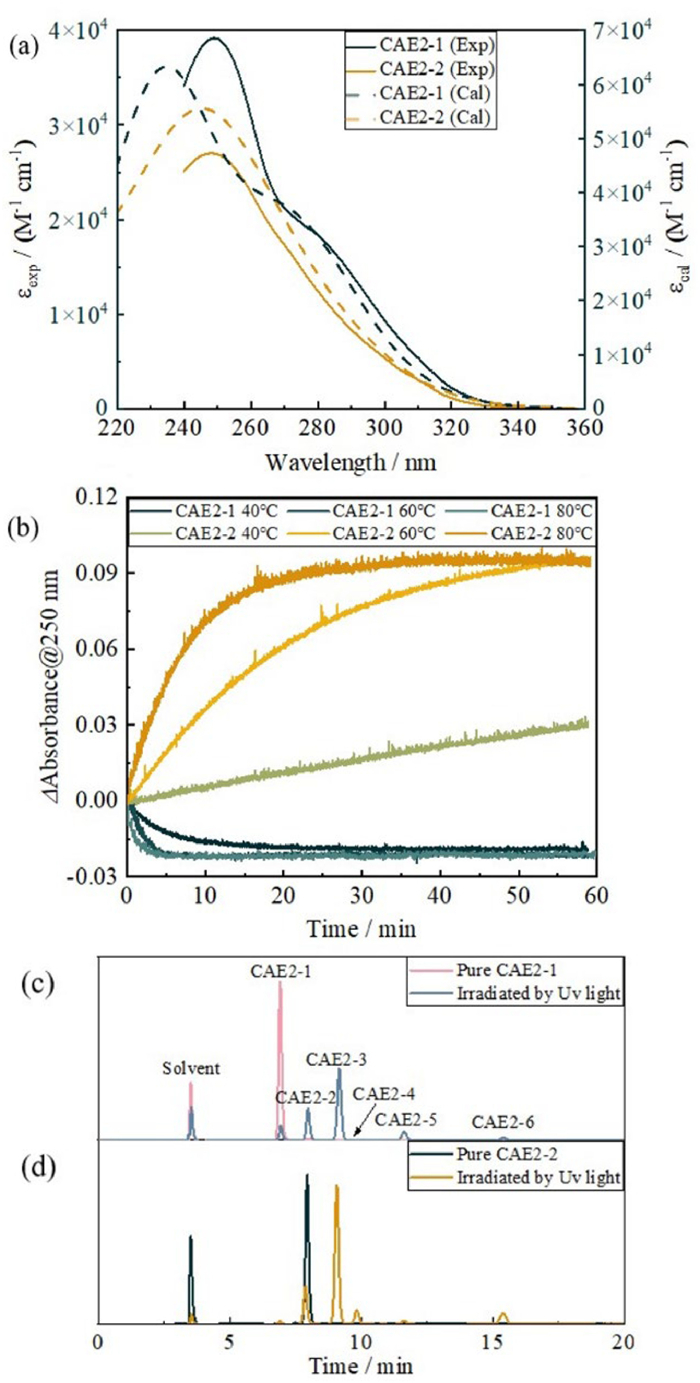
On the other hand, isomerization occurred in the presence of a catalytic amount of Brønsted acid. For example, when CAE2–1 was heated in acetonitrile (ACN): H2O (9:1 with 0.05% HCl), a decrease in absorbance at 250 nm was observed with the time (Fig. 2b and Fig. S15 in Supporting information). HPLC analyses revealed the emergence of CAE2–2 peak, while other isomers remained negligible. The variation in absorption intensity over time was fitted using the first-order kinetic model to derive the apparent reaction rate constants (Table S3 in Supporting information), giving k values of 1.9 × 10⁻¹ min−1 at 40 ℃, 5.9 × 10⁻¹ min−1 at 60 ℃ and 8.3 × 10⁻¹ min−1 at 80 ℃, respectively. These reactions ultimately led to the formation of CAE2–2 with yields of approximately 10% at equilibrium, although CAE2–1 remained dominant.
Conversely, the reaction of CAE2–2 under the acid-catalyzed conditions led to a pronounced increase in absorbance at 250 nm (Fig. 2b and Fig. S16). Curve fitting following the first-order kinetic model yielded k values of 6.6 × 10⁻³ min−1 at 40 ℃, 4.7 × 10⁻² min−1 at 60 ℃ and 1.3 × 10⁻¹ min−1 at 80 ℃ in ACN: H2O (9:1 with 0.05% HCl) (Table S4 in Supporting information). HPLC traces showed that at the equilibrium, most of CAE2–2 converted into CAE2–1, with ca. 12% of CAE2–2 remaining and negligible peaks of other [2 + 2] isomers. These results demonstrated cis/trans isomerization of the double bond under the catalysis of a small amount of acid, contrary to most acid-catalyzed isomerization of C=C bond, such as stilbene, by concentrated mineral acids [25,26]. The above findings suggested that the all-trans isomer CAE2–1 is the most stable isomer, likely due to the much lower energy of the trans double bond compared to cis. This observation is consistent with DFT calculations, which indicated that the Gibbs free energy of CAE2–1 was 2.1 kJ/mol lower than that of CAE2–2 (Fig. S22 and Table S16 in Supporting information). The same procedure was also conducted in chloroform containing 0.05% trifluoroacetic acid (TFA), with UV–vis intensity changes monitored (Figs. S17 and S18). Single exponential fitting revealed that the k values obtained in ACN (aq.) are generally higher than those obtained in chloroform, for which the increased acidity of the catalyst in polar solvents should be responsible (Tables S1-S6 in Supporting information).
The above observations demonstrate an acid-catalysis approach to regulate the isomerization of crown aldoxime ether with a controllable rate dependent on acid concentration, temperature and solvent. However, this approach yielded other [2 + 2] macrocycles in negligible yield. Photoisomerization of CAE2–1 or CAE2–2 in chloroform was carried out by using a 310 nm LED light in a deoxygenated quartz tube. HPLC traces of the sample irradiated for 3 min revealed that CAE2–3 emerged as the major photoproduct (Figs. 2c and d). In addition, CAE2–4, CAE2–5 and CAE2–6 also appeared with low yields, while a small amount of CAE2–1 and CAE2–2 either remained or were produced. CAE2–3 to CAE2–6 were separated by chromatography, and NMR analyses revealed CAE2–3, CAE2–4, CAE2–5, and CAE2–6 have configurations of trans,cis,trans,cis; trans,cis,cis,trans; trans,trans,cis,cis and trans,cis,cis,cis, respectively (Scheme 1, Figs. S6-S9 in Supporting information). However, there was no evidence of the formation of [2 + 2] macrocycle with four cis double bonds (CAE2–7) obtained through photoisomerization.
The chemical structures of CAE2–1 and CAE2–2 were also studied through DFT calculations (Figs. S19-S21 in Supporting information). All theoretical calculations were performed using Gaussian 09, and the results were analyzed and refined using Multiwfn [27,28]. All thermodynamic data was calculated using Shermo [29]. Calculation of their cavity diameter revealed that CAE2–1 possessed a larger cavity having a van der Waals diameter of 1.79 Å, while the cavity diameter of CAE2–2 was 0.85 Å (Fig. S20), both of which are too small to accommodate threading of an alkyl chain. 1H NMR spectral titration of G1 (2 mmol/L) with CAE2–1 in deuterated chloroform at 25 ℃ (Fig. S23 in Supporting information) revealed that increasing the concentration of CAE2–1 led to upfield shifts of G1 peaks (Fig. S24 in Supporting information), in contrast to the distinct splits observed when a crown ether was threaded by the alkyl chain of a guest [12,30]. This revealed that the complexation/decomplexation was a fast exchange process at the NMR timescale. Nonlinear least squares fitting yielded an association constant Ka of 123.2 L/mol (Fig. S26b in Supporting information). These results implied that G1 complexed with CAE2–1 through the ammonium cation attached to the cavity of CAE2–1, rather than threading through it, similar to the complexation of a small-sized crown ether with a quaternary ammonium salt [31].
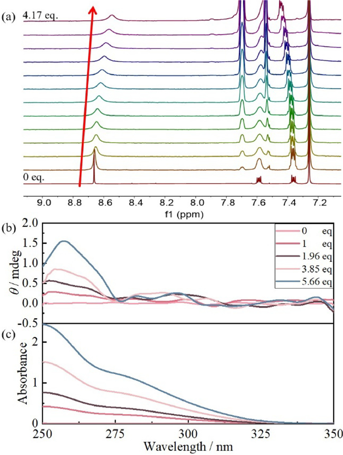
G2 was employed as a chiral guest to investigate the host-guest complexation with CAE2–1. G2 itself exhibited weak binding with the crown aldoxime ethers, while protonized G2 showed good complexation, demonstrating that the ion-dipole interactions dominate the complexation of crown aldoxime ethers. The 1H NMR spectral titration of CAE2–1 (2 mmol/L) with G2·BArF [32] showed a broadening and upfield shift of HC upon complexation (Fig. 3a and Fig. S25 in Supporting information). Nonlinear least squares fitting yielded a Ka of 366.7 L/mol (Fig. S26a in Supporting information). To explore chiroptical induction, the complexation of G2·PF6 with CAE2–1 was studied via circular dichroism (CD) spectroscopy (Fig. 3b). Adding CAE2–1 into the G2·PF6 solution led to a positive CD signal peaking at 257 nm, confirming the complexation and a chiroptical induction by guest binding. Other isomers could also form complexes with G2·PF6 and induced varied CD spectra (Figs. S27-S29 in Supporting information), demonstrating that the macrocyclic cis/trans isomers elicited different complexation and CD induction.
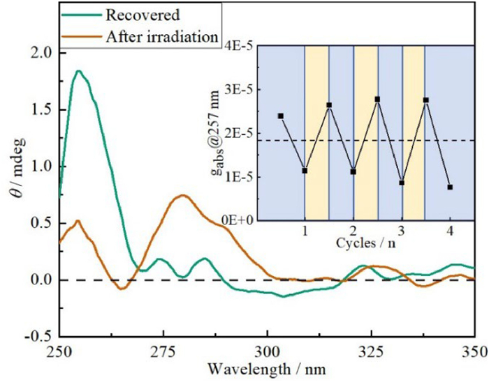
The acid-catalyzed and photo-induced isomerization of the macrocycles could provide a unique orthogonal handle for switching their complexation and chiroptical properties [10]. To manifest this, CAE2–1 (0.28 mmol/L) and G2·PF6 (1 mmol/L) were mixed in chloroform, resulting in an apparent positive Cotton effect at 257 nm (Fig. S30a in Supporting information). Upon irradiation with a 310 nm LED light for 3 min, the induced CD spectrum of the isomer mixture exhibited a decrease in the intensity at 257 nm, accompanied by the emergence of a positive Cotton effect at 280 nm (Fig. S30b in Supporting information). This signal should represent the sum of the CD signals generated by the multiple photoisomers complexing with G2·PF6 (Figs. 3b and 4, and Figs. S27-S31 in Supporting information). The mixture was then heated at 60 ℃ in the presence of 0.01% TFA to accelerate isomerization and recover the CD spectra. This entire procedure could be repeated multiple times, demonstrating good recyclability (Fig. 4).
In summary, we synthesized a series of crown aldoxime ethers with six cis-trans [2 + 2] isomers being separated via chromatography. Unlike common azo or imine, these crown aldoxime ethers demonstrate excellent thermal and hydrolysis stability. They undergo acid-catalyzed or photo-induced cis/trans isomerization, resulting in interconversion with varying isomer preferences. These crown aldoxime ethers can act as host molecules for complexation with organic ammoniums. The complexation of a chiral ammonium induces chiroptical effects at the transition band of the crown aldoxime ethers. By combining the isomerization processes, the complexation and chiroptical effects can be orthogonally controlled. The superior stability, isomerization properties, and complexation abilities render this new class of macrocycles promising for applications in host-guest chemistry, molecular assembly, and stimulus-responsive materials. Further chemical synthesis of aldoxime ether-based macrocycles and exploration of their binding and regulatory functions are currently underway.
The authors declare that they have no known competing financial interests or personal relationships that could have appeared to influence the work reported in this paper.
Yulin Mao: Writing – original draft, Investigation, Formal analysis, Data curation, Conceptualization. Jingyu Ma: Investigation, Data curation. Jiecheng Ji: Methodology, Data curation. Yuliang Wang: Supervision, Methodology. Wanhua Wu: Methodology, Data curation. Cheng Yang: Writing – review & editing, Supervision, Resources, Investigation, Conceptualization.
We acknowledge the support of the National Natural Science Foundation of China (Nos. 22271201, 92056116, 22171194, 22201194), the Science & Technology Department of Sichuan Province (Nos. 2022YFH0095 and 2021ZYD0052), the Fundamental Research Funds for the Central Universities (No. 20826041D4117). The Comprehensive Training Platform of Specialized Laboratory, College of Chemistry, Prof. Peng Wu and Dr. Pengchi Deng of the Analytical & Testing Center, Sichuan University.
Supplementary material associated with this article can be found, in the online version, at doi:
C.J. Pedersen, J. Am. Chem. Soc. 89 (1967) 7017–7036. doi: 10.1021/ja01002a035
B.L. Allwood, H. Shahriari-Zavareh, J.F. Stoddart, D.J. Williams, J. Chem. Soc., Chem. Commun. (1987) 1058–1061.
L. Yang, Y. Li, H. Zhang, et al., Chin. Chem. Lett. 34 (2023) 108108. doi: 10.1016/j.cclet.2022.108108
X. Yu, W. Wu, D. Zhou, et al., CCS Chem. 4 (2022) 1806–1814. doi: 10.31635/ccschem.021.202101036
D. Zhang, W. Liang, J. Yi, et al., Sci. China Chem. 65 (2022) 1149–1156. doi: 10.1007/s11426-022-1233-x
X. Liang, Y. Shen, D. Zhou, et al., Chem. Commun. 58 (2022) 13584–13587. doi: 10.1039/D2CC05690A
B. Li, Q. Xu, X. Shen, et al., Chin. Chem. Lett. 34 (2023) 108015. doi: 10.1016/j.cclet.2022.108015
K. Zhang, M. Hu, J. Luo, et al., Chin. Chem. Lett. 33 (2022) 1505–1510. doi: 10.1016/j.cclet.2021.08.072
H. Ma, R. Ye, L. Jin, et al., Chin. Chem. Lett. 34 (2023) 108355. doi: 10.1016/j.cclet.2023.108355
J. Yao, H. Mizuno, C. Xiao, et al., Chem. Sci. 12 (2021) 4361–4366. doi: 10.1039/D0SC06988D
J. Yao, W. Wu, W. Liang, et al., Angew. Chem. Int. Ed. 56 (2017) 6869–6873. doi: 10.1002/anie.201702542
S. Dasgupta, J. Wu, Chem. Sci. 3 (2012) 425–432. doi: 10.1039/C1SC00613D
S.J. Rao, Q. Zhang, J. Mei, et al., Chem. Sci. 8 (2017) 6777–6783. doi: 10.1039/C7SC03232C
T. Taga, S. Takaoka, S. Uemura, M. Funahashi, Mater. Chem. Front. 6 (2022) 880–890. doi: 10.1039/D1QM01542G
T. Tsuchiya, T. Shimizu, N. Kamigata, J. Am. Chem. Soc. 123 (2001) 11534–11538. doi: 10.1021/ja0102742
T. Amaya, H. Fujimoto, T. Tanaka, T. Moriuchi, Org. Lett. 20 (2018) 2055–2058. doi: 10.1021/acs.orglett.8b00598
C.H. Pollok, T. Riesebeck, C. Merten, Angew. Chem. Int. Ed. 56 (2017) 1925–1928. doi: 10.1002/anie.201610918
J. Yao, W. Wu, C. Xiao, et al., Nat. Commun. 12 (2021) 2600. doi: 10.1038/s41467-021-22880-z
H. Deng, G. Gong, S. Lv, et al., Org. Chem. Front. 10 (2023) 317–326. doi: 10.1039/D2QO01691E
Y. Liu, H. Wang, P. Liu, et al., Angew. Chem. Int. Ed. 60 (2021) 5766–5770. doi: 10.1002/anie.202015597
I. Lentin, A. Gorbunov, S. Bezzubov, et al., Org. Chem. Front. 10 (2023) 1470–1484. doi: 10.1039/D2QO01986H
J. Kalia, R.T. Raines, Angew. Chem. Int. Ed. 47 (2008) 7523–7526. doi: 10.1002/anie.200802651
K. Kanagaraj, R. Wang, M.K. Zhao, et al., J. Am. Chem. Soc. 145 (2023) 5816–5823. doi: 10.1021/jacs.2c12907
Y. Cui, X. Wang, G. Lin, et al., J. Agric. Food. Chem. 70 (2022) 13862–13872. doi: 10.1021/acs.jafc.2c05766
C.F. Bernasconi, W.J. Boyle, J. Am. Chem. Soc. 96 (1974) 6070–6077. doi: 10.1021/ja00826a019
T. Hao, Y. Yang, W. Liang, et al., Chem. Sci. 12 (2021) 2614–2622. doi: 10.1039/D0SC05213B
T. Lu, F. Chen, J. Comput. Chem. 33 (2012) 580–592. doi: 10.1002/jcc.22885
J. Zhang, T. Lu, Phys. Chem. Chem. Phys. 23 (2021) 20323–20328. doi: 10.1039/D1CP02805G
T. Lu, Q. Chen, Comput. Theor. Chem. 1200 (2021) 113249. doi: 10.1016/j.comptc.2021.113249
C. Zhang, S. Li, J. Zhang, et al., Org. Lett. 9 (2007) 5553–5556. doi: 10.1021/ol702510c
S.I. Kang, M. Lee, D. Lee, J. Am. Chem. Soc. 141 (2019) 5980–5986. doi: 10.1021/jacs.9b01002
F. Gao, X. Yu, L. Liu, et al., Chin. Chem. Lett. 34 (2023) 107558. doi: 10.1016/j.cclet.2022.05.072

Figure 1 Partial 1H NMR spectra of (a) CAE2–1 in chloroform-d and (b) CAE2–2 in acetone-d6 measured at room temperature. HA, HB, HC, and HD were marked with red, orange, green, and black squares, respectively. Front view of the single crystal structure of (c) CAE2–1 and (d) CAE2–2.

Figure 2 (a) UV–vis spectra of 40 µmol/L CAE2–1 (black) and 40 µmol/L CAE2–2 (yellow) in THF obtained by experimental measurement and theoretical calculation. (b) Time course of UV absorption at 250 nm upon heating CAE2–1 and CAE2–2 in ACN: H2O (9:1 with 0.05% HCl) at 40 ℃, 60 ℃ and 80 ℃, respectively. HPLC traces of (c) CAE2–1 before (pink) and after (blue) and (d) CAE2–2 before (black) and after (yellow) irradiating with a 310 nm LED light in chloroform.

Figure 3 (a) Enlarged 1H NMR spectral changes of CAE2–1 (2 mmol/L) upon titrating with G2·BArF in chloroform-d at 25 ℃; G2·BArF/CAE2–1 = 0, 0.05, 0.10, 0.15, 0.20, 0.30, 0.49, 0.73, 0.96, 1.42, 1.85, 2.68, 4.17. (b) CD spectra and (c) UV–vis spectra changes of CAE2–1 upon titrating G2·PF6 (0.1 mmol/L) with CAE2–1 in chloroform at 25 ℃; CAE2–1/ G2·PF6 = 0, 1, 1.96, 3.85, 5.66.

Figure 4 CD spectra of the mixture of CAE2–1 (0.28 mmol/L) and G2·PF6 (1 mmol/L) in the presence of TFA (0.01%) obtained after irradiation with a 310 nm LED light for 3 min and subsequent recovery at 60 ℃ for 10 h. An insert displays the plot of the gabs value at 257 nm versus the number of cycles (n).

 扫一扫看文章

扫一扫关注我们
 DownLoad:

 下载:
下载: