
Figure 1.
(A) X-ray powder diffraction patterns of (a) ZnTi-LDH, (b) AVB, (c) AVB@ZnTi-LDH. (B) FTIR spectra of (a) ZnTi-LDH, (b) AVB, (c) AVB@ZnTi-LDH. (C) Schematic illustration of AVB@ZnTi-LDH.
Synthesis of an AVB@ZnTi-LDH composite with synergistically enhance UV blocking activity and high stability for potential application in sunscreen formulations
Xiaomeng Hu , Jie Yu , Lijie Sun , Linfeng Zhang , Wei Zhou , Dongpeng Yan , Xinrui Wang

Ultraviolet (UV) irradiation could be classified into UVA (320–400 nm), UVB (290–320 nm), and UVC (200–290 nm) for biological purposes [1]. As UVC has been absorbed by the ozone layer and rarely comes onto human skin, UVA and UVB are the objects we need to focus on in our daily lives. UVA penetrates deep into the skin, reaching the epidermis and dermis, and is considered a major promoter of skin aging, known as photoaging [1–3]. UVA induces oxidative damage to the skin, inflammatory cells, increased expression of MMPs for collagen degradation and pro-elastin dissociation, and reduced expression of elastin, thus inducing skin aging in humans. UVB irradiation of the skin causes apoptosis, oxidative stress, and acute inflammatory responses, ultimately increasing the risk of various skin diseases [4–7]. UV exposure induces damage to the antioxidant defense system of keratin-forming cells through the production of excess reactive oxygen species (ROS). This stimulates an inflammatory response and cellular damage, leading to impaired function in keratin-forming cells. Since the first commercial sunscreen was introduced in 1928 in the United States, the use of sunscreens as an integral part of the photoprotection strategy has expanded worldwide. Concerns have been raised on the efficacy of ultraviolet filters (organic or inorganic) and the photostability of these filters.
Most of the UV absorbers used in sunscreens are photo-stable under the conditions of use. One exception is 1-(4-(1,1-dimethylethyl)phenyl)−3-(4-methoxyphenyl)−1,3-propanedione (Avobenzone/AVB). In the sunscreen regulation of the United States Food and Drug Administration (US FDA), AVB is approved within the concentration of up to 3% in over-the-counter sunscreen formulations [8]. In the European Union, Australia, and China, the concentration limit of AVB is 5% in sunscreen products [9]. AVB exists in two tautomeric forms: the enol form and keto form. UV irradiation leads to the formation of a triplet state of AVB, which photodegrades and loses its UV protection. Therefore, numerous studies have been dedicated to improving the photostability of AVB and the mechanisms of the photostability have been discussed. In commercial sunscreen formulations, avobenzone is often associated with Octocrylene or 2–hydroxy-4–methoxy benzophenone (BP-3) to avoid photodegradation when used individually [10,11]. Unfortunately, scientists have been looking into the impact of these chemicals on marine life, finding that at high concentrations in a laboratory setting, they can have toxic effects on both coral and fish. Based on this, certain countries have banned the sale and use of some sunscreen products that contain oxybenzone, Octocrylene, and octinoxate, which restricted the application of AVB consequentially [12,13]. Finding a reef-friendly way to stabilize AVB has captured a great deal of researchers' attention. Li and Yuan et al. have enhanced the water solubility, photostability, and thermal stability of avobenzone by the preparation of its inclusion complex with 2-HP-β-CD [14]. Wang et al. have found that avobenzone could be encapsulated into the hierarchically mesoporous silica powder because of its high specific surface area and pore volume; encapsulated avobenzone still maintained excellent UV protection abilities [15]. However, cyclodextrins or hierarchically mesoporous silica (HMS) and AVB are bound by van der Waals forces. The bonding is weak, thus leading to poor stability of the inclusion complex, affecting its practical application.
Layered double hydroxides (LDHs) are a class of two-dimensional layered anionic materials with exchangeable interlayer anions, biocompatible, and simple to prepare. LDHs have been widely used in catalysis, optoelectronic materials, drug carriers, etc. [16–21]. LDHs also have great potential for cosmetic applications due to their unique properties. Luana Perioli et al. inserted the UV absorber 5-benzoyl-4–hydroxy-2–methoxy-benzenesulphonate acid (4BHF) into the ZnAl-LDH using an anion exchange method and found that permitted a reduction of 4BHF release avoiding its direct contact and penetration in the skin, preventing thus cutaneous reactions and allergy problems that may occur [22]. Wang et al. prepared ZnTi-LDH by changing the ratio of Ti cations and found that the addition of Ti made the UV absorption of LDH superior to that of conventional ZnAl-LDH and MgAl-LDH, and the photocatalytic activity of ZnTi-LDH was lower compared to that of TiO2 and ZnO, which means ZnTi-LDH would be safer for the skin [23]. Li et al. introduced p-aminobenzoic acid and cinnamic acid into the ZnTi-LDH interlayer to enhance UV absorption and improve the thermal stability of the organic guest [24]. Organic sunscreens such as 2-phenylbenzimidazole-5-sulfonic acid, and 4-aminobenzoic acid are ionic sunscreens, which can be combined with hydrotalcite by anion exchange method. However, not all sunscreens are ionic. Little research has been performed on the intercalation of neutral organic UV filters, such as AVB and 3-Benzylidene camphor, which cannot be incorporated by traditional methods. Not to mention the possible mechanisms of interaction and characterization of properties.
In this paper, we prepared the organic-inorganic composite AVB@ZnTi-LDH by reconstruction method and investigated the structure and properties of the composite. The photostability, chemical stability, biological activity, and UV resistance of the loaded samples were investigated at the level of material, cellular, and zebrafish embryos. It was shown that AVB@ZnTi-LDH has potential applications as a raw material for sun protection products.
The XRD patterns of ZnTi-LDH precursor, AVB, and AVB@ZnTi-LDH are shown in Fig. 1A. The XRD pattern of the ZnTi-CO3-LDH precursor (Fig. 1A(a)) exhibits typical characteristics of the LDH phase, a series of narrow, symmetrical strong peaks appear at low 2θ values in (003), (006), and (009); weaker peaks appear at high 2θ values, including two (110) and (113) reflections between 60° and 63° The strong (003) peak reflection at 13.16° and the reflections of (006), (009) at 24.36°, and 34.85°, which correspond to the basal and higher-order reflections. The basal spacing value (d003) of the ZnTi-CO3-LDH is 0.672 nm, which is close to the value reported in the literature [25,26]. It should be noted that ZnTi-CO3-LDH shows several weak reflections at 2θ = 25.30°, 38.57°, 53.88°, 55.06°, 62.68°, 68.75°, which can be attributed to a small amount of titanium dioxide impurities (PDF card No. 99–0008). The X-ray diffraction pattern of AVB shows the many sharp peaks characteristic of its crystalline nature in Fig. 1A(b). The XRD pattern of AVB@ZnTi-LDH is shown in Fig. 1A(c). Two faint peaks appear at 10.41° and 11.38°, illustrating that AVB has been loaded on ZnTi-LDH. The lack of significant forward displacement in peak (003) indicates that the addition of AVB did not alter the interlayer spacing.
The FT-IR spectrum of the ZnTi-LDH precursor (Fig. 1B(a)) shows a strong and broad absorption band between 3500~3000 cm−1 due to -OH stretching vibration. The band at 1505 cm−1 and its accompanying band at 1385 cm−1 are attributed to the υ3 stretching vibration of the interlayer carbonate anion. The spectrum of AVB (Fig. 1B(b)) shows narrow absorption peaks at 1594 cm−1, 1544 cm−1, and 1504 cm−1 for the carbonyl stretching vibration and the C=C stretching vibration, respectively. Compared to AVB and ZnTi-LDH precursor, AVB@ZnTi-LDH (Fig. 1B(c)) shows new characteristic peaks at 3000~2900 cm−1, 1594 cm−1, 1531 cm−1, and 1493 cm−1 attributed to the C-H stretching vibration, C=O stretching vibration, C=C stretching vibrations respectively. C=C stretching vibration were red-shifted to 1531 and 1493 cm−1 after the confinement to the ZnTi-LDH complex, implying that C=C may be involved in the reaction. The schematic of AVB binding to ZnTi-LDH is shown in Fig. 1C.
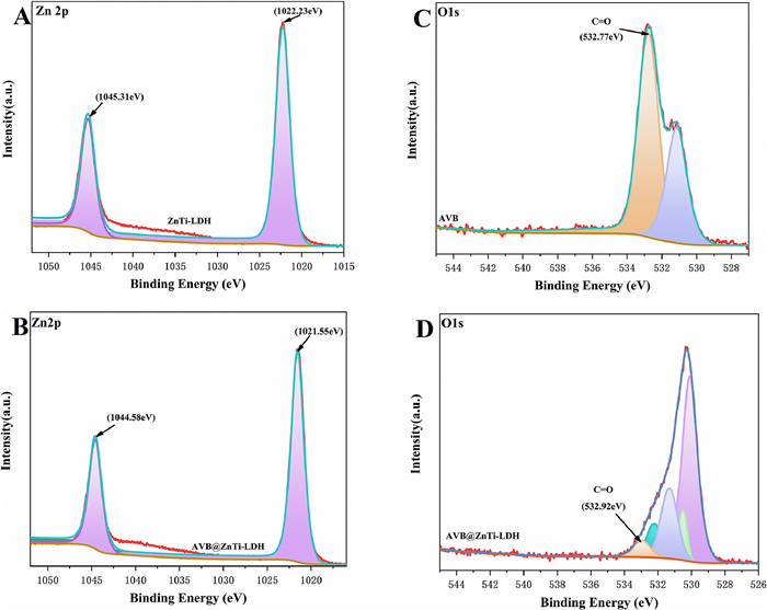
To further determine the bonding information between ZnTi-LDH and AVB, XPS analysis was performed on ZnTi-LDH precursor, pure AVB and AVB@ZnTi-LDH and the results are shown in Fig. 2. The Zn 2p XPS patterns of ZnTi-LDH and AVB@ZnTi-LDH showed characteristic peaks at 1045.31 eV, 1022.23 eV and 1044.58 eV, 1021.55 eV, respectively. The O 1s XPS patterns of AVB and AVB@ZnTi-LDH showed characteristic peaks at 532.77 eV and 532.92 eV, respectively. Compared with AVB, the binding energy of O in AVB@ZnTi-LDH samples was significantly reduced, which indicated that O tended to lose electrons gradually; compared with ZnTi-LDH, the binding energy of Zn in AVB@ZnTi-LDH samples was significantly reduced, which indicated that Zn tended to gain electrons gradually, suggesting that Zn was covalently coordinated with AVB, rather than ordinary drug loading. Therefore, this unique structure in AVB@ZnTi-LDH has the effect of stabilizing AVB.
DFT calculations were exerted to further understand the binding mode of AVB with ZnTi-LDH, and the adsorption energy (ΔE) calculations were carried out based on the model.
|
|
(1) |
E(slab) is the energy of ZnTi-LDH containing the vacancy, E(C20H22O3) is the energy after structural optimization of avobenzone, and E(total) is the energy after binding of avobenzone to ZnTi-LDH containing the vacancy.
The stability models for ZnTi-LDH, AVB, and AVB@ZnTi-LDH were obtained after optimization calculations as shown in Fig. 3. Due to computational cost considerations, the model simplifies the interlayer anions and water molecules in the LDH structure compared to the real LDH structure, retaining only the metal ion layer. This simplification is also justified in the calculations of the present system. Because AVB cannot enter the interlayer and the surface on which LDH reacts in contact with AVB is the metal ion layer. The normal ZnTi-LDH (Fig. 3A) structure has a layer of -OH on the surface and no metal exposure. In combination with this experiment, the hydroxyl group was removed from the metal surface after 200 ℃, so the hydroxyl group was chosen to be removed and a model of ZnTi-LDH containing vacancies was constructed (Fig. 3B).
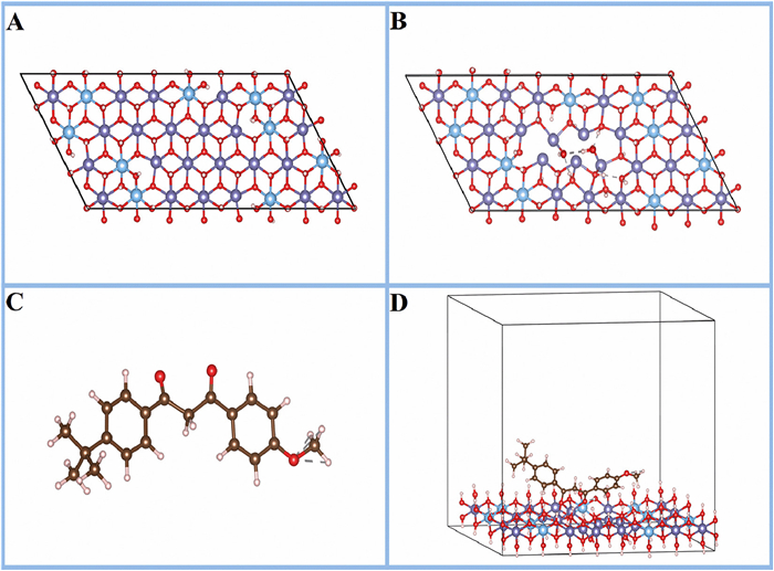
According to the adsorption energy equation, the ΔE of AVB is −1.38 eV after forming a complex with ZnTi-LDH. When the adsorption energy is negative, the adsorption process can proceed spontaneously and is accompanied by the release of energy, indicating that the model's binding model is spontaneously viable. At the same time, the C—C bond lengths in the β-diketone structure of AVB were reduced from 1.532 Å and 1.527 Å to 1.519 Å and 1.500 Å respectively, resulting in a more stable six-membered ring structure. Because of this, the photostability and chemical stability of AVB are enhanced, which is consistent with our experimental results.
To confirm the high biocompatibility and low cytotoxicity of AVB@ZnTi-LDH for further application in sunscreen formulations, composites at different concentrations were added to the cell and evaluated their effects on HSF and HaCaT cell viability.
Cytotoxicity assay helps to determine the effects of various drugs on cell viability. The toxicity of AVB, ZnTi-LDH, and AVB@ZnTi-LDH to HSF cells is shown in Fig. 4A. Cell survival of AVB-treated cells was significantly lower than that of ZnTi-LDH and AVB@ ZnTi-LDH, it was only about 30% at high concentrations. The cell viability of ZnTi-LDH and AVB@ZnTi-LDH-treated cells remained above 95%, indicating the excellent biocompatibility of AVB@ZnTi-LDH even at the high concentration of 100 µg/mL. In addition, in the study of the cytoprotective effects of AVB@ZnTi-LDH on UVA (Fig. 4B). Compared with the UVA damage model group, the cell death rate under AVB@ZnTi-LDH incubation was significantly reduced. Furthermore, the morphological changes of both HSF and HaCaT cells have been recorded by an inverted microscope. As revealed in Fig. 4C, in the control group, the normal cells were long pike-shaped with dense cells, the cells were closely interconnected and well defined. However, UVA irradiation at 10 J/cm2 resulted in a gradual decrease in cell density, a reduction in the characteristic fibrous ends of the cells, and the death of some cells into spherical aggregates. In contrast, the HSF cells appeared to be denser and more sharply defined, with less cellular debris when treated with AVB@ZnTi-LDH.
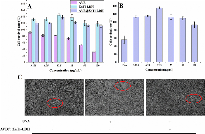
The toxicity of AVB, ZnTi-LDH, and AVB@ZnTi-LDH against HaCaT cells is shown in Fig. S1A (Supporting information). As with HSF cells, AVB was also much more toxic to HaCaT than AVB@ZnTi-LDH, and the cell viability decreased with the increase of AVB concentration. In addition, when the cytoprotective effect of AVB@ZnTi-LDH against UVB was investigated (Fig. S1B in Supporting information), the cell death rate under AVB@ZnTi-LDH incubation was significantly reduced compared with that in the UVB damage group. In the control group, the normal cells were flat, polygonal, and had round, central nuclei, the cells were closely connected to each other and their outlines were clear. However, 80 mJ/cm2 of UVB irradiation exposure has caused the cells to become round and shrunk in size, and parts of cells could be noticed floating, appearing to be dead. By comparison, it is clear from Fig. S1C (Supporting information) that treatment with AVB@ZnTi-LDH had significant effects on reducing the levels of UVB irradiation-induced injury. It appears that the HaCaT cells receiving AVB@ZnTi-LDH treatment showed less swelling, clearer contour, and reduced cell debris.
Through cell experiments, it is obvious that AVB@ZnTi-LDH has almost no toxic side effects on cells within the concentration range selected in this experiment, and can effectively prevent both UVA- and UVB-induced cell death.
Zebrafish were exposed to AVB@ZnTi-LDH and BP-4 and survival rates are shown in Fig. 5. When zebrafish were exposed to BP-4 at a concentration of 0.313 mg/mL (Fig. 5A), they died and were in poor condition compared to the normal group. In AVB@ZnTi-LDH, zebrafish showed mortality at high concentrations of 2.5 mg/mL (Fig. 5B). Composite AVB@ZnTi-LDH is much more compatible with zebrafish than BP-4, which has been reported to have toxic effects on aquatic organisms, so it can be said that AVB@ZnTi-LDH is relatively friendly.
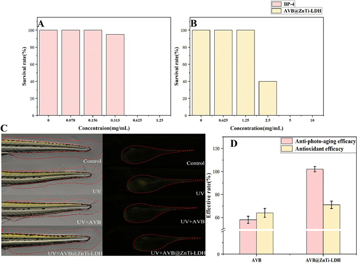
We then recorded fin morphology and yolk sac staining in a group of embryos. As shown in Fig. 5C, all mock control embryos (untreated with UV light) showed normal fins and fluorescence, but embryos exposed to UV light showed a higher incidence of aberrant fin phenotypes and higher fluorescence. We found that the coated sample group showed significantly less caudal fin wrinkling and diminished fluorescence. To further evaluate the protection of the samples against UV light, we introduced the concepts of anti-photo-aging efficacy and anti-oxidative stress efficacy by calculating the tail fin area and fluorescence intensity (Eqs. 2 and 3). The results are shown in Fig. 5D, both anti-photo aging efficacy and anti-oxidative stress efficacy we found that the composite AVB@ZnTi-LDH is superior to AVB.
|
|
(2) |
A: Zebrafish caudal fin area
|
|
(3) |
S: Fluorescence intensity of zebrafish yolk sac
The UV–vis spectra of AVB, ZnTi-LDH, and AVB@ZnTi-LDH were recorded. As shown in Fig. 6A, the addition of ZnTi-LDH produces a significant increase in the absorption intensity of AVB. The curve for ZnTi-LDH (Fig. 6A(a)) shows strong UV absorption between 200 nm and 350 nm that is attributed to the shielding effect of the LDH layers as well as the incorporation of Zn and Ti elements in the layers. The UV absorption curve of AVB (Fig. 6A(b)) shows that AVB has UV absorption between 200 nm and 450 nm. After the loading process, AVB@ZnTi-LDH exhibits better UV absorption than AVB and ZnTi-LDH at 200–450 nm (Fig. 6A(c)), while the UV absorption range is relatively extended. These observations are consistent with previous relevant literature.
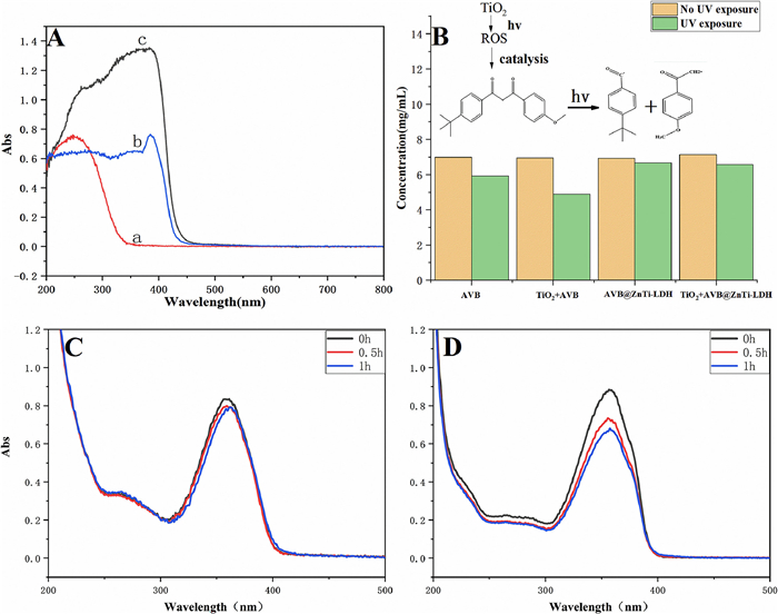
Avobenzone is one of the most commonly used UVA sunscreens, but it is very unstable, easily photolyzed to benzoyl methyl radicals and benzoyl radicals, and has no defensive effect against UV rays [27]. Especially when used together with titanium dioxide, the degradation rate of AVB was accelerated. [28] In this study, we prepared samples containing the same concentration of AVB and added equal amounts of TiO2 respectively. Then they were divided into two groups, one group did not receive light and the other group was irradiated with UV for 2 h, and then the content of AVB before and after UV irradiation was detected using HPLC, and the results are shown in Fig. 6B. After UV irradiation, AVB degradation rate was 15.06%, AVB@ZnTi-LDH degradation rate was 4.06% and AVB+TiO2 degradation rate was 29.75%, AVB@ZnTi-LDH +TiO2 degradation rate was 7.92%. There are two possibilities for this phenomenon, one is due to the laminar structure of LDH blocking part of the UV light; the other is due to the metal ions on the LDH laminate forming coordination bonds with C=O of AVB while forming a conjugated structure that makes AVB more stable. It is easy to see that the combination of AVB with ZnTi-LDH effectively reduces the decomposition of AVB when exposed to UV, and the results are even more pronounced with the addition of TiO2. When UV at wavelengths less than 387 nm is irradiated on titanium dioxide nanoparticles, electrons in the valence layer of the full band are excited into the conduction band to form negatively charged, highly reactive electrons (e−) and positively charged holes (h+) in the valence band, i.e., forming easily mobile and highly reactive e-h pairs (electron-hole pairs), which can recombine or migrate to the particle surface, participate in different redox processes, thus catalyzing the decomposition of avobenzone [29–31]. Therefore, titanium dioxide accelerates the degradation of AVB probably due to the photocatalytic activity, which upon illumination produces free radicals, resulting in accelerated decomposition of AVB.
In addition, we prepared solutions of AVB and AVB@ZnTi-LDH dissolved in ethanol to a certain concentration, and used a dark box UV analyzer to irradiate the solutions and detect the UV absorption at 0, 0.5, and 1 h respectively, and the results are shown in Figs. 6C and D. It is easy to see that with similar UV absorption, the UV absorption of both AVB@ZnTi-LDH and AVB decreased for the same time of UV exposure, but the rate of decrease of AVB@ZnTi-LDH was significantly lower than that of AVB itself. The results indicate that the UV resistance of AVB is more stable after loading under UV conditions. This is also consistent with the HPLC results.
In daily life, sunscreens containing AVB may accidentally get stained on clothes and turn them reddish brown, thus affecting the aesthetics. This may be mainly due to the complexation of carbonyl groups in AVB to higher valence ions commonly exist in sweat or washing water, forming a brownish-red complex that is likely to deposit on the surface of the clothes [32]. However, after loading onto the layered double hydroxide, the carbonyl groups in AVB are immobilized by Zn on the layered double hydroxide laminate. As the coordination with the iron ion is occupied by other metal ions, the complexation reaction with the trivalent iron ion is reduced and the stability of AVB itself is improved (Fig. S2 in Supporting information).
In summary, AVB@ZnTi-LDH was synthesized by reconstruction method and the confinement of AVB on the ZnTi-LDH can achieve the following advantages: (1) The AVB was considered as a long-wave UVA filter and further promote the UV blocking activity of ZnTi-LDH, due to the introduction of Zn and Ti elements with strong UV absorption and the unique lamellar structure of hydrotalcite. (2) AVB@ZnTi-LDH possessed high performances in photostability and chemical stability, attributed to the coordination between Zn on the layer and the oxygen atom of the carbonyl group of AVB. (3) For further application in sunscreen formulations, AVB@ZnTi-LDH composites at different concentrations confirmed the high biocompatibility and low cytotoxicity on HSF and HaCaT cell viability. (4) It is friendly to zebrafish, an aquatic organism, and has the potential to be used as an environmentally friendly sunscreen. All the findings suggest that LDH is a promising technique to improve the stability and enhance the UV absorption of AVB. This approach leads to a better use of avobenzone in sunscreen cosmetic formulations.
The authors declare that they have no known competing financial interests or personal relationships that could have appeared to influence the work reported in this paper.
The authors would like to thank Min Wang for her technical assistance. This work was supported by the China Scholarship Council (No. 202308110152).
Supplementary material associated with this article can be found, in the online version, at doi:
K. Tsuchida, N. Sakiyama, Y. Ogura, et al., Exp. Dermatol. 32 (2023) 146–153. doi: 10.1111/exd.14690
L.A. Schneider, K. Raizner, M. Wlaschek, et al., Exp. Dermatol. 26 (2017) 830–832. doi: 10.1111/exd.13286
C.C.E. Lan, Y.T. Hung, A.H. Fang, W. Ching-Shuang, J. Dermatol. Sci. 94 (2019) 220–228. doi: 10.1016/j.jdermsci.2019.03.005
H. Rahman, D. Kumar, T. Liu, et al., J. Invest Dermatol. 141 (2021) 132–141. doi: 10.1016/j.jid.2020.06.003
A. Moukova, L. Malina, H. Kolarova, R. Bajgar, IJMS 24 (2023) 9910. doi: 10.3390/ijms24129910
Y. Teng, Y. Yu, S. Li, et al., Front. Public Health. 9 (2021) 666528. doi: 10.3389/fpubh.2021.666528
L. Busch, M. Kröger, D.F. Zamudio Díaz, et al., Exp. Dermatol. 32 (2023) 1582–1587. doi: 10.1111/exd.14902
C. Yang, W. Lim, F.W. Bazer, G. Song, Reprod. Toxicol. 81 (2018) 50–57. doi: 10.1016/j.reprotox.2018.07.003
M.K. Matta, J. Florian, R. Zusterzeel, et al., JAMA 323 (2020) 256. doi: 10.1001/jama.2019.20747
K. Yamaguchi, R. Ohtsuka, K. Kaneko, et al., J. Oleo. Sci. 69 (2020) 1117–1124. doi: 10.5650/jos.ess19335
A.M. Cowden, A.L. Whittock, E.L. Holt, et al., RSC Adv. 13 (2023) 17017–17027. doi: 10.1039/D3RA02252H
R.B. Raffa, J.V. Pergolizzi, R. Taylor, et al., J. Clin. Pharm. Ther. 44 (2019) 134–139. doi: 10.1111/jcpt.12778
F. Seidel, Arch. Toxicol. 94 (2020) 3593–3594. doi: 10.1007/s00204-020-02865-5
L. Yuan, S. Li, D. Huo, et al., J. Photoc. Photobi. A 369 (2019) 174–180. doi: 10.1016/j.jphotochem.2018.09.036
W.H. Wang, H.T. Liang, Y.T. Yang-Wang, C.J. Shih, RSC Adv. 10 (2020) 15846–15852. doi: 10.1039/D0RA01837F
Z. Yang, J. Wei, G. Zeng, et al., Coordin. Chem. Rev. 386 (2019) 154–182. doi: 10.1016/j.ccr.2019.01.018
L.L. Liu, D. Zhu, L.L. Cao, D.W. Stephan, Dalton Trans. 46 (2017) 3095–3099. doi: 10.1039/C7DT00186J
A. Manna, S. Pramanik, A. Tripathy, et al., RSC Adv. 6 (2016) 25549–25561. doi: 10.1039/C6RA03093A
T. Hu, W. Shen, F. Meng, et al., Adv. Mater. 35 (2023) 2209692. doi: 10.1002/adma.202209692
Z. Lv, T. Hu, Y. Bian, et al., Adv. Mater. 35 (2023) 2206545. doi: 10.1002/adma.202206545
Y. Yang, T. Hu, Y. Bian, et al., Adv. Mater. 35 (2023) 2211205. doi: 10.1002/adma.202211205
L. Perioli, M. Nocchetti, V. Ambrogi, et al., Micropo. Mesopo. Mater. 107 (2008) 180–189. doi: 10.1016/j.micromeso.2007.02.021
X.R. Wang, Y. Li, L.P. Tang, et al., Chin. Chem. Lett. 28 (2017) 394–399. doi: 10.1016/j.cclet.2016.09.002
Y. Li, L. Tang, X. Ma, et al., J. Phys. Chem. Solid. 107 (2017) 62–67. doi: 10.1016/j.jpcs.2017.02.018
O. Saber, H. Tagaya, J. Incl. Phenom. Macro. 45 (2003) 109–111.
X.R. Wang, H.M. Cheng, X.W. Gao, et al., Chin. Chem. Lett. 30 (2019) 919–923. doi: 10.1016/j.cclet.2019.03.050
M. Kojić, M. Petković, M. Etinski, Phys. Chem. Chem. Phys. 18 (2016) 22168–22178. doi: 10.1039/C6CP03533G
R. Jansen, U. Osterwalder, S.Q. Wang, et al., J. Am. Acad. Dermatol. 69 (2013) 867. e1–867. e14.
T. Wang, Y. Zhu, Z. Xu, et al., J. Phys. Chem. C 120 (2016) 12293–12304.
X. Liu, W. Duan, Y. Chen, et al., Nanoscale Res. Lett. 11 (2016) 159. doi: 10.1186/s11671-016-1372-2
K. Lv, J. Yu, J. Fan, M. Jaronie, CrystEngComm 13 (2011) 7044–7048. doi: 10.1039/c1ce05907f
X.J. Zhang, Flavour Frag. J. 01 (2006) 12–14.

Figure 1 (A) X-ray powder diffraction patterns of (a) ZnTi-LDH, (b) AVB, (c) AVB@ZnTi-LDH. (B) FTIR spectra of (a) ZnTi-LDH, (b) AVB, (c) AVB@ZnTi-LDH. (C) Schematic illustration of AVB@ZnTi-LDH.

Figure 2 The Zn 2p XPS spectra of (A) ZnTi-LDH and (B) AVB@ZnTi-LDH, respectively. The O 1s XPS spectra of (C) AVB and (D) AVB@ZnTi-LDH, respectively.

Figure 3 (A) ZnTi-LDH, (B) ZnTi-LDH containing vacancies, (C) AVB, and (D) structures of AVB@ZnTi-LDH. Captions: white: H; brown: C; red: O; blue: Ti; purple: Zn.

Figure 4 Cell viability. (A) Cytotoxicity of AVB and AVB@ZnTi-LDH. (B) Protective effects of AVB@ZnTi-LDH on HSF with UVA. (C) Effects of AVB@ZnTi-LDH HSF cells with UVA.

Figure 5 Zebrafish toxicity of (A) BP-4, (B) AVB@ZnTi-LDH. (C, D) Effects of AVB@ZnTi-LDH zebrafish with UV.

 扫一扫看文章

扫一扫关注我们
 DownLoad:
 下载:
下载:
