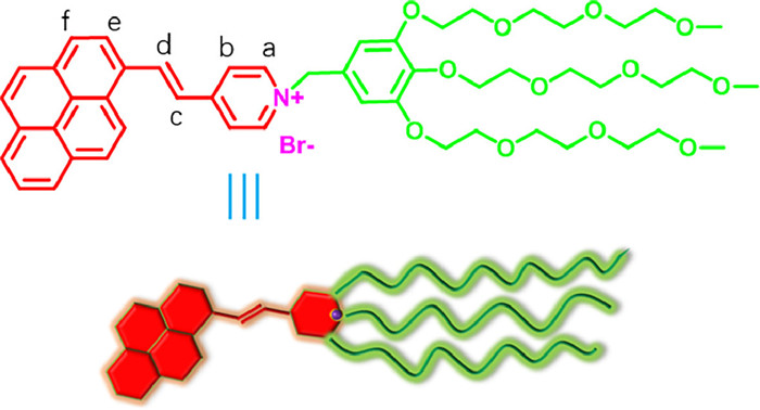
Figure 1.
The structure of amphiphilic ethylene-pyrene derivative 1.
Photo-induced tunable luminescence from an aggregated amphiphilic ethylene-pyrene derivative in aqueous media
Junying Zhang , Ruochen Li , Haihua Wang , Wenbing Kang , Xing-Dong Xu

Polychromatic luminescent materials with adjustable properties have gained significant attention for their potential applications in various fields such as fluorescent sensors [1,2], biological imaging agents [3], light-emitting diodes [4,5], and optoelectronic devices [6,7]. The conventional approach to obtaining these materials involves physically mixing or covalently bonding chromophores in specific proportions to achieve complementary fluorescence [8,9]. While this method is effective, it often entails complex and time-consuming organic synthesis steps, making it costly. Moreover, the hydrophobic nature of organic chromophores poses a significant challenge in preparing water-soluble luminescent materials [10-12].
Supramolecular assembly has emerged as a promising technique for the production of intelligent optical functional materials [13,14]. This approach involves the spontaneous formation of ordered structures with unique photophysical properties, achieved through non-covalent interactions or self-aggregation of a single chromophore component. In recent years, researchers have made progress in developing components that exhibit tunable multicolor emission characteristics in response to external stimuli [15,16]. For instance, Xu et al. introduced organic platinum fluorescent metallacycles, demonstrating high fluorescence quantum yield and adjustable emission wavelengths by modulating photoinduced electron transfer and intramolecular charge transfer properties [17]. Qu et al. combined pyrene fluorophore and acylhydrazone unit through amphiphilic self-assembly and γ-cyclodextrin-mediated host-guest recognition, resulting in a switchable multi-color emission system in different assembly states [18]. Tao et al. achieved diverse fluorescence emissions by manipulating the electron distribution of p-phenylenevinylene units within the hydrophobic cavity of cucurbit[8]uril [19]. Controlling polychromatic luminescence output through external stimuli provides a convenient approach for constructing novel photoluminescent materials [20,21]. Light is particularly interesting due to its clean, non-invasive, and remote-control characteristics [22,23]. However, there are limited reports on the adjustable, light-controlled fluorescent color conversion processes [24].
Pyrene units are commonly employed as fluorescent groups in building blocks for responsive supramolecular systems [25-27]. When stacked together in dimeric states, they exhibit strong redshift excimer emission, making them suitable for constructing stimulus-responsive fluorescent sensors [28-30]. However, these previous examples have limitations, as their emission wavelengths do not exceed 500 nm, resulting in a narrow spectral window for polychromatic fluorescence as a signal output. To overcome this limitation and broaden the emission window towards the red-shifted region, we introduced a pyridinium salt unit into a pyrene moiety connected with an alkenyl group (Fig. 1). This modification enhances the π-conjugated backbone and intramolecular charge transfer, potentially leading to a red-shift in fluorescence. Additionally, the other end of the pyridinium salt is covalently modified with three hydrophilic glycol chains, creating an amphiphilic molecule. This amphiphilic nature enables self-assembly in aqueous media, driving the ordered stacking of hydrophobic pyrene units in the inner membrane components.
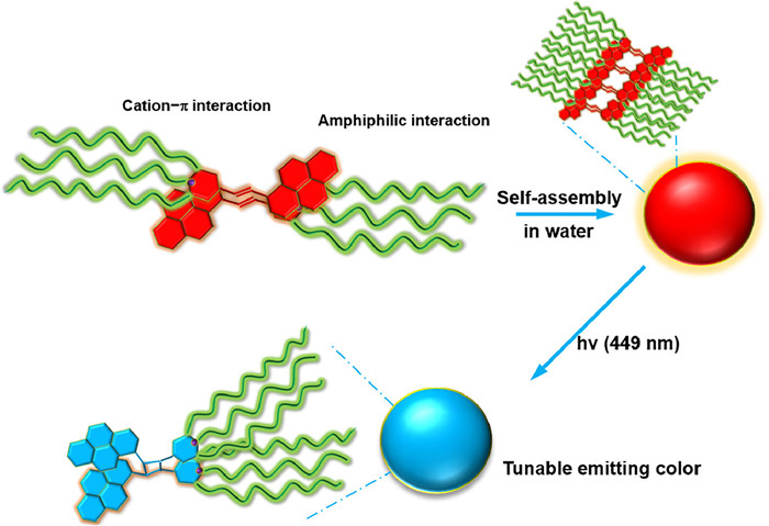
Scheme 1 presents a straightforward and promising photo-induced color-tunable material. Based on our previous research on fluorescent compounds [31-35], we designed amphiphilic compound 1. It incorporates a conjugated ethylene-pyrene fluorophore component, selected for its excellent planarity, strong fluorescence emission, and sensitivity to luminescence changes between the monomer and excimer states. The pyridinium salt serves as the terminal group, displaying intramolecular charge transfer properties with a noticeable Stokes shift. Additionally, a hydrophilic group based on glycol is appended to the fluorophore component, known for its ability to form amphiphilic compounds in solution. By controlling the irradiation time of a self-assembled solution of compound 1 in aqueous media, we achieved color tuning through [2 + 2] cycloaddition of the ethylene pyrene derivative. This process resulted in a wide Stokes shift (475−580 nm), enabling the formation of red, blue, and white emission assemblies. This operational versatility holds promise for the development of intelligent light-emitting materials.
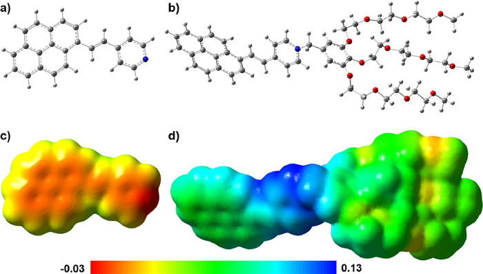
The synthesis of 1 is described in Scheme S1 (Supporting information). The structure of the product was confirmed using 1H and 13C NMR spectroscopy, as well as HR-ESI-MS. With the synthesized ethylene-pyrene compound in hand, we investigated its aggregation behavior both in solution and on surfaces. The aqueous solution of 1 showed an obvious red shift (around 35 nm) and significant broadening in its absorption spectrum compared to 1 in DCM (concentration: 1.0 × 10⁻⁵ mol/L, Fig. S1 in Supporting information). The observed shift and broadening in the spectrum can be attributed to J-aggregation-induced intramolecular charge-transfer (CT) [36]. To gain further insights into the aggregation behavior of compound 1, we performed DFT calculations. Our analysis focused on the molecular electrostatic potentials (MEP) and the changes in charge distribution around aromatic rings before and after alkylation [37]. For example, the pyridine ring, when alkylated by benzyl bromide, exhibited a significantly higher positive charge (0.85) compared to the free pyridine ring (0.08) upon the formation of the charged onium salt (Fig. 2). This alkylation also led to a slight decrease in electron density around the non-alkylated pyridine ring (0.25). These findings indicate a significant reduction in charge density in the alkylated compound compared to the free ethylene-pyrene hybrid. Furthermore, the analysis of MEP maps revealed the electron-deficient regions that facilitate charge transfer processes (Fig. S2 in Supporting information).
Given the amphiphilic properties of 1, we explored its self-assembly in aqueous solutions using SEM. As depicted in Fig. 3a, the self-assembled form of compound 1 displayed a spherical morphology, with diameters ranging between 150 nm and 200 nm. To gain further insights into the nature of these assembled nanostructures, we performed TEM measurements. Consistent with the SEM findings, TEM observations revealed the presence of spherical entities with a diameter of approximately 150 nm (Fig. 3b and Fig. S11 in Supporting information). Notably, these spherical structures exhibited a distinct contrast between the interior and periphery, indicative of vesicular structures. The corresponding thickness of the vesicle-like structures was calculated to be approximately 12 nm (Fig. 3b).
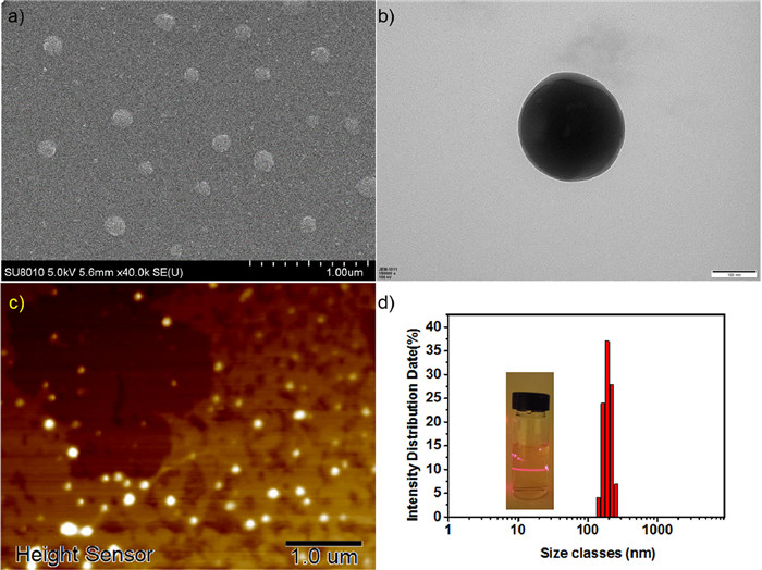
To further characterize the nanostructures obtained from compound 1, atomic force microscopy (AFM) was employed. AFM images revealed similar spherical structures (Fig. 3c and Fig. S12 in Supporting information), corroborating the findings from SEM and TEM studies. Moreover, the same solution of compound 1 exhibited the Tyndall effect, as evidenced by the inset in Fig. 3d, confirming the creation of nanoscale clusters. Dynamic laser scattering (DLS) analysis indicated a narrow size distribution of the nanospheres, with an average diameter of 200 nm at a scattering angle of 90° (Fig. 3d). These results strongly support the formation of vesicle assemblies of compound 1 in water.
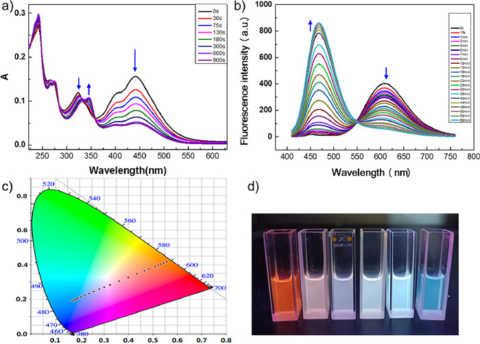
Typically, two π-bonds can undergo [2 + 2] photocycloaddition when they are nearly coplanar and positioned within a distance of 4.2 Å [38,39]. Therefore, the self-assembled ethylene-pyrene compound 1 represents an ideal candidate for photo-dimerization. To study the photo-reactivity of 1, its aqueous solution was irradiated with a 449 nm light source. The absorption peak at 450 nm gradually decreased, and nearly dropped to baseline after 15 min (Fig. 4a). This was accompanied by the appearance of two bands from free pyrene moiety at 330 and 345 nm. Similarly, the emission band around 608 nm gradually decreased and almost vanished after 60 min (Fig. 4b). In its place, new emission bands emerged at 467 nm, corresponding to excimer emission from free pyrene. Therefore, we conclude that the conjugated bond between the pyrene and pyridinium salt species has undergone photoreaction. Additionally, the presence of isosbestic point at 550 nm indicated selective photo-dimerization between the two unsaturated bonds, consistent with the findings of Barner-Kowollik and colleagues [40]. The ethylene-pyrene derivative 1 showed the quantum yield of 0.12 with nanosecond level lifetime (1.26 ns), while it showed a slightly higher quantum yield of 0.18 with 2.45 ns and 11.36 ns fluorescence decay (Fig. S6 in Supporting information). Considering the aggregation state often means fluorescence quenching, the fluorescence quantum yield is acceptable. The luminescence data were plotted in the Commission Internationale de L'Eclairage (CIE) 1931 chromaticity diagram at various exposure times, as shown in Fig. 4c. The CIE coordinates shifted from (0.56, 0.42) to (0.17, 0.19), revealing a transition from red to blue color. Notably, at a reaction time of 13 min, the CIE coordinates crossed the point (0.36, 0.31), indicating the emission of white light (Fig. 4c). Surprisingly, the expected dimeric complex was not formed when the photochemical reaction was conducted in pure dichloromethane solution at the same concentration, underscoring the significance of amphiphile aggregation. In addition, the morphology of the aggregate after illumination also showed nanosphere, but the diameter reduced from 150-200 nm to 50–70 nm (Fig. S7 in Supporting information).
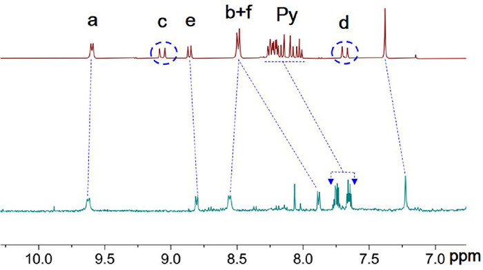
Additionally, 1H NMR spectroscopy evidences that the resonances of the protons of the conjugated system shifted or disappeared and the newly formed resonances are in good agreement with the formation of a cyclobutane moiety after [2 + 2]-cycloaddition (Fig. 5). Upon dimerisation, a shift can be observed for the aromatic resonances as well as new resonances appearing at 5.35 ppm, identified as the protons of the cyclobutane ring formed after cycloaddition.
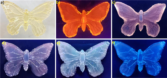
In addition, we have successfully demonstrated the potential of photo-induced emission color change for emission patterning, which is highly advantageous for multicolor display applications. To achieve this, a butterfly-shaped hydrogel was synthesized by polymerizing acrylamide in an aqueous solution with the adsorption of nano-scaled ethylene-pyrene hybrid 1 aggregates. Under daylight, the hydrogel appeared in a light yellow transparent state, while under ultraviolet lamp irradiation, it emitted bright red light, confirming the uniform dispersion of aggregates. To capture the color changes, we utilized a portable UV lamp, and the results are depicted in Fig. 6. Initially, the gel state exhibited a bright red fluorescence emission, which gradually transitioned to pink, then to white, and ultimately settled into a stable blue state, indicating the absence of further color changes. Remarkably, the hydrogel displayed high resolution and excellent durability when exposed to daylight, positioning it as a promising platform for multicolor display applications.
In conclusion, we have successfully synthesized an amphiphilic derivative with a large Stokes shift by introducing flexible hydrophilic long chains into a rigid ethylene-pyrene compound. The alkylated compound exhibited a notable change in charge distribution, facilitating cation-π interactions. Through the process of amphiphilic self-assembly, the formation of highly ordered aggregates enabled effective photo-dimerization under 449 nm LED irradiation. Our platform demonstrated its potential as a smart luminescent system capable of achieving a multi-color butterfly display. Notably, our photo-responsive technology not only exhibited advanced multi-color emission effects, including white light emission but also exhibited environmentally friendly behavior in the aqueous phase. This luminescent material represents a promising platform for the development of stimuli-responsive materials and multi-color display devices, surpassing previous reports in terms of versatility and eco-friendliness.
The authors declared that they have no conflicts of interest to this work. We declare that we do not have any commercial or associative interest that represents a conflict of interest in connection with the work submitted.
This work was supported by the National Natural Science Foundation of China (No. 21602124), Fluorine Silicone Materials Collaborative Fund of Shandong Provincial Natural Science Foundation (No. ZR2021LFG007), Key R&D Program of Shandong Province (No. 2019JZZY020229) and the Young Scholars Program of Shandong University (No. 2018WLJH40).
Supplementary material associated with this article can be found, in the online version, at doi:
Y. Lee, W. Cho, J. Sung, et al., J. Am. Chem. Soc. 140 (2018) 974–983. doi: 10.1021/jacs.7b10433
X. Jia, L. Zhu, Acc. Chem. Res. 56 (2023) 655–666. doi: 10.1021/acs.accounts.2c00818
W. Qian, M. Zuo, P. Niu, X.Y. Hu, L. Wang, Chin. Chem. Lett. 33 (2022) 1975–1978. doi: 10.1016/j.cclet.2021.09.070
T. Mori, Y. Yoshigoe, Y. Kuninobu, Angew. Chem. Int. Ed. 58 (2019) 14457–14461. doi: 10.1002/anie.201903408
F. Lu, T. Nakanishi, Adv. Opt. Mater. 7 (2019) 1900176. doi: 10.1002/adom.201900176
B.S. Santhosh, J. Aimi, H. Ozawa, et al., Angew. Chem. Int. Ed. 51 (2012) 3391–3395. doi: 10.1002/anie.201108853
L. Duan, Q. Zheng, T. Tu, Adv. Mater. 34 (2022) e2202540. doi: 10.1002/adma.202202540
D. Tu, P. Leong, S. Guo, et al., Angew. Chem. Int. Ed. 56 (2017) 11370–11374. doi: 10.1002/anie.201703862
C. Chen, X. Jin, X. Zhou, et al., J. Mater. Chem. C 3 (2015) 4563–4569. doi: 10.1039/C4TC02771J
H. Wu, Y. Chen, X. Dai, et al., J. Am. Chem. Soc. 141 (2019) 6583–6591. doi: 10.1021/jacs.8b13675
H. Sun, L. Zhu, Aggregate 4 (2023) 253. doi: 10.1002/agt2.253
Q. Xu, Z. Qin, Y. Bei, et al., Chin. Chem. Lett. 33 (2022) 4838–4841. doi: 10.1016/j.cclet.2022.01.079
K. Diao, D.J. Whitaker, Z. Huang, et al., Chem. Commun. 58 (2022) 2343–2346. doi: 10.1039/d1cc06647a
B.L. Zhang, X. Zhang, Chem. Commun. 58 (2022) 5261–5264. doi: 10.1039/D2CC01309F
R. Taniguchi, T. Yamada, K. Sada, et al., Macromolecules 47 (2014) 6382–6388. doi: 10.1021/ma501198d
N. Wang, L. Feng, X.D. Xu, et al., Macromol. Rapid Commun. 43 (2022) e2100885. doi: 10.1002/marc.202100885
J.L. Zhu, L. Xu, Y.Y. Ren, et al., Nat. Commun. 10 (2019) 4285. doi: 10.1038/s41467-019-12204-7
Q. Wang, Q. Zhang, Q.W. Zhang, et al., Nat. Commun. 11 (2020) 158. doi: 10.1038/s41467-019-13994-6
X.L. Ni, S. Chen, Y. Yang, et al., J. Am. Chem. Soc. 138 (2016) 6177–6183. doi: 10.1021/jacs.6b01223
X. Yan, F. Wang, B. Zheng, et al., Chem. Soc. Rev. 41 (2012) 6042–6065. doi: 10.1039/c2cs35091b
R. Fu, L. Yu, J. Zhang, et al., Chin. Chem. Lett. 33 (2022) 1993–1996. doi: 10.1016/j.cclet.2021.10.018
S. Chen, L.J. Chen, H.B. Yang, et al., J. Am. Chem. Soc. 134 (2012) 13596–13599. doi: 10.1021/ja306748k
D.H. Qu, Q.C. Wang, Q.W. Zhang, et al., Chem. Rev. 115 (2015) 7543–7588. doi: 10.1021/cr5006342
S. Chen, R. Costil, F.K. Leung, et al., Angew. Chem. Int. Ed. 60 (2021) 11604–11627. doi: 10.1002/anie.202007693
D. Niu, Y. Jiang, L. Ji, et al., Angew. Chem. Int. Ed. 58 (2019) 5946–5950. doi: 10.1002/anie.201900607
F. Camerel, L. Bonardi, M. Schmutz, et al., J. Am. Chem. Soc. 128 (2006) 4548–4549. doi: 10.1021/ja0606069
K.M. Chan, D.K. Kolmel, S. Wang, et al., Angew. Chem. Int. Ed. 56 (2017) 6497–6501. doi: 10.1002/anie.201701235
X. Chang, Z. Zhou, C. Shang, et al., J. Am. Chem. Soc. 141 (2019) 1757–1765. doi: 10.1021/jacs.8b12749
Q. Chen, K. Cheng, W. Wang, et al., J. Pharm. Anal. 10 (2020) 490–497. doi: 10.1016/j.jpha.2020.07.003
D. Kodura, L.L. Rodrigues, S.L. Walden, et al., J. Am. Chem. Soc. 144 (2022) 6343–6348. doi: 10.1021/jacs.2c00156
J. Zhang, S.X. Tang, R. Fu, X.D. Xu, S. Feng, J. Mater. Chem. C 7 (2019) 13786–13793. doi: 10.1039/c9tc03786a
M. Feng, J. Zhang, H. Ji, et al., Chem. Eng. J. 463 (2023) 142241. doi: 10.1016/j.cej.2023.142241
N. Wang, H. Feng, X. Hao, et al., Polym. Chem. UK 14 (2023) 1396–1403. doi: 10.1039/d3py00140g
N. Wang, J. Zhao, Q. Xu, et al., J. Mater. Chem. C: Mater. Opt. Electron. Devices 1 (2022) 1386–1387. doi: 10.3390/electronics11091386
R. Fu, J. Zhang, S. Liu, et al., Chem. Commun. 56 (2020) 6719–6722. doi: 10.1039/d0cc02214d
X.D. Xu, J. Zhang, X. Yu, et al., Chemistry 18 (2012) 16000–16013. doi: 10.1002/chem.201202902
B. Jiang, W. Wang, Y. Zhang, et al., Angew. Chem. Int. Ed. 56 (2017) 14438–14442. doi: 10.1002/anie.201707209
M.M. Gan, J.Q. Liu, L. Zhang, et al., Chem. Rev. 118 (2018) 9587–9641. doi: 10.1021/acs.chemrev.8b00119
Y. Xue, S. Jiang, H. Zhong, et al., Angew. Chem. Int. Ed. 61 (2022) e202110766. doi: 10.1002/anie.202110766
M. Van De Walle, K. De Bruycker, J.P. Blinco, et al., Angew. Chem. Int. Ed. 59 (2020) 14143–14147. doi: 10.1002/anie.202003130

Scheme 1 Schematic of the formation of nanovesicle from amphiphilic self-assembly based on ethylene-pyrene derivative 1 and photo-responsive controllable luminescence behavior.

Figure 2 Geometric structure of ethylene-pyrene derivative 1 optimized by the PM6 semiempirical molecular orbital method. Distribution of the electrostatic potential mapped onto the electron density surface of ethylene-pyrene derivative 1.

Figure 3 SEM image (a), TEM image (b), and AFM image (c) of ethylene-pyrene derivative 1 prepared in aqueous solution (2.5 mmol/L); the scale bars of the image are 1 µm, 200 nm, 1 µm, respectively.

Figure 4 The absorption spectrum (a) and emission spectra (b) of ethylene-pyrene derivative 1 in aqueous solution over 60 min upon exposure to 449 nm LED light. (c) CIE chromaticity diagram showing the time dependence of the (x, y) color coordinates of 1. (d) Photographs of solution of 1 (10.0 µmol/L) upon irradiation for different times under UV light at 365 nm.

Figure 5 1H NMR spectra (acetone-d6, 400 MHz) of compound 1 before and after irradiation.

 扫一扫看文章

扫一扫关注我们
 DownLoad:

 下载:
下载:
