
Figure 1.
Normalized absorbance (a) and fluorescence spectra (b) of DCITT in mixture of water and THF. (c) Relationship between FL intensity and polarity (Δf) of mixture (water and THF).
Water–soluble and polarity–sensitive near–infrared fluorescent probe for long–time specific cancer cell membranes imaging and C. Elegans label
Lixian Fu , Yiyun Tan , Yue Ding , Weixia Qing , Yong Wang

Cancer is one of the diseases threatening human health and many people die of cancer every day [1,2]. Therefore, it is urgent to find ways to diagnose cancer at an early stage [3]. However, some traditional detection methods have been unable to meet the needs of tumor diagnosis due to lack of sufficient sensitivity and selectivity at the cellular level [4,5]. It is imperative to development more effective early diagnosing methods. At present, with the development of fluorescence probe technology, it can provide reliable information for the visual detection of tumor cells due to its advantages of non–invasive, high selectivity and sensitivity [6-11]. And it is a promising method to design fluorescent probes based on microenvironmental differences (such as polarity and viscosity) between tumor and normal cells [12-14].
The cell membrane is located in the outermost layer of the cell, which prevents extracellular substances from entering the cell and ensures a stable intracellular environment [15]. Due to its special semi–permeability, it has physiological functions such as substance transport, signal transmission and cell growth [16,17]. There is a strong affinity between tumor cells and lipids and cholesterol [18,19], so cell membrane polarity of tumor cells is lower than that of normal cells, which can be used as a biomarker to differentiate tumor cells from normal cells. Fluorescent probes that specifically light up tumor cell membranes have clinical significance for early diagnosis. But it is extremely challenging to construct such fluorescent probes. So far, few near–infrared fluorescent probes have selectively light up tumor cell membranes [20-22].
Herein, we report a polarity sensitive and membrane targeting probe (DCITT, Scheme S1 in Supporting information). Due to its D–π–A structure, the fluorescence emission wavelength of DCITT reaches near–infrared region (NIR) through the push–pull electron effect, which can minimize photodamage and avoid cell autofluorescence [23]. DCITT targets cell membrane through hydrophobic and electrostatic interaction with the phospholipid bilayer of the cell membrane. Moreover, DCITT is sensitive to polarity. In high–polarity medium, DCITT and solvent have strong solvent effect, the excited energy is lost through non–radiation, resulting in the red shift of emission wavelength. Encouragingly, it can selectively images tumor cell membranes and shows bright and long–lasting red fluorescence, but fluorescence is almost invisible on normal cell membranes. Imaging of DCITT confirms that cell membrane polarity is lower in cancer cells than in normal cells, which is consistent with previous report [24]. The work provides an effective way for early diagnosis of cancer.
DCITT is a D–π–A structure, triphenylamine and thiophene are electron donors, π–bridge and dicyanoisoflurone are strong electron acceptors. And DCITT is characterized by polarity sensitivity due to the intramolecular charge transfer. The new compounds were confirmed by nuclear magnetic resonance (NMR) and high resolution mass spectrometry (HRMS) (Figs. S1−S9 in Supporting information).
The emission spectrum of DCITT is polar dependent, its wavelength redshifts with increasing solvent polarity, indicating that the excited state energy is released in non–radiative form [25,26]. However, its absorption spectrum does not change significantly in polar solutions (Fig. S10 in Supporting information), showing that the ground state dipole moment changes little in different solvents [27]. Besides, the fluorescence quantum yield (Φs) gradually decreases with the increase of solvent polarity (Table S1 in Supporting information). For instance, Φs is 6.82% in tetrahydrofuran (THF) but it decreases to 0.025% in H2O. The results show that DCITT is sensitive to solvent polarity.
The polarity response of DCITT was further studied by detecting its spectra in a mixture of THF and water. From Fig. 1a, ultraviolet–visible (UV–vis) spectra of DCITT do not change significantly with the increase of water content. However, the fluorescence spectra change significantly, the fluorescence intensity of DCITT sharply decreases by about 146 times when the solvent is converted from THF to H2O (Fig. 1b). What is more, the fluorescence intensity has a good linear relationship with Δf (Fig. 1c). Moreover, as shown in Figs. 1a and b, DCITT yields a Stokes shift greater than 300 nm, meaning that DCITT has strong penetration and little damage to biological samples. The results indicate that DCITT is a polar–dependent fluorescent probe.
Subsequently, fluorescence spectra of DCITT in glycerol (Gl) and MeOH were studied. With the increase of Gl content (0–100%), the viscosity of mixture also increases. In the intracellular viscosity range, as viscosity (η) increases from 0.59 (vMeOH/vGl = 100%/0%) to 55.0 cP (vMeOH/vGl = 40%/60%), fluorescence intensity of DCITT only increases by 1.33 times (Fig. S11 in Supporting information), indicating that the viscosity effect is negligible. Because the viscosity of THF and MeOH is similar, but the dielectric constant and polarity are obviously different. Next, the fluorescence intensity of DCITT in THF, MeOH and different proportions of Gl is compared to further explore the influence of polarity and viscosity on DCITT. From Fig. S11, the fluorescence intensity of DCITT in THF is obviously stronger than that in MeOH and Gl, indicating that the influence of viscosity on DCITT is far smaller than that of polarity. It has been reported that nystatin (Nys) can change ultrastructure of cells, resulting in increased cell viscosity [28]. So, we chose Nys as a stimulus to test the effect of cellular viscosity on DCITT. From Fig. S12 (Supporting information), the fluorescence intensity of HeLa cells treated with DCITT alone is the same as that of HeLa cells treated with DCITT and Nys, further showing DCITT is only regulated by polarity.
In view of the different pH of tumor and normal cells [27], we studied the fluorescence spectra of DCITT in different pH solutions. As a result, the emission fluorescence spectra do not change significantly (Fig. S13a in Supporting information). Then, we studied the effects of common ions and amino acids including Na+, K+, Ca2+, Mg2+, Zn2+, Cu2+, Fe3+, ClO−, OH•, 1O2, ONOO−, H2O2, lysine (Lys), leucine (Leu), methionine (Met), glycine (Gly), serine (Ser), valine (Val), cysteine (Cys) and glutathione (GSH) on the fluorescence of DCITT. From Fig. S13b (Supporting information), the fluorescence intensity of DCITT do not change significantly. It is worth noting that the low–polarity solvent (THF) is added to the test system, the florescence intensity is immediately enhanced, meaning that DCITT is unaffected by these molecules and ions.
We evaluated the toxicity of DCITT to tumor cells (MDA–MB–231, HeLa and HepG2) and normal cells (HL–7702 and RAW264.7). Even when DCITT reaches higher concentrations (50 µmol/L) and cell survival rate is more than 85%, indicating DCITT has low cytotoxicity (Fig. S14a in Supporting information). Next, DCITT solution (10 µmol/L) was continuously irradiated with a laser (535 nm) for 120 min, but the fluorescence intensity does not change significantly (Fig. S14b in Supporting information), showing that DCITT has good stability and it is suitable for long–term fluorescence imaging of cells.
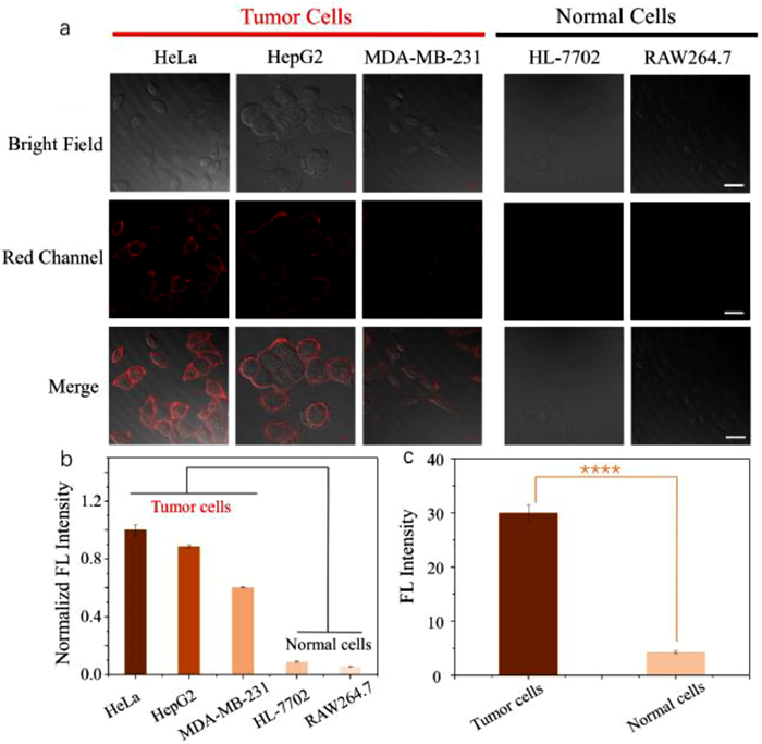
Then, we performed cell imaging experiments on cancer and normal cells. From Figs. 2a and b, it is very meaningful that strong red fluorescence signals of DCITT are observed only on the tumor cells membrane but there is little fluorescence elsewhere in the cells, indicating that DCITT can specifically target the tumor cell membranes. Because the distinction is statistically significant, DCITT can distinguish between tumor cells and normal cells by specifically lighting up tumor cells membrane (Fig. 2c).
Next, we examined the residence time of DCITT on the cell membrane. As shown in Fig. 3, after incubation for 2 h, DCITT still shows obvious red fluorescence on the cell membrane. However, for the commercial cell membrane green fluorescent probe Dio, after incubation for 1 h, part of Dio has penetrated the cell membrane and entered the cell. And after continued incubation for 1 h, almost all Dio has entered the cell, the green fluorescence is almost invisible on the cell membrane. Because Dio easily penetrates cell membrane and diffuses into the cell, it is not suitable for prolonged cell membrane imaging [29]. Meanwhile, the imaging of residence time on HeLa cell membrane was also monitored at the same location (Fig. S15 in Supporting information). All of the results prove that DCITT can realize long–time imaging of cell membrane.

Based on the unique transparent body and multicellular characteristics of C. elegans, the potential of DCITT in vivo imaging was examined. From Fig. 4, C. elegans fed with DCITT shows bright red fluorescence, suggesting that DCITT is ingested with food and accumulated in the body of C. elegans. Therefore, DCITT is expected to be a probe for labeling multicellular organisms.

In a word, we developed a polarity–sensitive and membrane–targeted near–infrared probe (DCITT). DCITT has a large Stokes shift (> 300 nm), high fluorescence quantum yield, low cytotoxicity and photostability. Importantly, DCITT is environmentally polar–dependent, can specifically light up cancer cells membrane and effectively distinguish between cancer cells and normal cells based on red fluorescence. And reasonable molecular structure enables it to be anchoring to the tumor cells membrane for a long time. In addition, DCITT can be used as a label probe in multicellular organisms. The work provides an effective tool for the study of cell membrane polarity and a new idea and method for the early diagnosis of cancer.
The authors declare that they have no known competing financial interests or personal relationships that could have appeared to influence the work reported in this paper.
We gratefully acknowledge Henan Provincial Science and Technology Research Project (No. 232102310369) for financial support.
Supplementary material associated with this article can be found, in the online version, at doi:
F. Bray, J. Ferlay, I. Soerjomataram, et al., CA Cancer J. Clin. 68 (2018) 394–424. doi: 10.3322/caac.21492
R.L. Siegel, A. Jemal, R.C. Wender, et al., CA Cancer J. Clin. 68 (2018) 329–339. doi: 10.3322/caac.21460
J.J. Zhang, L.L. Ning, J.G. Huang, et al., Chem. Sci. 11 (2020) 618–630. doi: 10.1039/c9sc05460j
D. Crosby, N. Lyons, E. Greenwood, et al., Lancet Oncol. 21 (2020) 1397–1399. doi: 10.1016/S1470-2045(20)30593-3
A.B. Chinen, C.M. Guan, J.R. Ferrer, et al., Chem. Rev. 115 (2015) 10530–10574. doi: 10.1021/acs.chemrev.5b00321
C. Duan, M. Won, P. Verwilst, et al., Anal. Chem. 91 (2019) 4172–4178. doi: 10.1021/acs.analchem.9b00224
J. Xu, J. Pan, X. Jiang, et al., Biosens. Bioelectron. 77 (2016) 725–732. doi: 10.1016/j.bios.2015.10.049
L. Jiang, T. Chen, E. Song, et al., Chem. Eng. J. 427 (2022) 131563. doi: 10.1016/j.cej.2021.131563
Y. Yang, H. Wu, B. Liu, Z. Liu, Adv. Drug Deliv. Rev. 179 (2021) 114004. doi: 10.1016/j.addr.2021.114004
Y. Li, G. Liu, J. Ma, et al., J. Control. Release 258 (2017) 95–107. doi: 10.1016/j.jconrel.2017.05.011
S. Wen, W. Wang, R. Liu, P. He, Int. J. Nanomed. 15 (2020) 3405–3414. doi: 10.2147/ijn.s233214
G. Peng, J. Dai, R. Zhou, et al., Anal. Chem. 94 (2022) 12095–12102. doi: 10.1021/acs.analchem.2c02077
Y.Q. Zhou, P. Li, X. Wang, et al., Chem. Sci. 11 (2020) 12149–12156. doi: 10.1039/d0sc02922j
A.K. Singh, A.V. Nair, N.D.P. Singh, Anal. Chem. 94 (2022) 177–192. doi: 10.1021/acs.analchem.1c04306
M. Collot, S. Pfister, A.S. Klymchenko, Curr. Opin. Chem. Biol. 69 (2022) 102161. doi: 10.1016/j.cbpa.2022.102161
R. Jahn, T. Lang, T.C. Südhof, Cell 112 (2003) 519–533. doi: 10.1016/S0092-8674(03)00112-0
S.J. Singer, G.L. Nicolson, Science 175 (1972) 720–731. doi: 10.1126/science.175.4023.720
R. Riscal, N. Skuli, M.C. Simon, Mol. Cell 76 (2019) 220–231. doi: 10.1016/j.molcel.2019.09.008
B. Huang, B. Song, C. Xu, Nat. Metab. 2 (2020) 132–141. doi: 10.1038/s42255-020-0174-0
H. Zhao, N. Li, C. Ma, et al., Chin. Chem. Lett. 34 (2023) 107699. doi: 10.1016/j.cclet.2022.07.042
Y.J. Reo, M. Dai, Y.J. Yang, K.H. Ahn, Anal. Chem. 92 (2020) 12678–12685. doi: 10.1021/acs.analchem.0c03013
L. Feng, Y. Xie, S.K. Au–Yeung, et al., Chem. Commun. 56 (2020) 8480–8483. doi: 10.1039/d0cc03069d
S. Feng, Y. Liu, Q. Li, Z. Gui, G. Feng, Anal. Chem. 94 (2022) 1601–1607. doi: 10.1021/acs.analchem.1c03685
Y. Zhang, Z. Li, W. Hu, Z. Liu, Anal. Chem. 91 (2019) 10302–10309. doi: 10.1021/acs.analchem.9b02678
Y. Chen, L. Yuan, L. Ding, et al., J. Mater. Chem. C 4 (2016) 8496–8505. doi: 10.1039/C6TC02945K
J. Yin, M. Peng, Y. Ma, et al., Chem. Commun. 54 (2018) 12093–12096. doi: 10.1039/c8cc07398h
N. Jiang, J. Fan, F. Xu, et al., Angew. Chem. Int. Ed. 54 (2015) 2510–2514. doi: 10.1002/anie.201410645
L. Fan, Q. Yang, Q. Zan, et al., Anal. Chem. 95 (2023) 5780–5787. doi: 10.1021/acs.analchem.3c00142
Y.N. Wang, B. Xu, L.H. Qiu, et al., Dyes Pigm. 185 (2021) 108883. doi: 10.1016/j.dyepig.2020.108883

Figure 1 Normalized absorbance (a) and fluorescence spectra (b) of DCITT in mixture of water and THF. (c) Relationship between FL intensity and polarity (Δf) of mixture (water and THF).

Figure 2 (a) Fluorescence imaging of tumor and normal cells incubated with DCITT (10 µmol/L) for 20 min. (b) Fluorescence quantization. (c) Significant difference in mean fluorescence intensity between tumor and normal cells. DCITT (λex = 561 nm, λem = 620–750 nm). Scale bar: 10 µm. Error bars: mean ± standard deviation (SD) (n = 3). ****P < 0.0001.

Figure 3 The residence time of DCITT (10 µmol/L) and Dio (10 µmol/L) on HeLa cell membrane. DCITT (λex = 561 nm, λem = 620–750 nm), Dio (λex = 488 nm, λem = 500–550 nm). Scale bar: 10 µm.

 扫一扫看文章

扫一扫关注我们
 DownLoad:
 下载:
下载:

 下载:
下载: