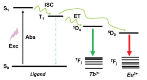
Figure 1.
Jablonsky energy level diagram to illustrate the ligand-to-metal energy transfer process.

Luminescent metal-organic frameworks (LMOFs) are important kinds of metal-organic frameworks (MOFs) [1]. Its light emission is generated after absorbing radiation excitation energy. Luminescence can be broadly defined as the emission of light from an excited electronic state after energy absorption. In the absorption process, the most common energy source is UV-light. While in a few other forms of excitation, luminescence can be divided into fluorescence and phosphorescence according to the pathway of excitation state relaxation accompanying photon emission [2]. Luminescence MOFs mainly include metal-centered luminescence (MC), ligand-centered luminescence (LC), metal-to-ligand charge transfer (MLCT), ligand-to-metal charge transfer (LMCT), ligand-ligand charge transfer (LLCT) and metal-to-metal charge transfer (MMCT) [3]. Luminescence characteristics of Ln-MOFs are suitable for metal-centered luminescence (MC).
Due to the narrow absorption cross section and limited absorption efficiency of lanthanide ions, it is difficult to directly photoexcite highly luminous lanthanide ions. This problem can be solved by the "antenna effect" [4]. The "antenna effect" is that an organic ligand acts as an antenna, completely absorbing optical energy and then transferring it to the coordinated central rare-earth ions through a molecular energy transfer mode. The specific process is usually that the organic ligand chromophore can be excited from the ground state (S0) to the first excitation singlet state (S1) after absorbing the excitation light with appropriate wavelength. The molecule in the excited state then returns to its ground state by non-radiative or radiative means (Fig. 1). The process of the first excited singlet state (S1) to the triplet excited state (T1) S1 → T1 is called inter-system crossing (ISC) [5]. Among them, the radiation mode leads to different forms of molecular luminescence, which can be divided into two kinds, namely fluorescence (S1 → S0) or phosphorescence (T1 → S0) [6]. If the ligand has good coordination ability with Ln3+, and the T1 state energy level of the ligand matches well with the energy level of Ln3+ ions, the energy of T1 state will be transferred to metal ions, resulting in highly sensitized luminescence of Ln3+, and the emission of the ligand will be completely quenched. Subsequently, the excited state of Ln3+ ion reduces the energy to the ground state through radiative transition, thereby emitting strong fluorescence with lanthanide characteristics. If the energy transfer from the ligand to the Ln3+ ion is not complete, the intramolecular transfer of the organic ligand may emit fluorescence in the form of radiation (π-π*, n-π*). In addition, due to the shielding effect of lanthanides, the f orbital is not disturbed by the external electronic environment, so it has sharp and narrow characteristic emission lines.
According to Reinhold's empirical rule, if the energy gap between S1 and T1 of the ligand is greater than 5000 cm−1, the ligand can be used for sensitization. According to the empirical rule proposed by Liu et al. [7, 8], when the energy gap between the T1 state of the ligand and the 5D0 state of the Eu ion is 2500–4000 cm−1 or the energy gap between the T1 state of the ligand and the 5D4 state of the Tb ion is 2500–4500 cm−1, lanthanide ion can be sensitized effectively by the "antenna effect" [4]. Combining the advantages of MOFs and the "antenna effect" of ligands, Ln-MOFs has been widely developed in many fields [9]. Ln-MOFs combines the advantages of MOFs with the intrinsic spectral properties of lanthanide compounds. Lanthanide ions are a kind of photoresponsive metal ions with good luminescence performance. The emission spectrum attributed to the 4f-4f transition is clear and insensitive to the environment [10]. Lanthanide metal ions usually have high coordination number and flexible coordination environment. Many new structures can often be obtained in the design and preparation of Ln-MOFs materials. In addition, due to the 4f internal electronic structure of lanthanide metal ions, Ln-MOFs have the particularity of large orbital coupling effect, strong internal magnetic anisotropy, clear emission band, wide emission range, long luminescence lifetime, large Stokes shift and high color purity. Ln-MOFs are considered as a treasure house of new materials [11, 12].
The sensing effect of Ln-MOFs usually takes advantage of reaction sites on the inner surface of the framework to have the host-guest interaction with the analyte, thereby changing the luminescence signal and realizing the sensing function [13]. Due to the energy/charge transfer process between Ln-MOFs and analyte, the luminescence characteristics of Ln-MOFs are sensitive to their structural characteristics, coordination environment, pores, surface properties, and interactions between host and guest through coordination bonds, π-π bonds, hydrogen bonds, etc., which provides a basis for the construction of Ln-MOFs sensors.
pH sensing usually results in luminescence intensity changes through hydrogen bonding or energy transfer between ligands and lanthanide ions. It is well documented that hydrogen bonding can generally enhance the luminescence in Ln-MOFs [14]. The hydrogen bond network in the system can give the material many fascinating properties. Zhu et al. designed a series of D-A molecules in which the phenolic hydroxyl group acts as the proton donor and the pyridine group acts as the acceptor [15]. Under the action of water-based molecular bridges, a network of ordered and alternate hydrogen bonds is formed in these crystal platforms to obtain macromolecular dipole-dipole interactions, endowed the material with multi-excitation and multi-emission characteristics. O and N atoms can act as an effective hydrogen bond acceptor, binding the proton/hydroxide to the hydrogen bond of the coordination environment, resulting in diminished luminescence [16]. Under high acidity conditions, the abundance of hydrogen bonds in the framework will be broken sporadically, or hydrogen protons will be trapped by ligands, resulting in the reduction of luminescence intensity. In basic solution, the ligand deprotonates, resulting in a change in the energy transfer between the ligand and the lanthanide ion, which affects the luminescence intensity.
Zang et al. synthesized {[Tb4(µ3OH)4L3·(H2O)7]Cl0.63·(NO3)4.37·3H2O}n material based on 1-(3, 5-dicarboxybenzyl)−4, 4′-bipyridinium chloride (H2L+Cl−) ligand [17]. It remains intact in aqueous solutions in the pH range of 2–7, which opens up the possibility of a pH sensor (Fig. 2a). The solid emission intensity of acid-treated samples gradually decreased with increasing acidity and reached the maximum value in the solution with pH 7. Due to the presence of pyridyl groups in the framework, protonation of these pyridyl nitrogen atoms will alter their electronic absorption capacity, which may be responsible for the reduced luminescence intensity at low pH. Rocha's group synthesized a new Eu3+ metal organic framework [Eu3(C14H6N2O4)4(OH)(H2O)4]·2H2O with 1, 10-phenanthroline-2, 9-dicarboxylic acid as ligand [18]. The covalency of the Eu-(O, N) bonds and the nephelauxetic effect can influence the energy of the 5D0 → 7F0 transition. It allows linear photoluminescence response in the range of 5–7.5 pH (Fig. 2b). This range is required for the use of biological fluids such as blood and culture media for cells. By combining this material and an optical fiber, a miniaturized pH sensor can be manufactured.
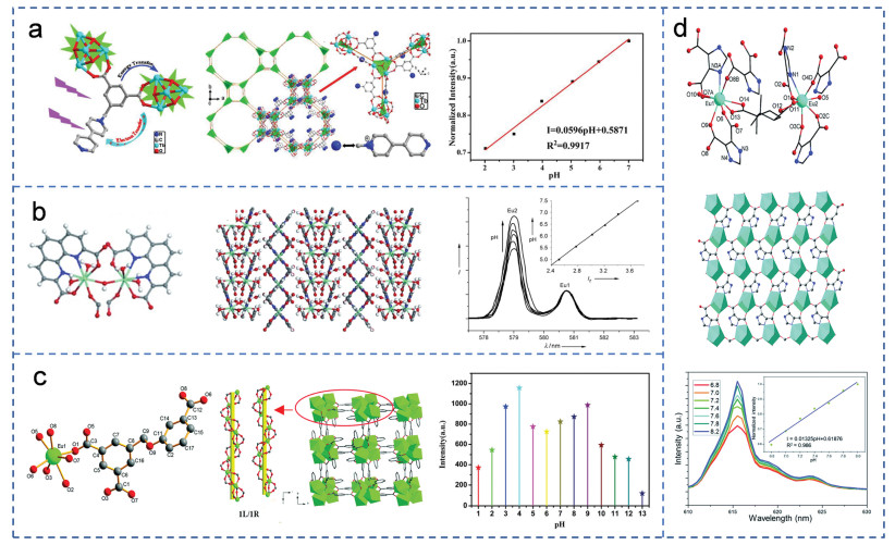
In addition, the abundance of hydrogen bonds also affects the luminous intensity of the framework. Fan et al. developed a new Ln-MOF, [Eu(L)(H2O)]⋅1.5H2O (H3L = 3-(3, 5-dicarboxylatoben-zyloxy)benzoic acid) [19]. It has left-handed and right-handed helical chains constructed from Eu3+ ions and three carboxylic acid groups (Fig. 2c). Due to the presence of ether bonds in the ligand, the oxygen atom of the ether bond can be used as an effective hydrogen bond acceptor to bind protons/hydroxides to the coordination environment. The luminescence intensity of the framework decreases in the pH range of 4–1, and reaches a maximum when the pH value is equal to 4. When the pH value exceeds 9, the luminescence intensity of framework decreases. Du's group prepared a new luminescent Ln-MOF, Eu2(D-cam)(Himdc)2(H2O)2 shows good chemical resistance to both acidity and alkalinity solutions with pH ranging from 2 to 13 (Fig. 2d), which make it potentially useful for wide-range pH sensors, especially in the physiological environment with pH 6.8–8.0 [20]. Under high acidity conditions, the abundance of hydrogen bonds in the framework will be destroyed, resulting in the reduction of luminescence intensity. In basic solutions, deprotonation of the imidazole ring occurs, which favors the energy transfer from ligand to lanthanide ions and hence increases the luminescent intensity.
The detection of metal cations or anions mainly through the optical signal transition can be attributed to the following factors: (1) the disintegration of the framework structure [21]; (2) interactions between ions and organic ligands [22]; (3) ion exchange between the central cations of the frameworks and the targeted cations [23]; (4) competition for energy absorption [24, 25].
Qian's group has designed a new electrochemical sensor (ZJU-27) based on MOF modified glassy carbon electrode, which can detect trace Cd2+ and Pb2+ ions with high sensitivity [26]. Hexagonal lanthanide MOF was synthesized by solvothermal method in the presence of Yb(NO3)3⋅5H2O and 1, 3, 5-benzenetriben acid (H3BTB) in 2-fluorobenzoic acid (2-FBA) (Fig. 3a). ZJU-27 was used to modify the vitrified carbon electrode. The high surface area of ZJU-27 enables rapid and full contact with analytes and high selectivity through size selection effects. The detection limit was 0.228 ppb. Hou's group successfully obtained two new 3D Ln-MOFs by solvothermal reactions of lanthanum nitrate(III) and semi-flexible carbazole tetracarboxylic acid linkers [27]. It can be used as a solid luminescence sensor for quantitative detection of Fe3+ or CrVI by luminescence quenching with high sensitivity, fast response, good selectivity and recyclability (Fig. 3b). The weak interaction of Fe3+ with carbazole nitrogen atoms and deprotonated carboxylic acids effectively quenched the luminescence of MOF. In addition, the competitive absorption of light source energy between Cr2O72− and the framework plays an important role in the fluorescence quenching of the framework, because the absorption spectra of Cr2O72− overlap extensively with the excitation spectra of the two materials. Yang and his coworkers prepared a highly selective and sensitive LMOF with 3, 3′, 4, 4′-biphenyltetracarboxylic acid as ligand, for rapid detection of Fe3+ from mixed metal ions (detection limit, 0.39 µmol/L) and Al3+ (detection limit, 0.084 µmol/L) [28]. Fluorescence quenching due to the exchange of Fe3+ with Eu3+ in the framework when added Fe3+. When Al3+ is added, the structure of Eu-MOF is destroyed, the luminescence intensity of ligand is enhanced, and the luminescence intensity of Eu3+ is weakened (Fig. 3c).
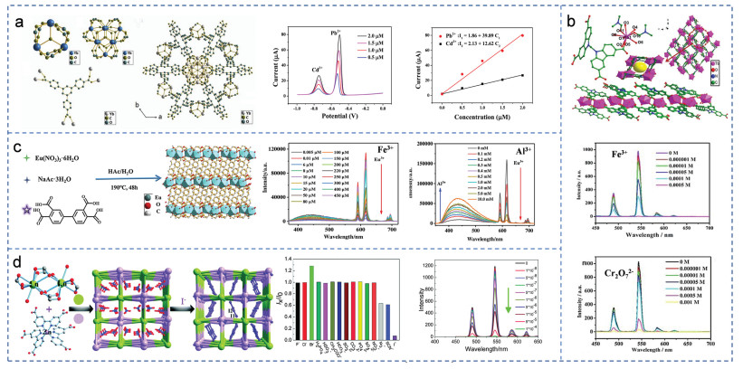
Except heavy metal ions, Ln-MOFs can also detect radioactive ions. Improper use and excessive release of iodide are harmful to human health and the environment. Zhao et al. first developed Eu-Zn (1·NO3−) and Tb-Zn (2·NO3−) for the detection of I− in aqueous solution [29]. 1·NO3− or 2·NO3− in the channel can rapidly oxidize I− to I3− (Fig. 3d). Due to the UV absorption wavelength of I3− overlaps with the excitation wavelength of the framework, leading to the luminescence quenching. The fast response time is only 10 s, and the detection limit is 0.001 ppm.
Temperature sensors play an important role in biology, chemistry and engineering, especially those that can work accurately in a noninvasive manner. The temperature sensors based on MOF are usually mixed with Eu3+ and Tb3+ MOF, and rely on the energy transfer process to build the proportional luminescence thermometer. There is an energy difference between Tb3+ and Eu3+, and the energy transfer of Tb3+ to Eu3+ ions is different at different temperatures. Then the ratio of the luminescence intensity of Eu3+ and Tb3+ at different temperatures is used to establish a relationship with temperature. In addition, a small number of articles reported that Ln-MOF composed of organic chromophore and Eu3+/Tb3+ realized temperature sensing through energy transfer between them at different temperatures.
Compared with other MOFs with one luminous center, binary hybrid Ln-MOF does not require additional luminous intensity calibration and is more instantaneous [30]. In 2020, Chen's group adopted a photoinduced synthetic copolymerization strategy to achieve a luminescence thermometer based on the emission intensity ratio of two lanthanide ions [31]. As the temperature increases, Tb3+ transfers energy to Eu3+ ions, and the luminescence intensity of Tb3+ ions decrease, while that of Eu3+ ions increase. Because of this different luminescence behavior, hybrid films are good candidates for luminescence thermometers. Therefore, taking a ratio of the luminescence intensity of Tb3+ and Eu3+, it can show a good linear relationship with the temperature (Fig. 4a). In addition, it exhibits excellent mechanical properties such as flexibility, elasticity and workability, and exhibits remarkable stability under harsh conditions of high humidity, strong acids and bases, which makes it possible to map temperature profiles under extreme conditions.
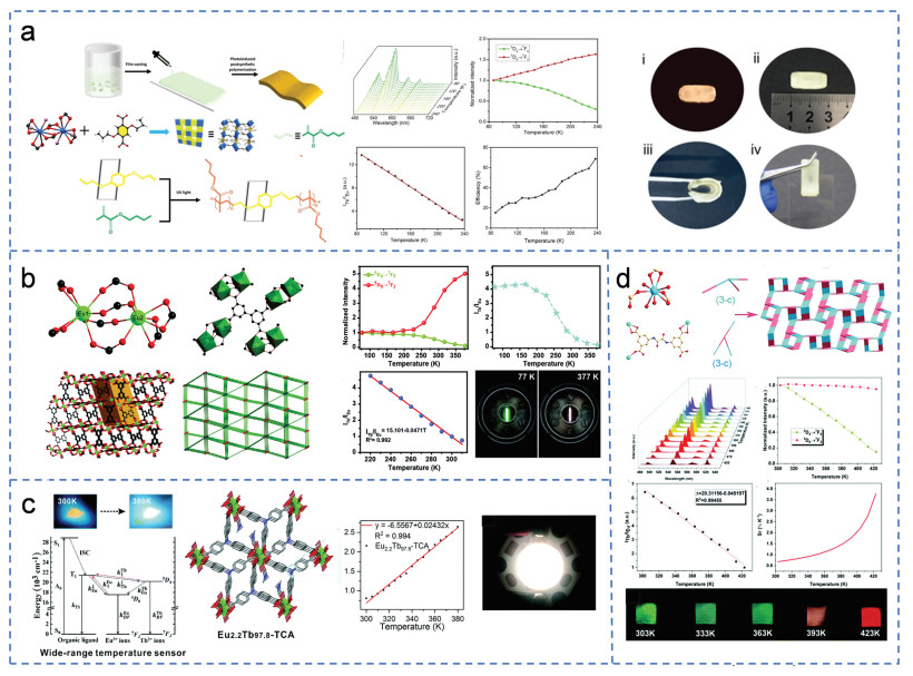
Similarly, Su's group synthesized an MOF ([(CH3)2NH2]Eu0.036Tb0.964BPTC) supplemented with Eu3+ and Tb3+, which could be developed as an excellent luminescence thermometer [32]. It exhibits a sensitive response to temperatures from 220 K to 310 K (Fig. 4b). In this temperature range, the ratio of the emission intensities of Tb3+ and Eu3+ is linearly dependent on temperature. By monitoring the lifetimes of 5D0 (Eu3+) and 5D4 (Tb3+) in the excited state of the mixed luminescent MOF at different temperatures, the lifetimes of Tb3+ ions are significantly shorter, while the lifetimes of Eu3+ ions are longer, indicating that Tb3+ to Eu3+ energy transfer occurs. Based on the same principle, the Eu2.2Tb97.8-TCA prepared by Tang's group can monitor the temperature in the range of 300 K to 380 K because of the energy transfer from Tb3+ to Eu3+ (Fig. 4c), with excellent linearity, high accuracy and good recoverability [33]. It exhibits white light emission under UV and raising temperature does not have much effect on white light emission, so it can be used as a promising white light phosphor to manufacture lighting devices. Sun et al. prepared temperature sensor Eu0.004Tb0.016-BDPO, as the temperature increases, energy shifts from Tb3+ to Eu3+ [34]. It has excellent temperature sensing performance in the temperature range of 303–423 K, and the color changes from green to red (Fig. 4d).
Although most work on Ln-MOFs thermometry is based on the Eu3+-to-Tb3+ emission ratio, a few papers explore the emission of both an organic chromophore and one of these two lanthanides. Yan's group reported another thermometer based on the ILigand/ILn ratio [35]. Eu3+ ions were introduced into a UiO-type MOF (Zr-bpydc) through post-synthetic modification. The ET and back ET process between the two emitters in the hybrid system enables the dual emitters to have opposite thermal dependence, thus providing highly sensitive proportional temperature sensing. The precision is ± 0.26 K. Qian and his coworkers developed a new ratio thermometer [36]. They synthesized the dual-emitting MOF⊃dye composite (ZJU-88⊃perylene) by encapsulating the luminescent perylene dye into the pores of the europium MOF ZJU-88. The ET between perylene molecules and Eu3+ enables this ZJU-88⊃perylene thermometer has highly temperature-sensitive property over the physiological temperature range.
The identification of biomolecules for disease diagnosis requires simple analytical techniques with high accuracy and reliability. The detection mechanism of Ln-MOFs based biosensors usually is: Biomolecules to be tested are bound to lanthanide MOFs by hydrogen bonding, polymerization or related reactions. The analyte can quench the luminescence or change the luminescence color by photo-induced electron transfer (PET) or energy transfer (ET). Fluorescence chromaticity shift value can be used as another response index and color change is also visually intuitive.
Tyrosinase (TYR) monophenolase activity plays a central role in the occurrence and development of diseases such as Parkinson's disease, melanoma and vitiligo [37, 38]. In addition, it also has great potential for industrial use in the preparation of o-diphenols [39]. Xiao et al. first developed a fluorescent on/off dual response biosensor for testing TYR monophenolase activity [40]. Dipicolinic acid (DPA) was selected as the ligand to synthesize Eu-DPA (Fig. 5a). L-Tyrosine (a specific substrate for TYR monophenolase) was first catalyzed to BA-levodopa. Then it was polymerized with Eu-DPA to form polymer spots with cross-linking agent. Compared with that of BA-levodopa, the LUMO energy levels of BA-leucodopachrome oligomers all appear significant decline, which can significantly promote the PET process from Eu-DPA to the BA-leucodopachrome polymers. Therefore, the polymer spots emit blue fluorescence (423 nm) and the red emission of Eu-DPA (622 nm) is quenched. The chromaticity changes showed a surprising linearity with TYR concentration.

8-oxo-2′-Deoxyguanosine (8-oxo-dG) is an early pathological feature of DNA lesions and various cancers [41, 42]. Li and his coworkers constructed a MOF sensor (Eu-ade-MOF) to detect 8-oxo-dG using a double ligand with adenine (ade) as the recognition unit, mellitic acid (H6mel) as the energy donor and Eu3+ as the signal source [43]. Because the maximum absorption wavelength of 8-oxo-dG at 295 nm is the same as the excitation wavelength of Eu-ade-MOF, thus severely hindering the energy transfer from the ligand to Eu3+ and the luminescence intensity decreases significantly. When excited at 309 nm, a hydrogen bond is formed between ade and 8-oxo-dG which can limit intramolecular rotation (IMR) in MOF and inhibit non-radiative attenuation and ET loss, and the emission intensity of Eu3+ is significantly enhanced (Fig. 5b). This material can be used as a dual detection platform for photoexcited dual response 8-oxo-dG. The above MOF suspension is solidified into agarose hydrogel, which can be used as a portable detector and the detection results can be visualized.
Lung cancer is a serious global health burden [44]. The urine biomarker of lung cancer, N-acetylneuraminic acid (NANA), is associated with the risk of lung cancer [45]. Yan's group proposed a molecular robot B-EuMOF that could be used as a ratio fluorescence sensor for the detection of NANA [46]. Molecular robot consists of three parts: actuator, processor and sensor (Fig. 5c). The boric acid group acts as the actuator to grab NANA. The fluorescence of B-EuMOF (I470 nm/I614 nm) was used as a sensor to detect NANA. After the boric acid group captures NANA, the antenna effect between the ligand and Eu3+ is destroyed by the interaction between Eu3+ and NANA. The processor consists of logic gates with λEx and NANA as dual inputs and λ614 nm and λ470 nm as dual outputs, and the sensing results are visible through the naked eye. This work opens new opportunities for the design of intelligent molecular robots based on fluorescent Ln-MOFs.
Ln-MOFs can also identify small gas molecules, solvent molecules and other small organic molecules, usually through the interaction of analyte and ligand (electrostatic interaction, hydrogen bonding, etc.) affects the energy transfer process between ligand and metal ions, causing the change of luminescence signal for sensing [47, 48].
The greenhouse effect caused by the rapid accumulation of CO2 in the atmosphere is attracting great attention. Zhang's group has developed an ultramicropore Ln-MOF, NKMOF-3-Ln (Ln = La, Nd, Eu, Dy, Lu) with high specific surface area, which can selectively bind CO2 under ambient conditions and play the functions of storage and fluorescence sensing [49]. CO2 molecules can be adsorbed into the framework through electrostatic interaction with nitric acid (NO3−). The adsorption capacity and binding energy of CO2 are gradually enhanced with the contraction of lanthanides and play a role of storage (Fig. 6a). The fluorescence intensity of the characteristic peak gradually decreases with the increase of CO2 and the fluorescence intensity of other gasses does not change significantly, so it is a potential fluorescence sensor. Li et al. developed a luminescent MOF JXNU-3(Tb) which can selectively adsorb CO2 in CH4 because of the strong interaction between CO2 and the framework [50].
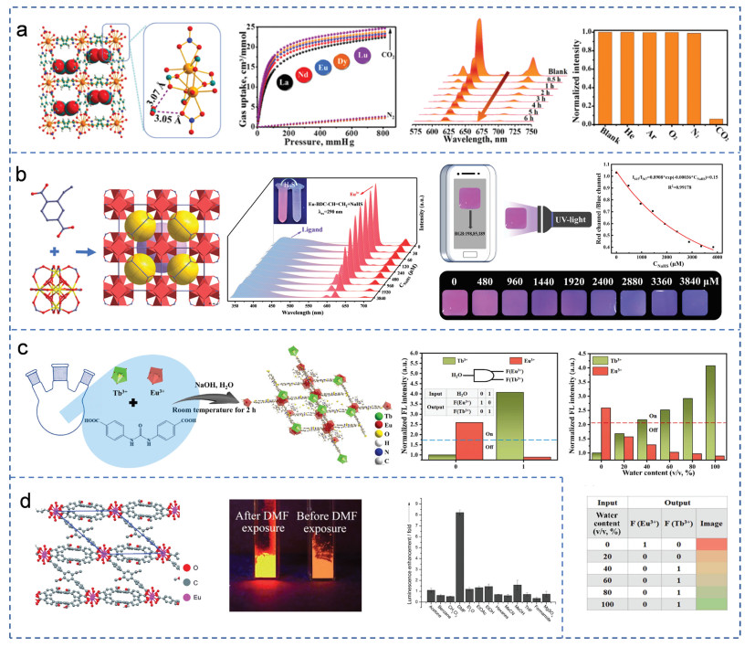
Recently, the detection of hydrogen sulfide in biological systems has attracted much attention. Han et al. prepared a ratio fluorescence sensor Eu-BDC−CH═CH2 (BDC−CH═CH2 = 2-vinylterephthalic acid) [51]. The vinyl in the ligand can not only regulate the "antenna effect" of the ligand on Eu3+ ions, but also act as the reaction site of H2S. The agarose hydrogel film based on the above MOF can visually detect hydrogen sulfide via a smartphone by recognizing Red, Green, Blue (RGB) values (Fig. 6b).
Solvent: Humidity sensing can be achieved by Ln-MOFs sensing H2O. It is usually because the groups in the ligand are protonated under the induction of water, which changes the highest occupied molecular orbital (HOMO) and the lowest unoccupied molecular orbital (LUMO) of the ligand, resulting in a significant reduction in the HOMO-LUMO gap and inhibiting the energy transfer of the ligand to Eu3+/Tb3+; or the ET between lanthanide ions and -OH of H2O adsorbed in the framework may cause luminescence changes.
Xiao et al. prepared a dual-Ln-MOF Eu0.2Tb0.8-UL (UL: urea-containing ligand) [52]. On the one hand, water inhibits the ligand ET to Tb3+ by protonating the ligand, on the other hand, it inhibits the ligand ET to Eu3+/Tb3+ and Tb3+ to Eu3+ by the -OH oscillation effect. With the increase of humidity, the luminescence color changes from red to green, realizing the high sensitivity ratio switch sensing of water. A "one to two" logic device with single input (water content) and double output (Eu3+ and Tb3+ emission) is designed for simple intelligent water detection, which is realized by three output results: YES (1, 0), PASS (0, 0) and NOT (0, 1) (Fig. 6c).
In addition, recent studies have shown that serious health problems are associated with N, N-dimethylformamide (DMF) through skin contact and inhalation in various industries. DMF has the risk of hepatotoxicity, embryotoxicity [53] and carcinogenesis [54]. Song et al. reported an Ln-MOF, [Eu2L3(H2O)4]·3DMF (L = 2′, 5′-bis(methoxymethyl)-[1, 1′: 4′, 1′′-terphenyl]−4, 4′′-dicarboxylate) for sensing in DMF steam [55]. The interaction of Eu3+ with the -OH bond of coordinated H2O quenches Eu luminescence. Therefore, when the channel H2O molecule is partially replaced by other solvent molecules, less -OH bonds are left around the Eu center. This interaction may change the excited state energy level of the ligand, thereby promoting the ligand-metal energy transfer (LMET) process and triggering the enhancement of Eu emission (Fig. 6d).
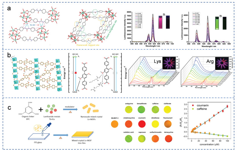
Nitro compound: Nitro compounds are extremely dangerous among many pollutants. They are not only highly toxic but also highly explosive, so their misuse can cause serious soil and water pollution problems [56]. Through the photoinduced electron transfer (PET) mechanism, nitro compounds are electron deficient molecules with strong quenching ability. After entering the pore, the nitro compound binds to the site on the ligand through hydrogen bond interaction, which promotes the PET process and causes luminescence quenching. Liu's group reasonably developed two Ln-MOF luminescent sensors based on urea recognition sites for quantitative detection of picric acid (PA), with two-dimensional layers of diamond shaped holes (Fig. 7a) [7]. Half of the hydrogen bond interactions between the urea unit and carboxylate groups accumulate into a 3D framework, while the other half is located in the pore and acts as a null recognition site. The nitro group binds to the urea site through hydrogen bond interaction, promotes the PET process, and exhibits a specific luminescence quenching response to PA, with high sensitivity, good selectivity and rapid response as well as excellent recyclability.
Nitrobenzene is one of the most important intermediates in industrial organic synthesis, such as dyes, perfumes and explosives, but it is highly toxic to humans, and excessive amounts of nitrobenzene can cause hyperhemoglobinemia [57, 58]. Wang's group obtained a new 3D isostructural Ln-MOF ({[Eu(L)1.5(H2O)2]·2H2O·2DMF}n, H2L = 4-(2-carboxyvinyl)benzoic acid) by solvothermal reaction [59]. It can be used as a promising potential photosensor with high detection performance for Fe3+ and nitrobenzene. Due to the electrostatic interaction between the ligands in Eu-MOF and nitrobenzene, and the energy competition between the fluorophores of Eu-MOF and nitrobenzene, luminescence quenching is caused.
Amino acids (AAs) are indispensable biomolecules in living systems [60]. Therefore, the analysis and detection of trace amino acids in food and living organisms are of great significance for nutritional analysis and disease diagnosis. Liu et al. reported a series of water-stabilized 3D Ln-MOFs, {[Ln(DMTP-DC)1.5(H2O)3]·DMF}n (Ln = Eu, Gd, Tb and Dy) [61]. It can be used as a fluorescence sensor for the ratio of arginine (Arg) and lysine (Lys) in aqueous solution (Fig. 7b). The detection limit of arginine and lysine is 24.38 µmol/L and 9.31 µmol/L, respectively. The interaction between the O atom of the methoxy group from the DMTP-DC ligand and Arg/Lys-inhibits the PET, which inhibits the single electron from the oxygen atom of the methoxy group to the fluorescence framework, and the inhibition of PET results in increased fluorescence of the ligand.
Pharmaceutical molecule: In contrast to sensors based on individual lanthanide ions, mixed crystal Ln-MOFs can amplify the relative emission ratio and the luminescence signal, allowing multiple molecules to be distinguished. Wang et al. first tried to use mixed crystal Ln-MOF film as luminescence indicator MLMOF-3 for drug detection [62]. It exhibits object-dependent luminescence through different host-guest interactions and different drug molecules glow in different colors from red to green (Fig. 7c). The emission intensity ratio of the transition from 5D0 → 7F2 (Eu3+, 619 nm)−5D4 → 7F5 (Tb3+, 547 nm) is different depending on the guest molecule, which enables MLMOF-3 film to recognize different drug molecules. In addition, it was observed that the intensity ratio was linearly related to the drug concentration.
Ln-MOFs assembled by lanthanide ions and organic bridging agents not only have the high stability of inorganic materials, but also ensure the excellent machinability of organic materials. The emission color, excitation band and pore can be fine-adjusted by changing lanthanide ions and organic linkers [63]. Multifunctional spectra of luminescent MOF derived from organic ligands or metal ions are capable of generating a large library of optical codes, which will provide a good platform for digital coding and multifunctional luminescent materials [64, 65].
Ln-MOFs, especially Eu/Tb-MOFs have clear emission band, wide emission range, long luminescence lifetime, large Stokes shift and high color purity [66]. Because of their attractive pore types and excellent luminescence properties, and the introduction of functional ligands or guest molecules, they are considered to be excellent candidates for fabrication of excellent light emitting devices.
Yan et al. prepared isostructured Ln-MOFs by self-assembly of 1, 3, 5-benzenetricarboxylic acid (BTC) and lanthanide ions which have one-dimensional microrod structures, high photoluminescence (PL) quantum yields and different emission colors (green, orange and red) [67]. Bright green, orange and red emission points were observed at both tips of each microrod, with relatively weak emission from the rod body. The microrods absorb the excitation light and propagate the PL emission to the tip, creating a low-loss optical waveguide, feature both linear and chiral polarized photoemission with high anisotropy (Fig. 8a).
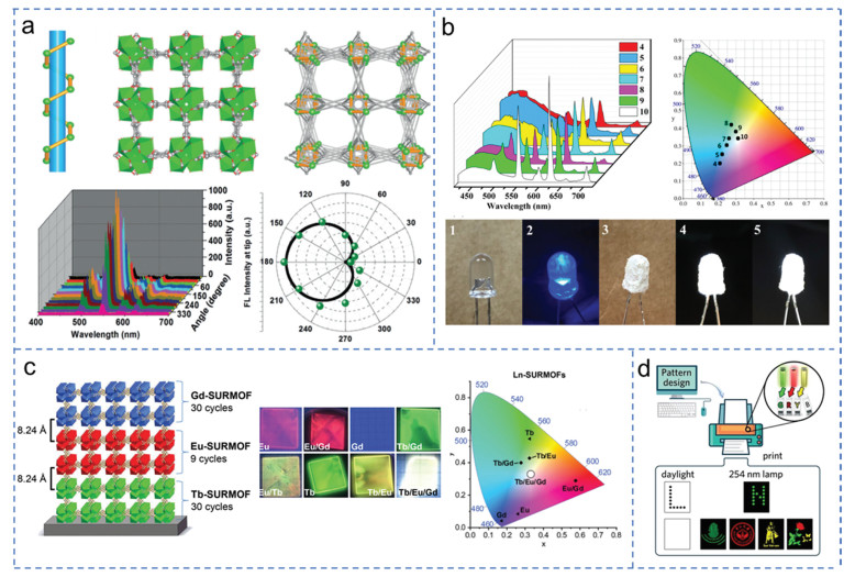
In addition, white light emission can be achieved by adjusting the ratio of lanthanide ions and ligands. Chen et al. synthesized a series of isometallic Ln-MOF ([TbxEu1−x(TCBA)(H2O)]2·DMF) by the blue emission of H3TCBA, the characteristic red light of Eu(III) and the green light of Tb(III) [68]. When x = 0.816, the excitation at 380 nm will emit bright white light, and may be used as a single-phase white light emitting diode (WLED) phosphor material (Fig. 8b). Simple WLED components are made by coating a thin powder layer of the above material on the light emitting diode (LED). The resulting WLED produces a bright white light at 3.8 V and can be lit continuously for a week without attenuation. Redel et al. developed a new set of Ln-MOF (lanthanide-metal-organic framework) thin films, known as Ln-SURMOFs (surface-supported MOFs), is fabricated with a layer-by-layer, in order to generate solid-state white-lighting devices [69]. Through a rational combination of red, green, and blue (RGB) emitting Eu3+, Tb3+ and Gd3+ containing layers in order to achieve white-light emission (Fig. 8c). Liang et al. adjusted the luminescence color of MOF by adjusting the proportion of (Tb3+, Eu3+ and Sm3+) doped framework to prepare uniform colorless safe ink [70]. An inkjet printer is designed to print complex computer patterns on filter paper with high precision inkjet printing and display beautiful patterns under UV light (Fig. 8d).
The excellent ability of MOFs to flexibly self-assemble into various 1D micro/nanostructures, such as wires, tubes, and rods, makes MOF a fundamental model for the design of novel optical barcodes [71]. Zhao et al. proposed a strategy to design micro-barcodes with high coding capacity based on 1D Ln-MOF multi-block heterostructures, prepared by epitaxial growth [72]. This heterostructure can realize the controlled emission color and the length of the heterogenous region. The spectral characteristics of the crystal and the length of each part as the intrinsic fingerprint to define the micro barcode, improving the coding ability (Fig. 9a). This design shows great potential in data logging and information tracking. In addition, except building heterostructures to edit photonic barcodes, Zhai's group reported a convenient method to design high-capacity photonic barcodes by manipulating the polarized emission of Eu3+ metal-organic frameworks [73]. Due to the highly anisotropic crystal structure of Eu3+ and the low symmetric splitting of energy levels, the large degree of polarization (almost 0.96) enables color-tunable emission. The chromaticity of the emission accordingly transforms from red to orange upon polarization angle switching (Fig. 9b), which demonstrates a polarization-responsive multicolor Ln-MOF crystal. This polarization-sensitive emission of crystals provides an intelligent coding strategy that can be further integrated into large coding libraries of photon barcodes.
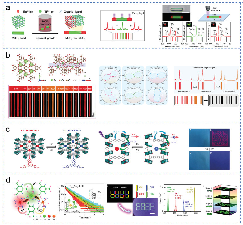
In addition, optical switch molecules are of great significance to construct intelligent anti-counterfeiting materials. Zhu et al. gained a self-restoring photoexcitation-controlled aggregation-induced luminescent organic gel by using AIEgens [74]. After withdrawing the UV light source in the dark, the bright phosphorescence can be self-recoverable which has the potential for anti-counterfeiting applications. The functional guest molecules such as optical switch molecules can be encapsulated into porous MOF to realize intelligent information storage with remote optical control and anti-counterfeiting MOF materials. Chen et al. implemented reversible information security by loading photoswitchable diarylethene derivative into a Ln-MOF [75]. Light-triggered open (OF-DAE) and closed (CF-DAE) isomerization of diarylethylene (DAE) units, which regulate the inactivation and activation of photochromic processes between diarylethylene acceptors and lanthanide donors (Fig. 9c). The OF-DAE is transformed into CF-DAE by UV irradiation. At this time, its UV absorption wavelength overlaps with the fluorescence emission peak of the framework to quench the framework's luminescence. Then, the configuration can be restored and the luminescence of framework can be restored by sunlight. The recorded information can be accurately hidden and read in a short time by alternating UV and visible light irradiation, making it an attractive next generation of intelligent information protection materials. Liang and his coworkers illustrated the potential application of Ln-MOFs for information storage and security in the spatial and temporal domains with a concept of inkjet printing and luminescence lifetime imaging and time-gated imaging [70]. The ET between ions in the framework with different ratios of lanthanide ions is different, which shows differential sensitization effect and can achieve color tunability and lifetime tunability (Fig. 9d). Tb0.70Smz-BTC (z = 0–0.10) can achieve the lifetime tunability of green emission in the range of 300–600 ms by changing the content of Sm3+. With the help of time-gated microscopy, different pattern information can be obtained by controlling the delay time.
Lanthanide metal ions in Ln-MOF can form coordination unsaturated sites as Lewis acid centers, which facilitate the use of Ln-MOFs as heterogeneous catalysts. In addition, Ln-MOF can introduce functional organic ligands that support metal nanoparticles and are converted to derivatives during design and synthesis, expanding the space of Ln-MOF in the catalyst environment [76].
Han et al. successfully constructed a series of Ln-cluster-based bifunctional MOFs Ln-BDC—NH2 (H2BDC—NH2 = 2-aminobenzenedicarboxylate; Ln = Yb, Dy, Sm) [77]. The presence of both Lewis acid and Brønsted base sites in these MOFs makes them a bifunctional catalyst for one-pot tandem deacetyl-Knoevenagel reactions (Fig. 10a). Among them, Yb-BDC—NH2 has high activity and excellent recyclability. The yield of the final product can reach 97%.
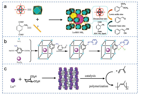
Sun et al. prepared a Fe3O4@La-MOF-Schiff base Pd for Suzuki-Miyaura reaction between bromobenzene or aryl iodine [78]. In the synthesis process, Fe3O4 was embedded in La-MOF (Fig. 10b). The amino group of the ligand reacted with the Schiff base of pyridine-2-formaldehyde to increase the content of nitrogen atoms, and finally palladium was introduced by coordination with nitrogen atoms. The catalyst can be used to obtain more than 99% yield and good cycling performance (repeated use more than 12 times). A synergistic cooperativity between Pd and the oxophilic La nodes improves the stability and excellent catalytic performance of the catalyst. Fe3O4 is loaded inside the catalyst, enabling the catalyst to be recovered using a magnet.
In addition, it can be used in polymerization of isoprene reaction. Numerous studies have shown that Nd-based carboxylate MOFs are active precatalysts for the stereoselective polymerization of conjugated dienes [79]. In 2016, Loiseau et al. synthesized new series of lanthanide MOFs Ln(1, 2-chdc)(form)(H2O) (Ln = Ce3+, Pr3+, Nd3+, Sm3+, Eu3+, Gd3+, Tb3+, Dy3+) with the 1, 2-cyclohexane dicarboxylate (1, 2-chdc) ligand [80]. Among these MOFs, the Nd compound was the most effective catalyst for isoprene polymerization with 87% yield and 93% cis-selectivity (Fig. 10c).
Currently, materials based on titanium dioxide, zirconium dioxide, zinc oxide, tin oxide and cadmium sulfide are the most popular photocatalysts [81]. However, these traditional photocatalysts have some problems, such as difficulty in activation, low light absorption ability, easy electron hole recombination and poor stability [82]. Recent studies have shown that MOF materials can be promising photocatalyst materials due to their designable structure and composition.
Huang et al. successfully developed a series of porphyrin-based two-dimensional lanthanide MOF (Ln-TCPP) (Ln = Ce, Sm, Eu, Yb) using a microwave-assisted strategy and successfully obtained nanosheets with different thicknesses by controlling acetic acid concentration [83]. It can be used to synthesize juglone by photooxidation of 1, 5-dihydroxynaphthalene (1, 5-DHN) (Fig. 11a). Thinner nanosheets have better photocatalytic activity. Due to the effective energy and electron transfer between TCPP and Yb3+, the constructed 2D Yb-TCPP nanosheets showed a higher photodynamic activity to produce O2− and 1O2 than the other synthesized 2D Ln-MOF, resulting in a greatly improved photocatalytic activity. Wang et al. prepared a large porous and stable metal-organic framework featuring dinuclear Eu(III)2 clusters as connecting nodes and Ru(phen)3-derived ligands as linkers is constructed to catalyze visible-light-driven CO2 reduction [84]. The photo-initiated electron transfers from Ru photocenters to dinuclear Eu2 oxo-clusters in Eu-Ru(phen)3−MOF lead to the photo-reduction of CO2 to HCOOH (Fig. 11b). In addition, it also has potential applications in dye degradation. Abdelhameed et al. used Ln (NO3)3 (Eu3+ and Tb3+) as a metal source and 1, 2, 4, 5-benzenetetracarboxylic dianhydride as an organic linker to prepare Ln-MOF into viscose fabric by in-situ growth as a high-efficiency photocatalyst for RhB dye degradation [85]. When Ln-MOF was added to the fabric, the dye degradation increased significantly by 55%−67%. Compared with Tb-MOF@viscose fabrics, Eu-MOF@viscose fabrics showed higher photocatalytic reactivity (Fig. 11c) and the RhB degradation rate of Eu-MOF was 96% after 120 min irradiation with fluorescent lamp (500 W).

Over the past few decades, the features of Ln-MOFs open the door to a range of incredibly important applications. Due to its excellent luminescence performance, unique chemical groups and high porosity, Ln-MOFs are widely used as various chemical sensors, gas storage, light emitting devices, information storage and anti-counterfeiting, catalysts, etc. [86].
Although these applications based on Ln-MOFs have achieved rapid progress and excellent results, there are still some aspects that need to be further explored. (1) The corresponding structure-function relationship is only explained by conjecture and hypothesis, rather than perfect theory. Accurate synthesis methods, in situ monitoring techniques and theoretical calculations are needed to determine the topological structure and the role of functional groups, metal centers and pore sizes in detecting guest molecules. (2) Ln-MOFs can be further integrated with electronic devices to be closer to practical applications. The optical signal is transformed into an electrical signal, and the results are visually displayed through the screen, reducing the need for professional knowledge of relevant personnel. (3) The mechanical properties of Ln-MOFs also need to be systematically studied to improve the objective use value.
In conclusion, Ln-MOFs can be considered as a material with great potential applications. Although many challenges remain and need to be addressed, Ln-MOFs have made great progress in structural design, controlled modification and functional applications over the past two decades. We believe that Ln-MOFs will not only have a perfect structure-function theory, but also have a wider application range in the future.
The authors declare that they have no known competing financial interests or personal relationships that could have appeared to influence the work reported in this paper.
This work was sponsored by Shanghai Sailing Program (No. 20YF1432400), and National Natural Science Foundation of China (NSFC, No. 22105128).
Y.P. Wang, F. Wang, D.F. Luo, et al., Inorg. Chem. Commun. 19 (2012) 43–46. doi: 10.1016/j.inoche.2012.01.033
S. Shen, G. Baryshnikov, B. Yue, et al., J. Mater. Chem. C 9 (2021) 11707–11714. doi: 10.1039/d1tc01605a
G.L. Yang, X.L. Jiang, H. Xu, et al., Small 17 (2021) 2005327. doi: 10.1002/smll.202005327
J. González, P. Sevilla, G. Gabarró-Riera, et al., Angew. Chem. Int. Ed. 60 (2021) 12001–12006. doi: 10.1002/anie.202100507
G. Wang, Z. Wang, B. Ding, et al., Chin. Chem. Lett. 32 (2021) 3039–3042. doi: 10.1016/j.cclet.2021.03.054
Q. Xu, L. Ma, X. Lin, et al., Chin. Chem. Lett. 33 (2022) 2965–2968. doi: 10.1016/j.cclet.2021.12.097
W. Liu, X. Huang, C. Chen, et al., Chemistry (Easton) 25 (2019) 1090–1097. doi: 10.1002/chem.201805080
M. Latva, H. Takalob, V.M. Mukkala, et al., J. Lumin. 75 (1997) 149–169. doi: 10.1016/S0022-2313(97)00113-0
Y. Zhao, D. Li, J. Mater. Chem. C 8 (2020) 12739–12754. doi: 10.1039/d0tc03430d
S. Petoud, G. Muller, E.G. Moore, et al., J. Am. Chem. Soc. 129 (2007) 77–83. doi: 10.1021/ja064902x
X. Lin, E. Ning, X. Li, et al., Chin. Chem. Lett. 31 (2020) 813–817. doi: 10.1016/j.cclet.2019.05.055
T. Xia, J. Wang, K. Jiang, et al., Chin. Chem. Lett. 29 (2018) 861–864. doi: 10.1016/j.cclet.2017.10.038
J. Othong, J. Boonmak, F. Kielar, et al., Sens. Actuators B 316 (2020) 128156. doi: 10.1016/j.snb.2020.128156
D. Wu, W. Mi, M. Ji, et al., Spectrochim. Acta A 97 (2012) 589–593. doi: 10.1016/j.saa.2012.07.014
Y. Xing, Z. Li, G.V. Baryshnikov, et al., Chem. Sci. 13 (2022) 6067–6073. doi: 10.1039/d2sc00908k
J. Wang, Y. Li, M. Jiang, et al., Chem. Eur. J. 22 (2016) 13023–13027. doi: 10.1002/chem.201602974
H.Y. Li, Y.L. Wei, X.Y. Dong, et al., Chem. Mater. 27 (2015) 1327–1331. doi: 10.1021/cm504350q
B.V. Harbuzaru, A. Corma, F. Rey, et al., Angew. Chem Int. Ed. 48 (2009) 6476–6479. doi: 10.1002/anie.200902045
Y. Tao, P. Zhang, J. Liu, et al., New J. Chem. 42 (2018) 19485–19493. doi: 10.1039/c8nj04601h
Y.H. Han, C.B. Tian, Q.H. Li, et al., J. Mater. Chem. C 2 (2014) 8065–8070. doi: 10.1039/C4TC01336K
W. Yan, C. Zhang, S. Chen, et al., ACS Appl. Mater. Interfaces 9 (2017) 1629–1634. doi: 10.1021/acsami.6b14563
P. Li, H. Li, J. Mater. Chem. C 4 (2016) 2165–2169. doi: 10.1039/C5TC04377H
Z. Chen, Y. Sun, L. Zhang, et al., Chem. Commun. 49 (2013) 11557–11559. doi: 10.1039/c3cc46613b
C.F. Huebner, R.D. Roeder, S.H. Foulger, Adv. Funct. Mater. 19 (2009) 3604–3609. doi: 10.1002/adfm.200900473
E. Oh, A.L. Huston, A. Shabaev, et al., Sci. Rep. 6 (2016) 35538. doi: 10.1038/srep35538
W. Ye, Y. Li, J. Wang, et al., J. Solid State Chem. 281 (2020) 121032. doi: 10.1016/j.jssc.2019.121032
X. Li, J. Tang, H. Liu, et al., Chem. Asian J. 14 (2019) 3721–3727. doi: 10.1002/asia.201900936
H. Guo, N. Wu, R. Xue, et al., Colloids Surf. A 585 (2020) 124094. doi: 10.1016/j.colsurfa.2019.124094
P.F. Shi, H.C. Hu, Z.Y. Zhang, et al., Chem. Commun. 51 (2015) 3985–3988. doi: 10.1039/C4CC09081K
K. Chen, C. Wu, Chin. Chem. Lett. 29 (2018) 823–826. doi: 10.1016/j.cclet.2017.09.040
T. Feng, Y. Ye, X. Liu, et al., Angew. Chem. Int. Ed. 59 (2020) 21752–21757. doi: 10.1002/anie.202009765
Y. Pan, H.Q. Su, E.L. Zhou, et al., Dalton Trans. 48 (2019) 3723–3729. doi: 10.1039/c9dt00217k
X. Zhai, P. Feng, N. Song, et al., Inorg. Chem. Front. 9 (2022) 1406–1415. doi: 10.1039/d2qi00093h
S. Wang, B. Sun, Z. Su, et al., Inorg. Chem. Front. 9 (2022) 3259–3266. doi: 10.1039/d2qi00682k
Y. Zhou, B. Yan, J. Mater. Chem. C 3 (2015) 9353–9358. doi: 10.1039/C5TC02004B
Y. Cui, R. Song, J. Yu, et al., Adv. Mater. 27 (2015) 1420–1425. doi: 10.1002/adma.201404700
I. Carballo-Carbajal, A. Laguna, J. Romero-Giménez, et al., Nat. Commun. 10 (2019) 973. doi: 10.1038/s41467-019-08858-y
M. Wang, J.L. Xie, J. Li, et al., ACS Sens. 5 (2020) 1634–1640. doi: 10.1021/acssensors.0c00122
M. Goldfeder, M. Kanteev, N. Adir, et al., Biochim. Biophys. Acta Proteins Proteom. 1834 (2013) 629–633. doi: 10.1016/j.bbapap.2012.12.021
L. Yu, Z. Gao, Q. Xu, et al., Biosens. Bioelectron. 210 (2022) 114320. doi: 10.1016/j.bios.2022.114320
Y. Lv, S. Chen, Y. Shen, et al., J. Am. Chem. Soc. 140 (2018) 2801–2804. doi: 10.1021/jacs.8b00515
C.P. Liang, P.Q. Ma, H. Liu, et al., Angew. Chem. Int. Ed. 56 (2017) 9077–9081. doi: 10.1002/anie.201704147
J.N. Hao, Y. Li, Adv. Funct. Mater. 29 (2019) 1903058. doi: 10.1002/adfm.201903058
G. Saranya, M.M. Joseph, V. Karunakaran, et al., ACS Appl. Mater. Interfaces 10 (2018) 38807–38818. doi: 10.1021/acsami.8b15583
Z. Xia, D. Li, W. Deng, Anal. Chem. 93 (2021) 4924–4931. doi: 10.1021/acs.analchem.0c05348
Y. Zhang, H. Lu, B. Yan, Sens. Actuat. B: Chem. 349 (2021) 130736. doi: 10.1016/j.snb.2021.130736
F. Chen, Y. Ling, C. Song, et al., Eur. J. Inorg. Chem. 2016 (2016) 503–508. doi: 10.1002/ejic.201501113
C.Y. Wang, C.C. Wang, X.W. Zhang, et al., Chin. Chem. Lett. 33 (2022) 1353–1357. doi: 10.1016/j.cclet.2021.08.095
L. Han, T. Pham, M. Zhuo, et al., ACS Appl. Mater. Interfaces 11 (2019) 23192–23197. doi: 10.1021/acsami.9b04619
Y.J. Li, Y.L. Wang, Q.Y. Liu, Inorg. Chem. 56 (2017) 2159–2164. doi: 10.1021/acs.inorgchem.6b02811
X. Han, J. Liu, K. Yu, et al., Inorg. Chem. 61 (2022) 5067–5075. doi: 10.1021/acs.inorgchem.2c00019
L. Yu, Q. Zheng, L. Xiong, et al., Sens. Actuat. B: Chem. 356 (2022) 131328. doi: 10.1016/j.snb.2021.131328
A.M. Saillenfait, J.P. Payan, D. Beydon, et al., Fundam. Appl. Toxicol. 39 (1997) 33–43. doi: 10.1093/toxsci/39.1.33
H. Senoh, S. Aiso, H. Arito, et al., J. Occup. Health 46 (2004) 429–439. doi: 10.1539/joh.46.429
Y. Li, S. Zhang, D. Song, Angew. Chem. Int. Ed. Engl. 52 (2013) 710–713. doi: 10.1002/anie.201207610
F. Zhang, H. Yao, Y. Zhao, et al., Talanta 174 (2017) 660–666. doi: 10.1016/j.talanta.2017.07.007
C. Nanthamathee, J. Environ. Sci. 81 (2019) 68–79. doi: 10.1016/j.jes.2019.02.002
X. Mi, D. Sheng, Y.E. Yu, et al., ACS Appl. Mater. Interfaces 11 (2019) 7914–7926. doi: 10.1021/acsami.8b18320
K.P. Bai, L.J. Zhou, G.P. Yang, et al., ChemistrySelect 4 (2019) 12794–12800. doi: 10.1002/slct.201902890
Y. Zhao, M.Y. Wan, J.P. Bai, et al., J. Mater. Chem. A 7 (2019) 11127–11133. doi: 10.1039/c9ta00384c
J.J. Hu, Y.G. Li, H.R. Wen, et al., Inorg. Chem. 61 (2022) 6819–6828. doi: 10.1021/acs.inorgchem.2c00121
Y. Gao, G. Yu, K. Liu, et al., Sens. Actuat. B: Chem. 257 (2018) 931–935. doi: 10.1016/j.snb.2017.10.180
R.B. Lin, S.Y. Liu, J.W. Ye, et al., Adv. Sci. 3 (2016) 1500434. doi: 10.1002/advs.201500434
L. Wang, H. Xu, J. Gao, et al., Coord. Chem. Rev. 398 (2019) 213016. doi: 10.1016/j.ccr.2019.213016
M. Pan, Y.X. Zhu, K. Wu, et al., Angew. Chem. Int. Ed. 56 (2017) 14582–14586. doi: 10.1002/anie.201708802
B. Yan, Inorg. Chem. Front. 8 (2021) 201–233. doi: 10.1039/d0qi01153c
X. Yang, X. Lin, Y. Zhao, et al., Angew. Chem. Int. Ed. 56 (2017) 7853–7857. doi: 10.1002/anie.201703917
W. Xu, H. Chen, Z. Xia, et al., Inorg. Chem. 58 (2019) 8198–8207. doi: 10.1021/acs.inorgchem.9b01008
D.H. Chen, A.E. Sedykh, G.E. Gomez, et al., Adv. Mater. Interfaces 7 (2020) 2000929. doi: 10.1002/admi.202000929
Y. Ou, W. Zhou, Z. Zhu, et al., Angew. Chem. Int. Ed. 59 (2020) 23810–23816. doi: 10.1002/anie.202011559
R.C. Arbulu, Y.B. Jiang, E.J. Peterson, et al., Angew. Chem. Int. Ed. 57 (2018) 5813–5817. doi: 10.1002/anie.201802694
Y. Yao, Z. Gao, Y. Lv, et al., Angew. Chem. Int. Ed. 58 (2019) 13803–13807. doi: 10.1002/anie.201907433
K. Pei, J. Wu, M. Zhao, et al., Adv. Opt. Mater. 10 (2021) 2102143.
T. Weng, Q. Zou, M. Zhang, et al., J. Phys. Chem. Lett. 12 (2021) 6182–6189. doi: 10.1021/acs.jpclett.1c01535
Z. Li, G. Wang, Y. Ye, et al., Angew. Chem. Int. Ed. 58 (2019) 18025–18031. doi: 10.1002/anie.201910467
M. Yoon, R. Srirambalaji, K. Kim, Chem. Rev. 112 (2012) 1196–1231. doi: 10.1021/cr2003147
Y. Zhang, Y. Wang, L. Liu, et al., Inorg. Chem. 57 (2018) 2193–2198. doi: 10.1021/acs.inorgchem.7b03084
G. Xiong, X.L. Chen, L.X. You, et al., J. Catal. 361 (2018) 116–125. doi: 10.1016/j.jcat.2018.02.026
M.J. Vitorino, T. Devic, M. Tromp, et al., Macromol. Chem. Phys. 210 (2009) 1923–1932. doi: 10.1002/macp.200900354
S.E. Russell, C. Gosset, X. Agache, et al., CrystEngComm 18 (2016) 3594–3605. doi: 10.1039/C6CE00101G
Y. Zhang, S. Liu, Z.S. Zhao, et al., Inorg. Chem. Front. 8 (2021) 590–619. doi: 10.1039/d0qi01191f
Q. Zhang, F. Yang, Z. Xu, et al., Nanoscale Horiz. 4 (2019) 579–591. doi: 10.1039/c8nh00373d
Z.W. Jiang, Y.C. Zou, T.T. Zhao, et al., Angew. Chem. Int. Ed. 59 (2020) 3300–3306. doi: 10.1002/anie.201913748
Z.H. Yan, M.H. Du, J. Liu, et al., Nat. Commun. 9 (2018) 3353. doi: 10.1038/s41467-018-05659-7
H.E. Emam, H.N. Abdelhamid, R.M. Abdelhameed, Dyes Pigm. 159 (2018) 491–498. doi: 10.1016/j.dyepig.2018.07.026
J. Castells-Gil, S. Mañas-Valero, I.J. Vitórica-Yrezábal, et al., Chem. Eur. J. 25 (2019) 12636–12643. doi: 10.1002/chem.201902855

Figure 1 Jablonsky energy level diagram to illustrate the ligand-to-metal energy transfer process.

Figure 2 (a) Structure of {[Tb4(µ3-OH)4L3⋅(H2O)7]Cl0.63⋅(NO3)4.37⋅3H2O}n and the linear variation of the emission intensity for Tb-MOF treated by HCl aqueous solutions with pH ranging from 7 to 2. Copied with permission [17]. Copyright 2015, American Chemical Society. (b) Structure of ITQMOF-3. Intensity variation of the Eu2 5D0→7F0 transition from pH 7.5 to pH 5 and the linear variation of Ir with the pH value. Copied with permission [18]. Copyright 2009, Wiley. (c) Structure of [Eu(L)(H2O)]⋅1.5H2O. The luminescence intensity of the framework at 616 nm in aqueous solutions with pH range from 1 to 13. Copied with permission [19]. Copyright 2018, Royal Society of Chemistry. (d) Structure of [Eu2(D-cam)(Himdc)2(H2O)2] and its emission spectra in 1 mol/L HEPES aqueous solution with various pH values from 6.8 to 8.0 and linear correlation between the fluorescent response and pH. Copied with permission [20]. Copyright 2014, Royal Society of Chemistry.

Figure 3 (a) Structure of ZJU-27. SWASV responses for the simultaneous detection of Cd2+ and Pb2+ on ZJU-27/GCE and the corresponding calibration curve. Copied with permission [26]. Copyright 2020, Elsevier. (b) Structure of [Tb(Hmcd)(H2O)(DMF)2]n. Luminescence spectra of [Tb(Hmcd)(H2O)(DMF)2]n in different concentration of Fe(NO3)3 and Cr2O72− solution. Copied with permission [27]. Copyright 2019, Wiley. (c) Schematic illustration of the synthetic process of Eu-MOFs. Emission spectra of Eu-MOF in aqueous solution with different concentrations of Fe3+ ions. Emission spectra of Al3+ ion at low concentration. Copied with permission [28]. Copyright 2020, Elsevier. (d) The process of Ln-Zn (2·NO3−) detecting the I−. Luminescence spectra in varying anionic solutions. The liquid PL spectra of 2·NO3− under different concentrations of KI aqueous solution (mol/L). Copied with permission [29]. Copyright 2015, Royal Society of Chemistry.

Figure 4 (a) Preparation of polyMOF hybrid membrane. The relationship between the ratio of luminous intensity of Tb3+ and Eu3+ and temperature (from 90 K to 240 K). Pictures of luminescent color, flexibility and adhesion test of the Ln-BABDC-PBMA membrane. Copied with permission [31]. Copyright 2020, Wiley. (b) Structure of [(CH3)2NH2]Eu0.036Tb0.964BPTC. The fluorescence spectrum and digital photograph of the temperature sensor (from 220 K to 310 K). Copied with permission [32]. Copyright 2019, Royal Society of Chemistry. (c) Schematic illustration of the structure and design strategy of Eu2.2Tb97.8-TCA used for temperature sensing. Luminescence intensity ratio of Tb3+ to Eu3+ and the fitting curve for Eu2.2Tb97.8-TCA at different temperatures. The luminescent sheet on a four-bead 310 nm LED in the on state. Copied with permission [33]. Copyright 2022, Royal Society of Chemistry. (d) Structure of Eu0.004Tb0.016-BDPO. The fluorescence spectrum and digital photograph of the temperature sensor (from 303 K to 423 K). Copied with permission [34]. Copyright 2022, Royal Society of Chemistry.

Figure 5 (a) The simplified pathway of fluorescence on/off dual-response sensing of tyrosinase monophenolase activity. The proposed dual-response mechanism for TYR monophenolase activity. Copied with permission [40]. Copyright 2022, Elsevier. (b) Structure of the Eu-ade-MOF probe and the mechanism of the recognition of 8-oxo-dG with switch-on/off effect. Copied with permission [43]. Copyright 2019, Wiley-VCH Verlag. (c) The integration and sensing process of B-EuMOF. Luminescent emission spectra of B-EuMOF and the histogram of I470 nm/I614 nm towards in different concentrations of NANA. Scheme for the molecular robot based on fluorescent B-EuMOF and the electronic equivalent circuitry of the combinational logic gate system. Copied with permission [46]. Copyright 2021, Elsevier.

Figure 6 (a) CO2 molecules adsorbed in the single-crystal structure and adsorption isotherms for NKMOF-3-Eu. Relationship between the fluorescence intensity of NKMOF-3-Eu and the bubbling time of CO2 and its good selectivity. Copied with permission [49]. Copyright 2019, American Chemical Society. (b) Structure of Eu-BDC−CH═CH2. Fluorescence spectra of Eu-BDC−CH═CH2 after adding different concentrations of NaHS. Schematic of H2S detected visually via a smartphone. Copied with permission [51]. Copyright 2022, American Chemical Society. (c) Simplified preparation process of Ln-UL MOF. Logic gate system of emission intensity and water content of Eu3+ and Tb3+. Copied with permission [52]. Copyright 2022, Elsevier. (d) Structure of [Eu2L3(H2O)4]⋅3DMF. Photos under UV lamp before and after adsorption of DMF. Selective adsorption of DMF by framework. Copied with permission [55]. Copyright 2013, Wiley.

Figure 7 (a) Structure of {[Ln2L2(DMF)(H2O)3(µ3-O)]⋅H2O}n (Ln = Eu, Tb). Luminescence responses of EuL/TbL toward different concentrations of PA in DMF. Copied with permission [7]. Copyright 2019, Wiley. (b) Structure of {[Ln(DMTP-DC)1.5(H2O)3]⋅DMF}n. Mechanism for the self-calibrated detection of Arg and Lys. Fluorescence emission spectra of framework with the increase in the concentration of Arg and Lys. Copied with permission [61]. Copyright 2022, American Chemical Society. (c) Preparation process of mixed-crystal Ln-MOF thin film. The optical photographs of Ln-MOF thin film in the presence of different analytes. Intensity ratio changes of Ln-MOF thin film in the presence of different concentrations of coumarin/caffeine. Copied with permission [62]. Copyright 2018, Elsevier.

Figure 8 (a) Structure of Ln-BTC. Polarized PL of Tb-BTC microrod at variant angles. Copied with permission [67]. Copyright 2017, Wiley. (b) Emission spectra of 4–10. CIE chromaticity diagram of 4–10. Photographs of the LEDs. Copied with permission [68]. Copyright 2019, American Chemical Society. (c) Scheme and CIE chromaticity diagram of the SURMOF device composed by RGB layers. Copied with permission [69]. Copyright 2020, Wiley. (d) Schematic illustration of inkjet printing process with green, red, and yellow inks. Copied with permission [70]. Copyright 2020, Wiley.

Figure 9 (a) Epitaxial growth process of heterogeneous crystals. Illustration of a multicolor barcode heterostructure. The corresponding PL spectra collected from three different areas. Copied with permission [72]. Copyright 2019, Wiley. (b) Structure of Eu-BTC. The digital photographs and intensity polar plots of PL of Eu-BTC microrod under different polarization angles. Concept demonstration of polarization-responsive emission for photonic barcodes application. Copied with permission [73]. Copyright 2022, Wiley. (c) Schematic of photochromic DAE chromophores incorporated into ZJU-88 with the photo-triggered luminescent on-off switching behavior. Copied with permission [74]. Copyright 2019, Wiley. (d) Structure of Y-BTC. Luminescence decay curves of TbxSmz-BTC doped with different proportion of Sm. Demonstration of information protection with high load. Copied with permission [70]. Copyright 2020, Wiley.

Figure 10 (a) Representation of Ln-BDC—NH2 (yellow: C; red: O; green: Ln) catalysts in the deacetalization-Knoevenagel condensation reaction. Copied with permission [77]. Copyright 2018, American Chemical Society. (b) Synthesis route of Fe3O4@La-MOF-Schiff base-Pd. Copied with permission [78]. Copyright 2018, Elsevier. (c) Synthesis route of Ln(1, 2-chdc)(form)(H2O) and its applications. Copied with permission [80]. Copyright 2016, Royal Society of Chemistry.

Figure 11 (a) Crystal structure of Tb-TCPP. Illustration for the thickness- and metal-node-dependent photocatalytic activity. Copied with permission [83]. Copyright 2020, Wiley. (b) Crystal structure of Eu-Ru(phen)3−MOF. Catalytic mechanism of photocatalytic CO2 reduction to HCOOH. Copied with permission [84]. Copyright 2018, Nature. (c) The formation mechanism of Ln-MOF@viscose. Photographic images of Ln-MOF@viscose fabric under UV-lamp. Kinetic linear function of photo-catalytic degradation of RhB in the daylight lamp (500 W) over Ln-MOF@viscose fabrics. Copied with permission [85]. Copyright 2018, Elsevier.

 扫一扫看文章

扫一扫关注我们
 DownLoad:
 下载:
下载: