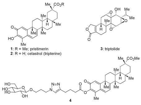
Figure 1.
The structures of anticancer terpenoids pristimerin, celastrol (tripterine), and triptolide (1–3) and the devised structure of a glucose conjugate of pristimerin (4).

Most cancer cells rely on aerobic glycolysis, rather than mitochondrial oxidative phosphorylation favored by normal cells, to generate energy for cellular processes. This phenomenon is known as the Warburg effect [1-3]. The high rate of aerobic glycolysis in cancer cells requires a significant increase in glucose uptake [2]. Glucose transporters (GLUTs), a family of membrane proteins that facilitate the transport of glucose across the plasma membrane, are overexpressed in a wide range of cancer cells [4]. Of note, some glucose analogues can also be recognized by GLUTs [5]. For instance, 2-deoxy-2-[18F]fluoro-d-glucose (18F-FDG), a radiolabeled glucose analogue, is taken up by cancer cells via GLUTs (in particular GLUT1 and GLUT3) at a markedly higher rate than by normal cells, which forms the basis of tumor imaging by positron emission tomography (PET) in clinical oncology [3]. Based on a similar mechanism of action, glucose conjugation has been considered a promising strategy for targeted delivery of anticancer agents [6]. The glucose conjugates of a number of cytotoxic compounds, including those of ifosfamide [7], duocarmycin SA [8], and paclitaxel [9, 10], exhibited improved target selectivity and water solubility.
Tripterygium wilfordii is a Chinese herb known natively as "Thunder God Vine". A series of hydrophobic terpenoids with potent but non-selective cytotoxicity have been identified from this species, including triterpenoids pristimerin [11, 12] and celastrol (tripterine) [13] (1 and 2, Fig. 1) and diterpenoid triptolide [14] (3). Despite considerable in vitro and in vivo studies, the lack of selectivity toward cancer cells, coupled with the poor water solubility, hampered the evolution of these natural products into anticancer drug candidates [12]. Of note, Liu, Pomper, and co-workers developed a glucose conjugate of 3, which displayed significantly improved selectivity and water solubility compared to the natural product itself [15]. Inspired by this work and in accordance with our continued interest in triterpenoids [16-18], we devised a glucose−pristimerin conjugate (4, Fig. 1) to overcome the problems of 1. This structure embodied the following considerations. (a) From the payload perspective, esterification of the enol could be a suitable method for the modification of 1. The C3 hydroxyl group of 1 had proved critical for its biological activities [19, 20]. Capping this hydroxyl group might help to reduce the side effects of 1 in the extracellular environment. Upon entry into cells, the conjugate would be cleaved at the ester linkage by cellular esterases, releasing the payload. Therefore, this conjugate was expected to serve as a prodrug to some content [10, 15]. (b) From the saccharide perspective, the β-glucoside should remain recognizable for GLUT1 [5]. (c) From the linker perspective, the 1, 4-disubstituted 1, 2, 3-triazole moiety would arise from an azide fragment and an alkyne fragment; copper-mediated 1, 3-dipolar cycloaddition [21-25] could be used for convergent and modular assembly of this type of conjugate. Herein, we report the synthesis of 4 and evaluation of its cytotoxicity in cancer cells and normal cells; however, the selectivity toward the former was not significantly improved. The unexpected cleavage of the ester linkage of 4 in the cell culture medium highlights the importance of extracellular stability of the conjugate.
Following our experience with the synthesis of hybridaphniphylline B [26], we first prepared tetraacetate 5 as a plausible precursor of 4 (Scheme 1). On one hand, β-d-glucose pentaacetate (6) underwent a known two-step sequence of glycosylation and substitution to give compound 7, via 3-bromopropyl β-d-glucoside tetraacetate [27] as an intermediate. On the other hand, treatment of 1 with acyl chloride 8 in the presence of Et3N provided enol ester 9 with excellent efficiency [28]. Under the 1, 3-dipolar cycloaddition conditions (CuI, Et3N) modified from those used in our one-pot triflate elimination−cycloaddition protocol [29], 5 was obtained from 7 and 9 in good yield. However, global deacetylation of 5 resulted in undesired cleavage of the enol ester, even under the mild conditions (Et3N, MeOH, water) used for our synthesis of hybridaphniphylline B [26, 30]. We then had recourse to fully deacetylated precursor 10 derived from 6 (Scheme 1) [31]. To our delight, the [3 + 2] cycloaddition reaction of 10 and 9 proceeded smoothly to afford target molecule 4. From a broad perspective, these efforts established a convergent and modular approach to preparation of glucose−payload conjugates.

We then evaluated the cytotoxicity of 4 in a number of human cancer cell lines and a human fetal hepatocyte line (L-02), using 1 as a control (Table 1 and Fig. S1 in (Supporting information). This glucose conjugate displayed potent inhibitory activity against triple-negative breast cancer (TNBC) MDA-MB-231 and cisplatin-resistant MDA-MB-231 (MDA-MB-231/DDP) cells and hepatocellular carcinoma (HCC) SMMC-7721 and QGY-7703 cells (Table 1, entries 1–4). However, its cytotoxicity against L-02 cells (entry 5) was not reduced as expected. The significant similarity between the cytotoxicity profiles of 4 and 1 implied that the glucose conjugation did not improve the selectivity toward cancer cells. Intrigued by the cause of this unsatisfactory selectivity, we investigated whether 4 could be effectively delivered into cancer cells via GLUTs. Frommer and colleagues reported that HCC Hep G2 cells rely predominantly on GLUT1 for glucose uptake [32]. If 4 enters Hep G2 cells via GLUT1, WZB117 [33], a GLUT1 inhibitor, should be able to antagonize the anticancer effect of this glucose conjugate [15]. However, when Hep G2 cells were co-treated with 4 and WZB117, the potency of the former remained unaffected (entry 6 versus entry 7). These results suggested that 4 was not effectively taken up by cancer cells and might have been hydrolyzed extracellularly to release 1.
We continued to examine the extracellular stability of 4, 5, and 9 (Table 2), all of which possess enol ester motifs. A series of media containing these compounds were incubated at 37 ℃ for 4 h. Not surprisingly, all the compounds were stable in water (Table 2, entry 1), which verified the intrinsic stability of their ester linkages. However, Dulbecco's Modified Eagle's Medium (DMEM) with and without Fetal Bovine Serum (FBS) both resulted in the release of 1 from the compounds (entries 2 and 3). This may be attributable to the ester cleavage mediated by amine nucleophiles in the media. Interestingly, the inorganic components in DMEM (including CaCl2 and MgCl2; see Supporting information for details) were found to be sufficient to induce the cleavage of the ester linkage of 4 but not to affect 5 and 9 (entry 4). We postulated that aquated Ca2+ or Mg2+ might be transiently enriched by the glucose moiety of 4 via its multiple hydroxyl groups and then serve as a local promoter of the enol ester hydrolysis.
In summary, we developed a convergent and modular approach to constructing glucose conjugates of anticancer agents and prepared glucose−pristimerin conjugate 4 through this approach. Compared to the payload (1), this conjugate did not exhibit significantly improved selectivity toward cancer cells as expected. Further investigations revealed that the inorganic salts in DMEM were sufficient to induce the cleavage of its ester linkage and the release of 1 in the extracellular environment. This work points to the need for proper evaluation of extracellular stability in the development of glucose−payload conjugates as well as other types of target-selective conjugates. The study of the next-generation glucose conjugates of 1 is underway in our laboratories.
The authors declare that they have no known competing financial interests or personal relationships that could have appeared to influence the work reported in this paper.
This paper is dedicated to Prof. Guo-Qiang Lin. This work was supported by Ministry of Science and Technology (National Key Research and Development Program of China, No. 2018YFA0901900), National Natural Science Foundation of China (Nos. 21931014, U2002221, 21772225, 21572064, 81502956 and 21621002), Chinese Academy of Sciences (Strategic Priority Research Program, No. XDB20000000; International Partner Program, No. 121731KYSB20190039; Key Research Program of Frontier Sciences, No. QYZDB-SSW-SLH040), Science and Technology Commission of Shanghai Municipality (Nos. 20430713400, 17XD1404600 and JCYJ-SHFY-2022–005).
Supplementary material associated with this article can be found, in the online version, at doi:10.1016/j.cclet.2022.04.036.
O. Warburg, Science 123 (1956) 309–314. doi: 10.1126/science.123.3191.309
S.Y. Lunt, M.G. Vander Heiden, Annu. Rev. Cell Dev. Biol. 27 (2011) 441–464. doi: 10.1146/annurev-cellbio-092910-154237
S.J. Bensinger, H.R. Christo, Semin. Cell Dev. Biol. 23 (2012) 352–361. doi: 10.1016/j.semcdb.2012.02.003
M.L. Macheda, S. Rogers, J.D. Best, J. Cell Physiol. 202 (2005) 654–662. doi: 10.1002/jcp.20166
S.S. Yuan, M.L. Li, J.S. Chen, L. Zhou, W. Zhou, ChemMedChem 13 (2018) 764–778. doi: 10.1002/cmdc.201700762
E.C. Calvaresi, P.J. Hergenrother, Chem. Sci. 4 (2013) 2319–2333. doi: 10.1039/c3sc22205e
J. Pohl, B. Bertram, P. Hilgard, et al., Cancer Chemother. Pharmacol. 35 (1995) 364–370. doi: 10.1007/s002800050248
L.F. Tietze, F. Major, I. Schuberth, Angew. Chem. Int. Ed. 45 (2006) 6574–6577. doi: 10.1002/anie.200600936
T. Mandai, H. Okumoto, T. Oshitari, et al., Heterocycles 54 (2001) 561–566. doi: 10.3987/COM-00-S(I)34
Y.S. Lin, R. Tungpradit, S. Sinchaikul, et al., J. Med. Chem. 51 (2008) 7428–7441. doi: 10.1021/jm8006257
A.B. Kulkarni, R.C. Shah, Nature 173 (1954) 1237–1238. doi: 10.1038/1731237b0
J.J. Li, Y.Y. Yan, H.M. Sun, et al., Front. Pharmacol. 10 (2019) 746. doi: 10.3389/fphar.2019.00746
G. Lan, J. Zhang, W. Ye, et al., Sci. China Chem. 62 (2019) 409–416. doi: 10.1007/s11426-018-9404-9
Z.L. Zhou, Y.X. Yang, J. Ding, Y.C. Li, Z.H. Miao, Nat. Prod. Rep. 29 (2012) 457–475. doi: 10.1039/c2np00088a
Q.L. He, I. Minn, Q. Wang, et al., Angew. Chem. Int. Ed. 55 (2016) 12035–12039. doi: 10.1002/anie.201606121
J. Li, P. Yang, M. Yao, J. Deng, A. Li, J. Am. Chem. Soc. 136 (2014) 16477–16480. doi: 10.1021/ja5092563
P. Yang, M. Yao, J. Li, Y. Li, A. Li, Angew. Chem. Int. Ed. 55 (2016) 6964–6968. doi: 10.1002/anie.201601915
P. Yang, J. Li, L. Sun, et al., J. Am. Chem. Soc. 142 (2020) 13701–13708. doi: 10.1021/jacs.9b09699
W.G. Shan, H.G. Wang, Y. Chen, et al., Bioorg. Med. Chem. Lett. 27 (2017) 3450–3453. doi: 10.1016/j.bmcl.2017.05.083
Z. Chen, D. Zhang, S. Yan, et al., Eur. J. Med. Chem. 177 (2019) 171–187. doi: 10.1016/j.ejmech.2019.05.009
V.V. Rostovtsev, L.G. Green, V.V. Fokin, K.B. Sharpless, Angew. Chem. Int. Ed. 41 (2002) 2596–2599. doi: 10.1002/1521-3773(20020715)41:14<2596::AID-ANIE2596>3.0.CO;2-4
C.W. Tornøe, C. Christensen, M. Meldal, J. Org. Chem. 67 (2002) 3057–3064. doi: 10.1021/jo011148j
M. Meldal, C.W. Tornøe, Chem. Rev. 108 (2008) 2952–3015. doi: 10.1021/cr0783479
J.E. Hein, V.V. Fokin, Chem. Soc. Rev. 39 (2010) 1302–1315. doi: 10.1039/b904091a
W.Y. Lu, X.W. Sun, C. Zhu, J.H. Xu, G.Q. Lin, Tetrahedron 66 (2010) 750–757. doi: 10.1016/j.tet.2009.11.044
W. Zhang, M. Ding, J. Li, et al., J. Am. Chem. Soc. 140 (2018) 4227–4231. doi: 10.1021/jacs.8b01681
J.A.F. Joosten, V. Loimaranta, C.C.M. Appeldoorn, et al., J. Med. Chem. 47 (2004) 6499–6508. doi: 10.1021/jm049476+
H. Sun, L. Xu, P. Yu, et al., Bioorg. Med. Chem. Lett. 20 (2010) 3844–3847. doi: 10.1016/j.bmcl.2010.05.066
Y. Chen, L. Liu, D. Wu, Y.P. He, A. Li, Chin. Chem. Lett. 30 (2019) 269–270. doi: 10.1016/j.cclet.2018.06.001
I.K. Mangion, D.W.C. MacMillan, J. Am. Chem. Soc. 127 (2005) 3696–3697. doi: 10.1021/ja050064f
A.K. Yadav, D.L. Shen, X. Shan, et al., J. Am. Chem. Soc. 137 (2015) 1181–1189. doi: 10.1021/ja5106738
H. Takanaga, B. Chaudhuri, W.B. Frommer, Biochim. Biophys. Acta 1778 (2008) 1091–1099. doi: 10.1016/j.bbamem.2007.11.015
W. Zhang, Y. Liu, X. Chen, S.C. Bergmeier, Bioorg. Med. Chem. Lett. 20 (2010) 2191–2194. doi: 10.1016/j.bmcl.2010.02.027

Figure 1 The structures of anticancer terpenoids pristimerin, celastrol (tripterine), and triptolide (1–3) and the devised structure of a glucose conjugate of pristimerin (4).

Scheme 1 Preparation of glucose conjugate 4 through a convergent and modular approach.

 扫一扫看文章

扫一扫关注我们
 DownLoad:


 下载:
下载: