
Scheme 1.
Schematic illustration of various types of H2O2 electrochemical sensor.
A review on recent advances in hydrogen peroxide electrochemical sensors for applications in cell detection
Yan Yu , Meng Pan , Jinrong Peng , Danrong Hu , Ying Hao , Zhiyong Qian

Hydrogen peroxide is a simple compound in nature, but as a highly oxidizing agent, it is mainly used as a disinfect, playing an important role in pharmaceutical, clinical, environmental, food, and other applications [1-3]. H2O2 is a member of the ROS family, and is relatively stable [4, 5]. Therefore, it not only plays a catalytic role, but also in cell toxicity [6]. Scientists report that H2O2 is an important metabolite, which plays critical role in the cellular metabolism in humans. As a messenger molecule, H2O2 participates in the initiation of a variety of cellular effects. Besides, it can interact with biological macromolecules in cells, and cause peroxidation of lipids of the cell membrane, proteins and enzyme denaturation in the cells, and DNA damage [4, 6, 7]. Therefore, maintaining normal levels of H2O2 concentration is key to realize the normal physiological activity of cells. Otherwise, if allowed to accumulate, it can lead to cardiovascular diseases, cancer, and neurodegenerative diseases and other major diseases [5, 6, 8]. Thus, considerable attention is being paid to be the biological role of H2O2 in vivo [9, 10]. Traditional hydrogen peroxide detection technologies, such as chemical titration [11], fluorescence [12, 13], chemiluminescence [14, 15], spectrophotometry [16] and high performance liquid chromatography [17, 18], are complex, expensive and time-consuming [19]. However, since H2O2 is an electroactive molecule, that can be oxidized or reduced on the sensor electrode [20], electrochemical sensors can provide a simple, rapid, sensitive and economically effective means of detecting H2O2.
Electrochemical sensors can be categorized into enzymatic sensors and nano-enzymatic sensors based on the presence or absence of enzymes [20, 21]. Enzymatic sensors are characterized by high catalytic activity and substrate specificity. However, they have low stability, can be easily denatured and high cost, which raises some serious difficulties, thus limiting their practical application [22, 23]. Therefore, with the rapid development of nanomaterials, nano-enzymatic sensors also known as non-enzymatic sensors have been in the research limelight, whereby the enzymes are replaced with low-cost and solid inorganic nano-catalysts [24-26]. Compared with natural enzyme sensors, non-enzymatic sensors have the advantages of simple preparation, convenient operation, good stability, high sensitivity, and low cost, and are widely used in environmental monitoring, food safety detection, and biomedical fields [25, 27-29]. However, there is currently no published review focusing on the application of electrochemical sensors in the detection of hydrogen peroxide in cells or tissues [30]. Therefore, this review focuses on the construction materials for using in enzymatic electrochemical sensing of H2O2 (Scheme 1). Besides, the review also pays attention to nano-enzymatic H2O2 electrochemical biosensors, including numerous new types of noble metal nanomaterials, metal oxides, polymers, carbon, and other materials, which can form nanocomposites. The types, advantages, and disadvantages of stimulants are also summarized in this review. Lastly, the problem associated with the development of H2O2 electrochemical sensors, including portability, reliability, miniaturization, and detection sensitivity are discussed. The future prospects of H2O2 electrochemical sensors are prospected.
Enzymatic biosensors were the first biosensors to be developed and they have widespread biomedical applications. Enzymes play a prominent role in biosensors because they are specific, efficient, and with high sensitivity. Horseradish peroxidase (HRP) [31], cytochrome c (Cyt c) [32, 33], and catalase (CAT) [34] are an important group of metalloproteins, with a catalytic active center, which is based on the structure of iron porphyrin [35]. Peroxidases have a wide range of sensing applications [36]. However, there are no more sensors that satisfy the H2O2 sensor in cells due to their instability and low reproducibility. Enzymes require to be immobilized on support materials [37, 38].
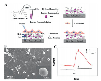
G. Bai et al. [39] fabricated a new electrochemical H2O2 biosensor by immobilizing HRP onto dendritic mesoporous silica nanoparticles (HRP/DMSNs) and reported a detection limit of 0.11 µmol/L. The HRP/DMSNs/GCE was found to detect H2O2 released from PC12 cells. Mesoporous materials were used to immobilize HRP, while chitosan-wrapped single-walled carbon nanotubes were used for the immobilization of HRP [40]. M. Murphy et al. [41] also prepared a carboxyl functionalized ionic liquid (TP-HA[TFSI]) through covalent immobilization of Cyt c on TPP-HA[TFSI]/MWCNT, which was found to enhance stability and conductivity. Enzymes can also be immobilized on support materials with different structures and functions. M. Lian et al. group [42] reported an enzymatic H2O2 sensor that was fabricated using self-assembled peptide nanofibrous hydrogel (N-fluorenylmethoxycarbonyl-diphenylalanine (Fmoc-FF)) embedded on HRP (Fig. 1A). The H2O2 released from HeLa cells was adhered to HPR/Fmoc-FF hydrogel modified GCE (Figs. 1B and C). The above studies mainly focused on immobilizing enzymes through covalent linking and polymer entrapment.
In summary, research on enzymatic H2O2 biosensors is fundamental. However, some of the disadvantages of enzymatic biosensors are the high cost, and that the enzymatic reaction is limited by diffusion. Besides, the interaction between enzymes and support materials has some influence on the sensing performance. Currently, few studies are reporting the role of enzymatic sensors in biological detection especially cell detection, which may be attributed to the low sensitivity and stability. Some strategies could be considered to overcome these drawbacks, for example, the size of the support materials and immobilization of enzymes may influence the catalytic effect of enzymatic H2O2 biosensors. Furthermore, the recent development of nanotechnology, and the use of nano-enzymes offers the possibility to overcome these limitations.
With the rapid development of nano-technology and nano-materials, nano-structural materials have been used in the development of nano-enzymatic biosensors. For example, the first Fe3O4 nanoparticles to be developed were peroxidase mimetics in 2007 [43]. For biosensor application, several nanomaterials, including noble metals, metal oxides, carbon nanomaterials, and nano-structured polymer have been used [44]. Here, we only discuss a few typical nanomaterials based on nano-enzymatic H2O2 biosensors for application in cell detection. Combined with the previous work [45], H2O2 electro-reduction mechanism for nano-enzymatic based H2O2 sensor on the surface of nanomaterial is generally summarized in following equations:
|
|
Compared with natural enzymatic biosensors, electrochemical sensors based on noble metal nanoparticles can amplify the sensing signal, which improves the sensitivity of the electrochemical sensor [46-48]. Therefore, we focus on the unique characteristics of a variety of noble metal nanoparticles and their bimetallic, trimetallic nanomaterials and nanocomposites, which are used to develop biomedical sensing platforms with high selectivity and sensitivity.
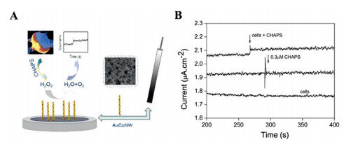
Gold nanoparticles have a high specific surface area, good stability, a simple preparation method, high catalytic activity, and excellent biological safety [49-51]. Therefore, the electrochemical sensors and biosensor platforms constructed using this method has higher stability [52]. These characteristics contribute to the construction of electrochemical sensors with stable comprehensive performance and high reliability [53, 54]. S.K. Maji et al. [55] presented a novel hybrid material AuNP-encapsulated periodic mesoporous silica (PMS) coated with reduced graphene oxide (RGO-PMS@AuNPs) for detection of H2O2 released from cells. A nonconductive PMS was formed in a sandwich-like structure on RGO, and processed semiconductor ability. RGO-PMS@AuNPs based H2O2 sensor showed satisfactory results, in human urine and living cells (HeLa and HepG2). H2O2 biosensor based on the AuCuNWs catalyst has also been reported to have unique catalytic properties with a low determination limit (2 nmol/L) due to the bimetallic mechanism. As shown in Fig. 2 [56], the sensor successfully detected H2O2 from Raw 264.7 cells, indicating possible biological applications. Y. Zhang et al. [57] synthesized gold nanocages using a simple one-pot method, and without the use of solid template or Au seed. AuNCs were found to have good electrochemical performance and a large specific surface area. Besides, electrochemical experiments showed that AuNCs/GCE had high electrocatalytic activity for H2O2 reduction. The sensor also had good anti-interference ability to uric acid, ascorbic acid, glucose, ethanol, and glycine. Gold nanocage material are simple to prepare and of low cost making them more attractive in sensor application. J. Hu et al. [58] reported H2O2 electrochemical sensors based on MoS2-Au-Ag. MoS2 as support material has a uniform lamellar structure which enhances the electrode conductivity. MoS2-Au-Ag-modified GCE can detect H2O2 from MCF-7 cells and RPE-1 cells. Y. Zhang et al. [59] synthesized AuNFs modified IL functionalized GF with 3D hierarchical porous structure. The nanohybrid paper sensors exhibit excellent sensing performance toward H2O2. Authors applied the sensor to track H2O2 from HBL-100, MDA-MB-231 and MCF-7 cells for application in radiotherapy treatment, which was expanded application of H2O2 sensors. These studies indicate that materials with a larger specific surface area can be obtained by adjusting the micro morphology of gold nanoparticles, which is conducive to improving the space and efficiency of electron transfer. In addition, gold nanoparticles have high molar absorptivity, which can also be used to develop a visual hydrogen peroxide sensor.
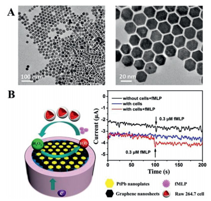
Pt-based nanomaterials have attracted extensive interest in the field of electrochemical biosensors due to their unique electrical and electrocatalytic properties [60]. Pt-based nanocomposites provide effective electrode materials for the expansion of their novel characteristics, and can be used to develop reliable, rapid, and accurate biosensors for the detection of various biomarkers and early detection of diseases [61-63]. J.X. Liu et al. [64] reported a dendrimer-encapsulated Pt nanoparticle and carbon nanotubes (Pt-DENs/CNTs) non-enzymatic H2O2 sensor. The sensor was found to have outstanding catalytic activity under the applied voltage (–0.15 V). Pt-DENs/CNTs sensors can be used to monitor H2O2 released from MCF-7 cells. A highly sensitive H2O2 sensor was constructed using similar synergetic mechanism is utilized to prepare graphene-Pt nanocomposites for measuring the H2O2 released from living cells [65]. A highly sensitive H2O2 sensor was constructed with Pt-Au bimetallic nanoparticles on reduced graphene sheets hybrid films [66]. The proposed sensor was found to have a low detection limit of 0.31 µmol/L in PC12 cells, thus providing a promising alternative H2O2 detection method. H2O2 sensor is modified by PtNPs@polyazure that can detect H2O2 from the bGB5 plant cells in real-time [67]. This sensor consists of a screen-printed carbon electrode (SPCE), and not a traditional GCE. Furthermore, this sensor can be reused after a simple cleaning, thus suitable for the development of an efficient and cheap commercial H2O2 biosensor. Y. Sun et al. [68] reported a H2O2 sensor based on novel graphene-supported intermetallic PtPb nanoplates nanocomposite, which has a very low detection limit of 2 nmol/L with a wide linear detection range of 2 nmol/L to 2.5 mmol/L. From Fig. 3, H2O2 detection was realized in Raw 264.7 cells. Pt nanomaterials can therefore be considered as outstanding electroanalytical material for nonenzymatic H2O2 sensors. Furthermore, our group developed PtNi nanoparticle-doped N-reduced graphene oxide (PtNi-N-rGO) nanocomposite modified GCE which exhibits the lower potential (−0.6 V) [69]. This sensing can capture H2O2 from the cancer cells and tissues. From the application point of view, H2O2 sensor will be used as a complementary pathology laboratory for tumor diagnosis.
Ag nanomaterials have the characteristics of high conductivity, the amplified response signal, and biological safety [70-72]. Therefore, electrochemical sensors based on silver nanoparticles (AgNPs) have great potential for biomedical applications [73]. The sensitivity and stability of the electrochemical biosensor platform mainly depend on the dispersion of AgNPs in the network or matrix [70, 73].
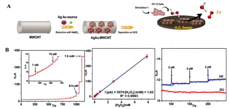
D. Yang et al. [74] developed Ag NPs doped mesoporous silica nanoparticles (mSiO2)-based H2O2 sensor for simultaneous detection of H2O2 in PC12 cells. Y. Yang et al. [75] developed 3D foam networks Ag-wire that not only exhibited efficient electronic delivery but also satisfactory sensitivity, due to the Ag-wire foam with hierarchical porous-structure. Besides, a microfluidic chip H2O2 sensor was constructed based on this foam for simultaneous detection of K562 cancer cells. In comparison with monometallic noble metal, bimetallic nanomaterials have used as electrocatalysts for detection H2O2 duo to their higher electronic effect. P. Balasubramanian et al. [76] prepared AgAu-MWCNTs nanohybrid materials and constructed a novel amperometric sensor. They successfully applied to monitor H2O2 expelled from the PC12 cells. Bimetallic AgAu nanoparticles distributed on the MWCNTs which caused broader electroactive surface area and greater electron transfer rate. This sensor had the lowest detection limit (0.23 nmol/L) for H2O2 in-situ monitoring (Fig. 4).
Pd nanoparticles have attracted much attention due to their high catalytic and sensing properties [60]. Compared with precious metals such as platinum and gold, the abundance of palladium is relatively high, which makes palladium a cheap alternative for various electrochemical and biosensor platform applications [77]. Due to the unique electronic properties of palladium, palladium nanocomposites and bimetallic nanoparticles have been widely studied, to improve catalytic performance and sensing selectivity.
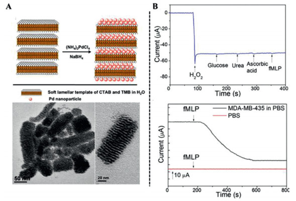
L. Yao et al. [78] prepared a novel porous Pd nanostructure using the soft micellar template of TMB interacting with surfactant CTAB. As shown in Fig. 5 [78], the average size of the porous Pd nanoparticles was 2 nm, which was evenly dispersed on the template. The sensor was used for the highly sensitive H2O2 electrochemical detection. The detection limit was 0.25 µmol/L, and the linear range was 0.25-900 µmol/L. Finally, the sensor was used to detect the H2O2 released by MDA-MB-435 cells under the voltage of −0.34 V. K.G. Nikolaev et al. [79] prepared a PdAu bimetallic electrochemical sensor by directional electrochemical nanowire assembly (DENA). The sensor was used to test H2O2 released from HL-1 cells utilizing the synergistic catalytic capacity of Pd and Au. The new multi-sensor detection platform utilized different electrochemical components of DENA developed on a single chip. These studies demonstrate that Pd nanoparticles have excellent catalytic activity, and are cheaper than Pt and Au nanoparticles. At the same time, the detection limit of the sensor is lower, making them ideal electrode materials for the sensor. In summary, different noble metal nanoparticles based electrochemical sensors have different roles depending on their cost, application and impact on the environment.
Metal oxide nanomaterials are widely used in electrochemistry, soft magnetism, catalysis, sensors and other fields [80]. And they can significantly improve the sensitivity and overall performance of sensors due to their small particle size, large specific surface area, strong electrocatalytic activity, and low cost. Besides, they are widely used as effective electrocatalytic materials for various analyte sensors in biological and biomedical fields [80]. Various metal oxide nanoparticles have been used in bioelectrical catalysis, including cobalt oxide (Co3O4), nickel oxide (NiO), cerium oxide (CeO2), copper oxide (CuO), titanium oxide (TiO2), zinc oxide (ZnO) and manganese oxide (MnO).
Common magnetic nanomaterials, such as Fe3O4, Fe2O3, Co3O4, NiO, have high specific surface area and superior electron transfer behavior, thus, they are ideal nanomaterials for electrochemical biosensors [81-85].
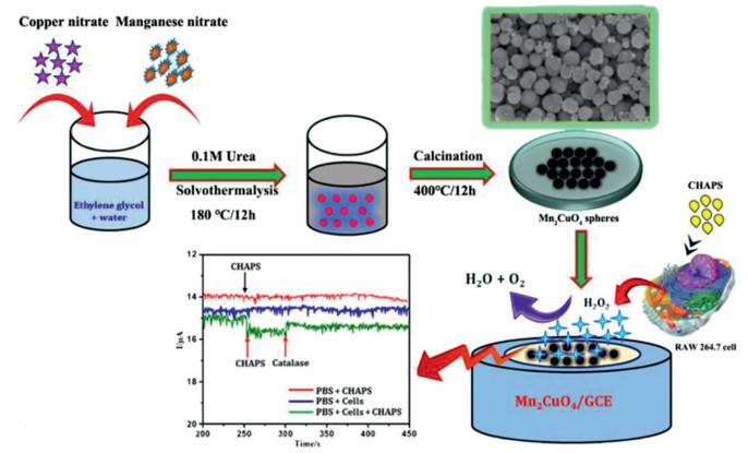
Y. Zhao et al. [86] prepared ultrasmall Fe3O4 decorated on three-dimensional graphene (3DG) nanocomposites by the hydrothermal method, and constructed as a nano-enzymic H2O2 sensor. The 3DG networks provided a relatively large surface area and high conductivity but Fe3O4 QDs with 5-7 nm size processed the catalyze roles. This biosensor could monitor H2O2 from A549 cells. J. Xie et al. [87] prepared 2D holey CuCo2O4 (2D HCCO) nanocomposites, and use them to construct the H2O2 electrochemical platform with CeO2. The H2O2 biosensor based CeO2/HCCO/MWCNTs was successfully fabricated and used to monitor H2O2 released from Hela cells. The holey HCCO-based biosensors have shown great potential in early cancer diagnosis. L.H. Nonaka et al. [88] prepared rGO-MnFe2O4 composites using an aerosol-assisted capillary compression process to construct H2O2 sensors. A. Ding et al. [89] developed MnO nanoparticles on the surface of carbon nanofibers (CNF) which were found to offer more active sites and higher electrocatalytic activity. P. Balasubramanian [90] reported an H2O2 electrochemical sensor based on Mn2CuO4 (MCO) microspheres prepared by the solvothermal method. This sensor could assay cellular H2O2 in the presence of CHAPS (Fig. 6). This sensor was used to detect H2O2 from A375 cells, which demonstrated potential biomedical application. These H2O2 sensors have the best performance due to the synergistic effect between different components.
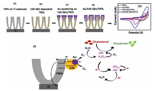
Other metallic oxides, including ZnO, SnO2, and TiO2, can be used to improve the physical and chemical properties of nanocomposites [91-94]. For example, a biocompatible C-dot-wrapped ZnO nanoparticle based H2O2 sensor has been reported [95]. This biosensor has reliable reproducibility and low detection limit (2.4 nmol/L), possesses sensitive and selective photoelectrochemical performance, due to the wrapping with C-dots. Similarly, it can be used for detecting H2O2 from HeLa cells. Titanium dioxide nanotubes (TNTs) are chemically stable, biocompatible, and with strong adhesion to the Ti substrate. Although TNTs have bio-sensing performance, the large band gap can weaken electron transfer. N. Khaliq et al. [96] designed and synthesized Au/CdS QDs/TNTs, which has a hybrid structure with fast electron transfer, high selectivity, and high specific surface area due to TNTs decorated with dispersed CdS QDs and Au NPs layer (Fig. 7). The different redox potential results in a voltage-induced switchable biosensor, which can detect H2O2 in human blood serum.
Polymer-based electrochemical sensors and biosensor platforms are widely used in detection because they are easy to realize biomolecule functionalization and long-term stability. Polymer-based sensors based on conducting polymers (CPs) and dendrimers have high sensitivity and selectivity, and can be used to detect DNA, enzymes, proteins, antigens, and metabolites [80, 97]. The main advantages of polymers are the encapsulation of metal nanomaterials, which can increase conductivity. Different kinds of polymers are discussed in the development of H2O2 electrochemical sensors.
CPs are a large class of polymer nano-materials that are doped by chemical or electrochemical methods to change the insulating state into a conductive state. A.J. Heeger et al. discovered the first conducting polymer, CIS polyacetylene in 1974 [98]. CPs widely used in sensing including polyaniline (PANI), polypyrrole (PPy), and poly(3, 4-ethylenedioxythiophene) (PEDOT) [99-102]. M.H. Naveen et al. [103] designed a simple method to prepare porous materials AuNi@pTBA dendrite catalyst, with conductive polymer tributyl acrylate (PtBA) as the base layer, electrodeposited on the conductive polymer, and finally performed dealloying treatment, to improve the performance of the catalyst. As shown in Fig. 8 [103], the porous dendrite morphology was uniform. The sensor was used to monitor H2O2 released by normal and tumor cells. In vitro studies showed that the composite had no obvious cytotoxicity to cells. However, the conductive polymer had some defects such as poor processability and solubility, thus, needed to be modified, but it is a promising sensor material.

Dendritic macromolecules are synthetic polymers with a highly branched molecular structure, which is considered as a nanostructure. Electrochemical sensors/biosensors platform based on dendrimers are widely used due to their unique structural characteristics, including structural consistency, chemical stability, and low cytotoxicity. Different classifications of dendrimers are used in the fabrication of electrochemical sensors [104]. Notably, M. Elancheziyan et al. [105] synthesized PAMAM dendrimer encapsulated with AuNPs. The PAMAM dendrimer act as supporting materials in sensor fabrication of Hb modified electrode using nanocomposite. The PAMAM biosensor was used in the detection of H2O2 at a broad linear range of 20 µmol/L to 950.22 µmol/L. M. Baghayeri et al. [106] researched and designed a nanocomposite graphene oxide composite Fe3O4 to form magnetic graphene. Graphene and polyamide dendrimer with an active terminal amine group formed GO-Fe3O4-PAMAM through covalent binding. The existence of amine groups on the surface can provide amply binding sites for Pd nanoparticles. The unique hyperbranched structure of PAMAM was also favorable to the uniform dispersion of Pd nanoparticles. The electrocatalytic activity of the modified electrode was effectively improved. However, the sensors typically detect H2O2 in real water samples but not in cells, which may limit their application in disease diagnosis. PPI dendrimer also acts as a nano-mediator and it is used as an electrochemical biosensor [104]. E. Murugan et al. [107] reported poly(propylene imine) as supporting materials encapsulated with ruthenium nanoparticles for detecting H2O2 in the range of 100 µmol/L to 5 mmol/L. Few studies have reported the use of PPI dendrimer-based sensors in H2O2 detection, and this indicates the great application potential of PPI dendrimer-based sensors in H2O2 electrochemical sensors for use in biological detection.
Molecularly imprinted polymer (MIP) is a polymer obtained by polymerizing template molecules, functional monomers, crosslinkers and initiators in solvent, and then removing the template molecules using certain methods [108, 109]. The obtained polymer is referred to as MIP and has the characteristics of stable molecular shape and spatial structure [110, 111]. The biosensor platform based on MIPs has been used to detect a variety of biological and organic molecules, including drugs, pesticides, amino acids and peptides [112-117]. These applications have confirmed that it is conducive to the selectivity of functional groups in the process of molecular imprinting. Y.H. Cao et al. [118, 119] used the microemulsion method and molecularly imprinted technology to prepare imprinting Hb on the surface of Fe3O4@SiO2 magnetic imprinted nanoparticles (MMIPS NPs) and constructed a Hb based enzyme hydrogen peroxide sensor. But this sensor was not used furtherly systematic research in cells detection. Interestingly, compared with non-magnetic imprinted materials, the catalytic current of the magnetic imprinted materials increased by 14.3%. This was due to the use of magnetically imprinted nanoparticles to make Hb and produce O2 through the decomposition of H2O2. The result is that the sensor can enhance the H2O2-sensitizing effects.
Carbon-based nanomaterials have emerged in recent years, such as carbon nanotubes (CNTs), graphene, carbon quantum dots (CDs) and ordered mesoporous carbon (OMCs) [120-122]. Carbon-based electrochemical sensors have been widely developed due to their low cost, chemical stability, and good electron transfer kinetics.
Carbon nanotubes (CNTs) were firstly reported in 1991 [123] and are characterized by many incredible properties in terms of their structure, function, and morphology. The structure is a hollow cylindrical tube with a special length diameter ratio composed of a sp2 hybrid graphite sheet [124]. Therefore, CNTs can be divided into SWNTs and MWNTs based on the number of graphite layers [125] and have been widely used in many fields, such as electronics [126], energy [127], aerospace [128], military [127], biomedicine [129] and biosensors [130, 131]. Therefore, several scholars have carried out exploratory studies on how to improve the performance of electrochemical sensing and biosensor platforms using CNTs [132-134]. M. Eguílaz et al. [135] reviewed H2O2 electrochemical sensing-based CNTs developed between 2016 and 2018. However, this review did not include all recent biological detection studies. J. Bai group [136] prepared H2O2 sensors comprising of CDs and MWCNTs. More importantly, CDs/MWCNTs/GCE exhibited significant synergistic effect, and with a better detection limit. The H2O2 released from HeLa cells was tracked with satisfactory results. This is a general strategy for the fabrication of carbon-based H2O2 biosensors. Similarly, H. Tavakkoli et al. [137] designed an H2O2 electrochemical sensor using multiwalled carbon nanotubes and zein nanoparticles. CNTs can be intermixed with zein proteins to form GCE, to provide higher electrocatalytic performance for an H2O2 sensor. This sensor can respond to H2O2 in the 0.049 µmol/L to 22 µmol/L concentration range and can distinguish between cancer cells (HepG2) and normal cells (HDF). J. Li group [138] reported a novel electrochemical platform based on sandwich-like nanostructured nano-Au/CNTs/PDMS film. This sensor had excellent stretchable and flexible properties attributed to the transfer CNTs on flexible PDMS and then deposition of nano-Au on the CNT surface (Fig. 9). These H2O2 mechanical flexible sensors can monitor H2O2 released from HeLa and HUVEC cells.

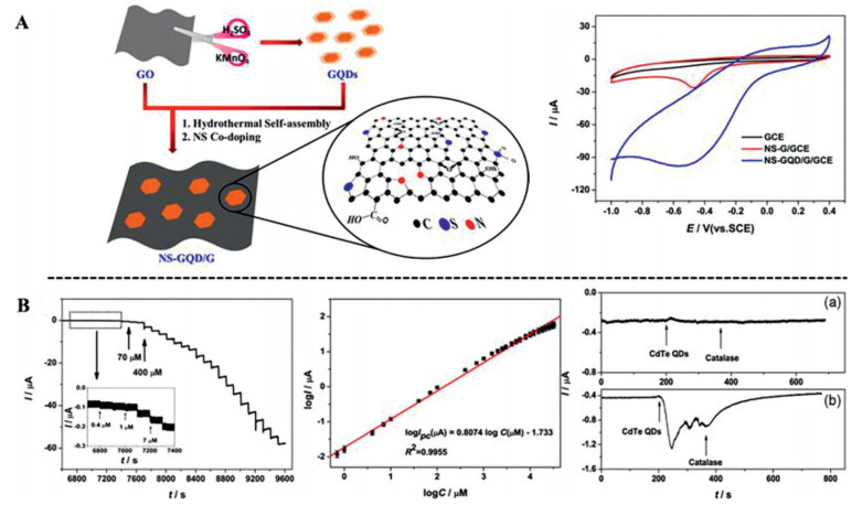
Graphene is one kind of two-dimensional carbon material with infinite extension and has a hexagonal lattice similar to a honeycomb structure [139, 140]. The unique structure makes it to be highly sensitive, selective and with excellent electrocatalytic activity [141]. Compared with carbon nanotubes, graphene has the advantages of easy processing and low cost [142]. Numerous studies have been devoted to the applications of graphene and graphene-based nanocomposites in electrochemical sensing and biosensors [143, 144]. Y. Sun et al. [145] developed a highly sensitive and selective H2O2 sensor for monitoring normal cell activity. PtNi NWS/rGO composites were fabricated through ultrasonic physical self-assembly using rGO and synthesized PtNi nanowires. The nanowires were loaded on graphene network structure and assembled into PtNi NWS/rGO composites. The linear range of the electrochemical response of the material was very wide (1 nmol/L–5.3 mmol/L), and the detection limit was as low as 0.3 nmol/L. The sensor performance was superior compared with conventional sensors because PtNi nanowires in PtNi NWS /rGO composites provided high-density active centers. The amperometric potential was −0.6 V (vs. Ag/AgCl) and can be used to detect H2O2 released from RAW 264.7 cells. B. Patella et al. [146] developed an H2O2 sensor by electro co-deposition methods, using a solution containing GO and KAuCl4 for electrodeposition rGO/Au-NPs-based on poly-ethylene-terephthalate foil (ITO-PET). The developed sensor could detect H2O2 from THP-1 cells and be verified by flow cytometry. The detection correlation shows that an H2O2 sensor is valuable approach in telemedicine for H2O2 monitoring. T. Zhang [147] used a two-step process to prepare N and S co-doped NS-GQD/G hybrid nanosheets (Fig. 10). The developed H2O2 sensor was designed based on the hybrid nanosheets with high specific surface area and numerous doping sites. The highlighted studies reveal the high performance of H2O2 sensors in detection of H2O2 in cells.
The first mesoporous carbon (OMCs) was discovered by professor Ryoo in 1999 [148]. Since then, a lot of basic research work has been done on the applications of mesoporous carbon [149-151]. Due to their special ordered structure and remarkable properties, such as periodic mesoporous structure, high specific surface area and large pore volume, OMCs have been widely used in electrocatalysis [152-154]. T. Wang et al. [155] reported a facile H2O2 sensor based on MnO nanoparticles on mesoporous carbon (MnO@C), prepared by the thermal calcination method. 10 nm MnONPs provided efficient catalytic properties in nanocomposites, while the mesoporous carbon served as a matrix for enhancing charge transport. A H2O2 sensor was constructed using MnO@C nanocomposites on Ti foil and was successfully used for cellular H2O2 detection in normal (293T) and tumor cells (HeLa).
Carbon quantum dots (CQDs) are known as spherical nanoparticles with less than 10 nm in size [156, 157]. CQDs are newly discovered carbon nanomaterials, which have attracted great attention over recent years due to their excellent properties in physical and chemical capabilities, such as good water solubility, low-toxicity, photostability, and good stability [158-160]. With further research on CQDs, they are considered a good choice for the development of sensors due to their stronger catalytic activity, which is suitable for improving the electrocatalytic activity of nanocomposites [161]. J.S. Kumar et al. [162] prepared CuO@CQDs@CHNS with a core shell structure through facile hydrothermal method. This nanocomposite was used for H2O2 sensing with detection limit of 2.4 nmol/L. Similarly, H2O2 non-enzymatic electrochemical sensor based CQDs/octahedral cuprous oxide (Cu2O) nanocomposites were designed by Y. Li et al. [163]. The amount of CQDs was usually decorated on the supporting materials, which has exhibited a significant synergistic electrocatalytic activity for H2O2 sensing. However, the vast majority of studies have focused on fluorescent sensors or probes, due to their unique optical properties and easy surface functionalization [164].
With the rapid development of other 2D materials, including metal organic frameworks (MOF) [165], Bi2Te3 [166], MoS2 [167], and Ti3C2, they have also shown great potential in sensing applications [168, 169].
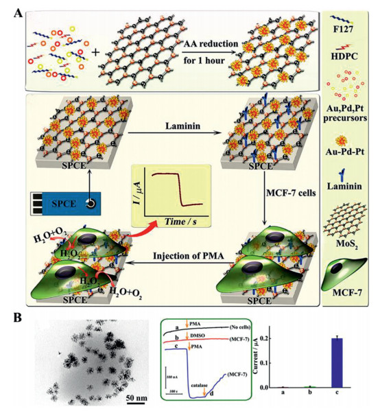
Transition metal disulfide compounds (TMDC) are a large class of two-dimensional crystal materials. The general chemical formula is MX2, where M is a transition metal (molybdenum, tungsten, niobium, etc.), and X is a chalcogenide element. This material presents an X-M-X sandwich structure, and the layers are connected by van der Waals force. MoS2 is a member of TMDC, and the Mo layer (S-Mo-S) sandwiched between the two S layers is laminated by weak van der Waals forces interactions, to form a unique graphene-like structure. The indirect band gap of MoS2 is 1.30 eV, therefore, it is widely used as active material in energy storage, solar cells, catalysis, and other fields [170-172]. H2O2 biosensor-based ultrasmall MoS2 nanoparticles have an extremely sensitive ability to detect H2O2 [172, 173]. This biosensor has a low limit determination at 2.5 nmol/L. Under the stimulation of fMLP, it can detect H2O2 from Raw 264.7 cells. L. Zhu et al. [174] prepared PtW/MoS2 nanocomposites by in-situ growth method, with a good catalytic effect on H2O2. Therefore, a combination of PtW nanocrystals and MoS2 nanosheets enhances the adsorption and catalysis of H2O2 on the surface of sensing materials and further improves comprehensive performance of the sensor. Finally, the H2O2 sensor constructed using this material was applied to detect the H2O2 released by 4T1 breast cancer cells. P. Wei et al. [175] investigated H2O2 sensing of Pt/cMIL-68/MoS2/GCE. They selected MoS2 nanosheets as the supporting materials because of the large surface area and electrical conductivity. Calcined MOF and PtNPs have better electrocatalytic performances. The developed sensor was found to be suitable for monitoring H2O2 released in MCF-7 cells. MoS2 are of interest because of their easy preparation and relatively large surface area. B. Dou et al. [176] developed a trimetallic hybrid Au-Pd-Pt nanoflower-decorated MoS2 nanosheet-based H2O2 biosensor (Fig. 11). The Au-Pd-Pt/MoS2 nanocomposites were synthesized via a simple wet-chemistry technique. The superior performance of the sensor was attributed to the synergistic effect of the highly dispersed Au-Pd-Pt nanoflowers on MoS2 nanosheets. The sensor had the ability to in situ monitor of H2O2 from MCF-7 cancer cells.
Two-dimensional (2D) transition metal carbides or nitrides (MXenes) are mainly used in optical, energy storage and sensor application [177, 178]. Ti3C2 has been extensively researched due to its high conductivity and surface properties [179, 180]. Y. Dang et al. [181] fabricated the non-enzymatic H2O2 biosensor based on Prussian blue nanoparticles (PB NPs) intercalated Ti3C2 nanosheets. PB/Ti3C2/GCE showed the satisfactory sensitivity and stability, due to the synergistic effect between PB and Ti3C2. PB/Ti3C2 achieved real-time detection of H2O2 from HeLa cells. S. Neampet et al. [182] prepared Pt/PANI/MXene nanocomposites and constructed an H2O2 sensor with a modified a SPCE. MXene as the supporting materials can enhance the specific surface area, incorporation of PANI in MXene can improve conductivity, while, Pt particles effectively accelerate the electron transfer. This synergistic effect enhances the stability and reliability of the H2O2 sensor.
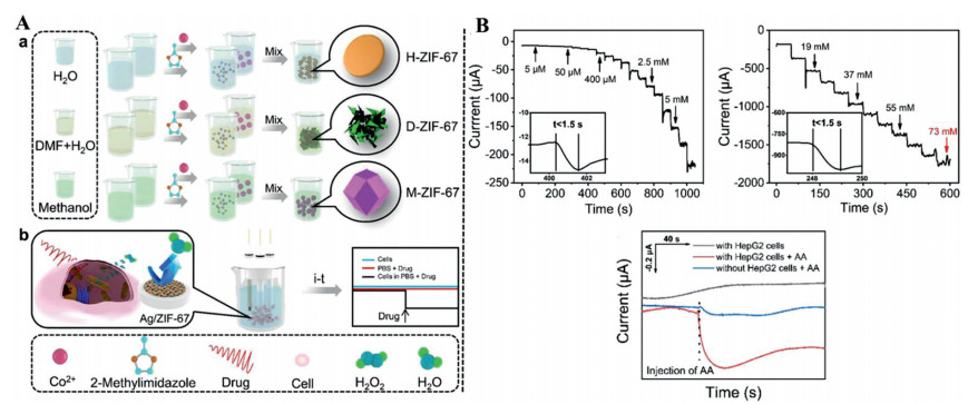
MOFs are a new class of materials prepared through self-assembly of organic ligands and metal ions [183]. In recent years, the applications of MOFs in ion exchange, separation science, sensors, gas storage, energy storage, and other fields, has been extensively studied [184-186]. Indeed, MOFs with good electrocatalytic properties for electroanalysis have been designed and synthesized [185, 187]. J. Lu research group [188] fabricated a H2O2 biosensor based on gold nanoflowers-encapsulated molybdenum disulfide and magnetic metal-organic framework nanozymes (AuNFs/Fe3O4@ZIF-8-MoS2). They found that incorporation of MoS2 nanosheets and AuNFs enhanced the stability, conductivity and catalytic performance of Fe3O4@ZIF-8. Moreover, this biosensor was able to detect H2O2 in H9C2 cells under AA stimulation. Elsewhere, a novel Cu-based metal-organic framework (Cu-MOF) has been prepared for the construction of AuNPs-NH2/Cu-MOF composite [189]. The framework was used to modify GCE to detect H2O2 in HeLa cells. Some scholars have designed more convenient H2O2 sensor and are suitable for cell biological detection [190]. The D. Sun research group [191] studied the morphology and performance of co-containing zeolite imidazolate framework-67 (ZIF-67). They build an electrochemical sensor based on the one-step electrodeposition of Ag nanostructures on ZIF-67 (Fig. 12). This sensor showed good performance in detection of H2O2 in HepG2 cells.
In summary, nano materials have stimulated the development of nano-enzymatic H2O2 sensor quickly in recent years. Different kind of nano materials have different roles. The selection of suitable supported materials and loading method are key points for sensor sensing materials. Two-dimensional nano materials such as carbon, polymer, MoS2, MOFs and MXenes nanomaterials mainly play good roles as supported materials. But these materials have agglomeration bottleneck and their synthesis is time consuming and demand special conditions. Therefore, more effective synthesis methods could be needed. Different noble metal and metallic oxide nanoparticles have different morphologies, such as dots, tubes, fibers, and spheres, which have different the surface area and conductivity. Modifying the morphology of nanoparticles can achieved high selectivity and sensing performance. However, noble metal nanocomposites have limitation of higher cost, so this limitation could be overcome by designing bimetallic or trimetallic nanomaterials. Metal oxides also have some disadvantages like poor conductivity and structural instability. For example, metal oxides doped with noble metal, which can give better conductivity and electro-catalytic activity. In spite of the bottleneck that remain, we expect that future innovative research on nanomaterials for sensors will improve their shortcomings.
Whether it is an enzymatic sensor or nano-enzymatic sensor, H2O2 in cells must be stimulated to form respiratory bursts and release H2O2 from cells [192]. No review has been conducted to summarize the stimulant agents of H2O2 sensors. The A. Ding research group [89] used Zymosan (0.5–2 g/L) as a stimulant to trigger the generation of H2O2 in cancer cells. Another research group [64] used ascorbic acid (AA, 2 × 10⁻⁶ mol/L) as a stimulant to initiate H2O2 from living cancer cells. AA is water- and fat-soluble and can move to specific places to cause occur oxidative damage in cells [193]. K+ also was selected as a stimulant for H2O2 sensor based on AgAu-MWCNTs [76]. CdTe QDs was used as a stimulus, which can induce cell death by generating H2O2 due to CdTe QDs as toxic molecules [136]. The N. Wang research group [56] used 0.3 × 10⁻⁶ mol/L CHAPS (3-[(3-cholamidopropyl) dimethylammonio]-1-propanesulfonate) as a stimulant for H2O2 released from Raw 264.7 cells due to CHAPS as one the important ingredients of lysis buffer. G. Yu group [66] utilized 5 µg/mL lipopolysaccharide (LPS) to induce H2O2 released from cells. Because some paper reported that LPS induced oxidative burst was found to be of mitochondrial origin [194, 195]. Phorbol myristate acetate (PMA) as a H2O2 inducer was employed to stimulate cells [97]. Although PMA can activate PKC directly [196], it is a strong poisonous chemical agent. N-Formylmethionyl-leucyl-phenylalanine (fMLP) is used as a stimulant for detection of H2O2 in cells [197, 198]. fMLP is considered as chemotactic peptide because it induces various responses in cells, including adhesion, migration and phagocytosis of cells. However, fMLP also has its shortcoming of being expensive. Collectively, different kinds of stimulants have similar effect in these studies (Table 1). Researchers should aim to develop safe, cheap, and environmentally-friendly stimulants that will be useful for real-time analysis of H2O2 sensors.
 DownLoad:
CSV
DownLoad:
CSV

|
In summary, we reviewed advances in the application of hydrogen peroxide electrochemical sensors in the last five years. Progress achieved in sensor development has been spurred by the use of many nanomaterials. In terms of enzyme-based H2O2 electrochemical sensor, some problems are key impediments to the development of this field, such as high costs and immobilization of enzymes. This review discussed nano-enzymatic H2O2 electrochemical biosensors, including nanomaterials of noble metal, metal oxide, polymer, carbon, and other materials, which tend to form nanocomposites. Nano-composites are widely applied in sensor development because they have synergistic effect and cause more defect sites. Finally, a summary of stimulants of H2O2 sensors is provided. In addition, the principle and application of different stimulant agents are highlighted.
Several challenges exist which hinder the design of more reproducible H2O2 electrochemical sensors. For instance, conventional glassy carbon electrodes are not suitable for practical application and convenient batch testing. Nevertheless, for clinical cells or tissues samples, they need disposable and ensure the reproducibility of the results. In the future, Screen printed electrodes should be developed. In addition, the structure, morphology and composition of nanocomposites influence the performance of sensors. In the future, it is necessary to integrate a sensor material evaluation platform to determine whether the nanocomposites meet the application standards. Furthermore, there is need to apply miniaturization and intelligent systems to design electrochemical instruments that require little operational skills which will improve disease diagnosis. Finally, simple and cheap nanomaterials with good performance for sensors should be developed. In conclusion, future research should aim to develop effective diagnostic methods for cancer and other diseases.
The authors declare that they have no known competing financial interests or personal relationships that could have appeared to influence the work reported in this paper.
This work was financially supported by the National Natural Science Fund (NSFC, Nos. U21A20417, 31930067, 31525009), and the 1·3·5 Project for Disciplines of Excellence, West China Hospital, Sichuan University (No. ZYGD18002). The authors also thank the support by the Fundamental Research Funds for the Central Universities.
S. Zhao, G.C. Zang, Y.C. Zhang, et al., Biosens. Bioelectron. 179 (2021) 113052. doi: 10.1016/j.bios.2021.113052
C.C. Hsu, Z.Q. Xu, Y.C. Xu, et al., Sens. Actuat. A 322 (2021) 112630. doi: 10.1016/j.sna.2021.112630
Y. Wandee, D. Uttapap, P. Mischnick, V. Rungsardthong, Food Chem. 348 (2021) 129078. doi: 10.1016/j.foodchem.2021.129078
C. Lennicke, J. Rahn, R. Lichtenfels, et al., Cell Commun. Signal. 13 (2015) 39. doi: 10.1186/s12964-015-0118-6
C.R. Reczek, N.S. Chandel, Annu. Rev. Cancer Biol. 1 (2017) 79-98. doi: 10.1146/annurev-cancerbio-041916-065808
C. Lismont, I. Revenco, M. Fransen, Int. J. Mol. Sci. 20 (2019) 3673. doi: 10.3390/ijms20153673
M. Schieber, N.S. Chandel, Curr. Biol. 24 (2014) R453-R462. doi: 10.1016/j.cub.2014.03.034
G. Calabrese, E. Peker, P.S. Amponsah, et al., EMBO J. 38 (2019) e101552.
C.H. Reid, N.J. Finnerty, Biosensors (Basel) 17 (2017) 1596. doi: 10.3390/s17071596
O.M. Subach, T.A. Kunitsyna, O.A. Mineyeva, et al., Int. J. Mol. Sci. 20 (2019) 3138. doi: 10.3390/ijms20133138
N.V. Klassen, D. Marchington, H.C.E. McGowan, Anal. Chem. 66 (1994) 2921-2925. doi: 10.1021/ac00090a020
C. Yik Sham Chung, G.A. Timblin, K. Saijo, C.J. Chang, J. Am. Chem. Soc. 140 (2018) 6109-6121. doi: 10.1021/jacs.8b02279
J. Xu, Y. Zhang, H. Yu, et al., Anal. Chem. 88 (2016) 1455-1461. doi: 10.1021/acs.analchem.5b04424
M. Haddad Irani-Nezhad, J. Hassanzadeh, A. Khataee, Y. Orooji, Molecules 24 (2019) 689. doi: 10.3390/molecules24040689
G. Zambrano, F. Nastri, V. Pavone, et al., Sensors (Basel) 20 (2020) 3793. doi: 10.3390/s20133793
D. Mukhopadhyay, D. Sinha Dasgupta, et al., Free Radicals Antioxid. 6 (2015) 123-131.
A.S. Ivanova, A.D. Merkuleva, S.V. Andreev, K.A. Sakharov, Food Chem. 283 (2019) 431-436. doi: 10.1016/j.foodchem.2019.01.051
S.M. Steinberg, Environ. Monit. Assess. 185 (2013) 3749-3757. doi: 10.1007/s10661-012-2825-4
W.X. Wang, W.L. Jiang, G.J. Mao, et al., Anal. Chem. 93 (2021) 3301-3307. doi: 10.1021/acs.analchem.0c05364
W. Chen, S. Cai, Q.Q. Ren, et al., Analyst 137 (2012) 49-58. doi: 10.1039/C1AN15738H
X.Y. Yang, P.P. Qiu, J.P. Yang, et al., Small 17 (2021) 1904022. doi: 10.1002/smll.201904022
G. Rocchitta, A. Spanu, S. Babudieri, et al., Biosensors (Basel) 16 (2016) 780. doi: 10.3390/s16060780
J.N. Tiwari, V. Vij, K.C. Kemp, K.S. Kim, ACS Nano 10 (2016) 46-80. doi: 10.1021/acsnano.5b05690
S.J. Rowley-Neale, E.P. Randviir, A.S. Abo Dena, C.E. Banks, Appl. Mater. Today 10 (2018) 218-226. doi: 10.1016/j.apmt.2017.11.010
C.Z. Zhu, G.H. Yang, H. Li, et al., Anal. Chem. 87 (2015) 230-249. doi: 10.1021/ac5039863
D. Thatikayala, D. Ponnamma, K.K. Sadasivuni, et al., Biosensors (Basel) 10 (2020) 151.
C.R. Zhao, X.L. Li, S.X. An, et al., Sci. Bull. 64 (2019) 1272-1279. doi: 10.1016/j.scib.2019.07.015
N. Joshi, T. Hayasaka, Y. Liu, et al., Microchim. Acta 185 (2018) 213. doi: 10.1007/s00604-018-2750-5
Y.B. Hou, K. Sheng, Y. Lu, et al., Microchim. Acta 185 (2018) 397. doi: 10.1007/s00604-018-2925-0
H.Y. Liu, L.Y. Weng, C. Yang, Microchim. Acta 184 (2017) 1267-1283. doi: 10.1007/s00604-017-2179-2
X.W. Huang, S. Xu, W. Zhao, et al., ACS Appl. Nano Mater. 3 (2020) 9158-9166. doi: 10.1021/acsanm.0c01800
L. Zanetti-Polzi, I. Daidone, C.A. Bortolotti, S. Corni, J. Am. Chem. Soc. 136 (2014) 12929-12937. doi: 10.1021/ja505251a
Y. Luo, H. Liu, Q. Rui, Y. Tian, Anal. Chem. 81 (2009) 3035-3041. doi: 10.1021/ac802721x
P.A. Walton, C. Brees, C. Lismont, et al., BBA -Mol. Cell Res. 1864 (2017) 1833-1843.
R. Qu, L.L. Shen, Z.H. Chai, et al., ACS Appl. Mater. Interfaces 6 (2014) 19207-19216. doi: 10.1021/am505232h
Y.F. Wang, J. Du, Y.Y. Li, et al., Colloids Surf. B 90 (2012) 62-67. doi: 10.1016/j.colsurfb.2011.09.045
X.Y. Yang, P.P. Qiu, J.P. Yang, et al., Small 17 (2019) 1904022.
W.P. Liu, H.H. Pan, C.X. Liu, et al., ACS Appl. Mater. Interfaces 11 (2019) 11466-11473. doi: 10.1021/acsami.8b22686
G. Bai, X. Xu, Q. Dai, et al., Analyst 144 (2019) 481-487. doi: 10.1039/C8AN01712C
H. Jiang, C. Du, Z. Zou, et al., J. Solid State Electrochem. 13 (2009) 791-798. doi: 10.1007/s10008-008-0612-5
M. Murphy, K. Theyagarajan, P. Ganesan, et al., Appl. Surf. Sci. 492 (2019) 718-725. doi: 10.1016/j.apsusc.2019.06.283
M. Lian, X. Chen, Y. Lu, W. Yang, ACS Appl. Mater. Interfaces 8 (2016) 25036-25042. doi: 10.1021/acsami.6b05409
L. Gao, J. Zhuang, L. Nie, et al., Nat. Nanotechnol. 2 (2007) 577-583. doi: 10.1038/nnano.2007.260
J. Wu, X. Wang, Q. Wang, et al., Chem. Soc. Rev. 48 (2019) 1004-1076. doi: 10.1039/C8CS00457A
K. Dhara, D.R. Mahapatra, J. Mater. Sci. 54 (2019) 12319-12357. doi: 10.1007/s10853-019-03750-y
S.M. Taghdisi, N.M. Danesh, P. Lavaee, et al., RSC Adv. 5 (2015) 43508-43514. doi: 10.1039/C5RA06326D
C. Jiang, S. Pang, J. Luo, et al., J. Anal. Chem. 74 (2019) 679-685. doi: 10.1134/S1061934819070049
Z. Huang, B. Liu, J. Liu, Nanoscale 12 (2020) 22467-22472. doi: 10.1039/D0NR07055F
J. Fan, Y. Cheng, M. Sun, Chem. Rec. 20 (2020) 1474-1504. doi: 10.1002/tcr.202000087
C.B. Huang, Y. Yao, V. Montes-García, et al., Small 17 (2021) 2007593. doi: 10.1002/smll.202007593
J. Hovancová, I. Šišoláková, P. Vanýsek, et al., Electroanalysis 31 (2019) 1680-1689. doi: 10.1002/elan.201900163
Y. Lai, H. Huang, Z. Xia, et al., Mater. Express 9 (2019) 444-450. doi: 10.1166/mex.2019.1524
R.A. Masitas, S.L. Allen, F.P. Zamborini, J. Am. Chem. Soc. 138 (2016) 15295-15298. doi: 10.1021/jacs.6b09172
A. Gupta, D.F. Moyano, A. Parnsubsakul, et al., ACS Appl. Mater. Interfaces 8 (2016) 14096-14101. doi: 10.1021/acsami.6b02548
S.K. Maji, S. Sreejith, A.K. Mandal, et al., ACS Appl. Mater. Interfaces 6 (2014) 13648-13656. doi: 10.1021/am503110s
N. Wang, Y. Han, Y. Xu, et al., Anal. Chem. 87 (2015) 457-463. doi: 10.1021/ac502682n
Y. Zhang, Y. Sun, Z. Liu, et al., J. Electroanal. Chem. 656 (2011) 23-28. doi: 10.1016/j.jelechem.2011.01.037
J. Hu, C. Zhang, X. Li, X. Du, Sensors 20 (2020) 6817. doi: 10.3390/s20236817
Y. Zhang, J. Xiao, Q. Lv, et al., ACS Appl. Mater. Interfaces 9 (2017) 38201-38210. doi: 10.1021/acsami.7b08781
C. Zhu, G. Yang, H. Li, et al., Anal. Chem. 87 (2015) 230-249. doi: 10.1021/ac5039863
G. Chen, I. Roy, C. Yang, P.N. Prasad, Chem. Rev. 116 (2016) 2826-2885. doi: 10.1021/acs.chemrev.5b00148
L. Wang, Y. Dong, Y. Zhang, et al., NPG Asia Mater. 8 (2016) e337. doi: 10.1038/am.2016.189
H. Liu, Z. Yan, X. Chen, et al., Research 2020 (2020) 9068270.
J.X. Liu, S.N. Ding, Sens. Actuat. B: Chem. 251 (2017) 200-207. doi: 10.1016/j.snb.2017.05.043
Y. Zhang, X. Bai, X. Wang, et al., Anal. Chem. 86 (2014) 9459-9465. doi: 10.1021/ac5009699
G. Yu, W. Wu, X. Pan, et al., Sensors 15 (2015) 2709-2722. doi: 10.3390/s150202709
R. Jiménez-Pérez, L. Almagro, M.I. González-Sánchez, et al., Bioelectrochemistry 134 (2020) 107526. doi: 10.1016/j.bioelechem.2020.107526
Y. Sun, M. Luo, X. Meng, et al., Anal. Chem. 89 (2017) 3761-3767. doi: 10.1021/acs.analchem.7b00248
Y. Yu, J.R. Peng, M. Pan, et al., Small Methods 5 (2021) 2001212. doi: 10.1002/smtd.202001212
C. Zhao, H. Zhang, J. Zheng, J. Electroanal. Chem. 784 (2017) 55-61. doi: 10.1016/j.jelechem.2016.12.005
R.M. Sarhan, G.A. El-Nagar, A. Abouserie, C. Roth, ACS Sustain. Chem. Eng. 7 (2019) 4335-4342. doi: 10.1021/acssuschemeng.8b06182
R. Han, J. Peng, Y. Xiao, et al., Chin. Chem. Lett. 31 (2020) 1717-1728. doi: 10.1016/j.cclet.2020.03.038
W. Hooch Antink, Y. Choi, K.D. Seong, Y. Piao, Sens. Actuat. B: Chem. 255 (2018) 1995-2001. doi: 10.1016/j.snb.2017.08.217
D. Yang, N. Ni, L. Cao, et al., Micromachines 10 (2019) 268. doi: 10.3390/mi10040268
Y. Yang, H. Zhang, Z. Wang, et al., ChemElectroChem 7 (2020) 2485-2492. doi: 10.1002/celc.202000570
P. Balasubramanian, S.B. He, A. Jansirani, et al., J. Electroanal. Chem. 878 (2020) 114554. doi: 10.1016/j.jelechem.2020.114554
A. Chen, C. Ostrom, Chem. Rev. 115 (2015) 11999-12044. doi: 10.1021/acs.chemrev.5b00324
L. Yao, Y. Yan, J.M. Lee, ACS Sustain. Chem. Eng. 5 (2017) 1248-1252. doi: 10.1021/acssuschemeng.6b02605
K.G. Nikolaev, V. Maybeck, E. Neumann, et al., J. Solid State Electrochem. 22 (2018) 1023-1035. doi: 10.1007/s10008-017-3829-3
G. Maduraiveeran, M. Sasidharan, V. Ganesan, Biosens. Bioelectron. 103 (2018) 113-129. doi: 10.1016/j.bios.2017.12.031
S.Z. Bas, C. Cummins, D. Borah, et al., Anal. Chem. 90 (2018) 1122-1128. doi: 10.1021/acs.analchem.7b03244
P. Wang, L. Cao, Y. Chen, et al., ACS Appl. Nano Mater. 2 (2019) 2204-2211. doi: 10.1021/acsanm.9b00165
H.L. Xu, W.D. Zhang, Chin. Chem. Lett. 28 (2017) 143-148. doi: 10.1016/j.cclet.2016.10.008
Y. Qin, Y. Sun, Y. Li, et al., Chin. Chem. Lett. 31 (2020) 774-778. doi: 10.1016/j.cclet.2019.09.016
H. Gong, C. Zhao, G. Niu, et al., Research 2020 (2020) 1-11.
Y. Zhao, D. Huo, J. Bao, et al., Sens. Actuat. B: Chem. 244 (2017) 1037-1044. doi: 10.1016/j.snb.2017.01.029
J. Xie, D. Cheng, Z. Zhou, et al., Microchim. Acta 187 (2020) 469. doi: 10.1007/s00604-020-04389-2
L.H. Nonaka, T.S.D. Almeida, C.B. Aquino, et al., ACS Appl. Nano Mater. 3 (2020) 4859-4869. doi: 10.1021/acsanm.0c01012
A. Ding, F. Liu, J. Zheng, et al., Macromol. Mater. Eng. 303 (2018) 1800079. doi: 10.1002/mame.201800079
P. Balasubramanian, M. Annalakshmi, S.M. Chen, et al., ACS Appl. Mater. Interfaces 10 (2018) 43543-43551. doi: 10.1021/acsami.8b18510
D. Zhen, F. Zhong, D. Yang, et al., Mater. Express 9 (2019) 319-327. doi: 10.1166/mex.2019.1499
D. Zhang, X. Fan, X. Hao, G. Dong, Ind. Eng. Chem. Res. 58 (2019) 1906-1913. doi: 10.1021/acs.iecr.8b04869
S. Shao, X. Chen, Y. Chen, et al., ACS Appl. Nano Mater. 3 (2020) 5220-5230. doi: 10.1021/acsanm.0c00642
R. Yu, C. Pan, J. Chen, et al., Adv. Funct. Mater. 23 (2013) 5868-5874. doi: 10.1002/adfm.201300593
F. Khan, N. Akhtar, N. Jalal, et al., Microchim. Acta 186 (2019) 127. doi: 10.1007/s00604-019-3227-x
N. Khaliq, M.A. Rasheed, M. Khan, et al., ACS Appl. Mater. Interfaces 13 (2021) 3653-3668. doi: 10.1021/acsami.0c19979
X. Jiang, H. Wang, R. Yuan, Y. Chai, Anal. Chem. 90 (2018) 8462-8469. doi: 10.1021/acs.analchem.8b01168
H. Shirakawa, E.J. Louis, A.G. MacDiarmid, et al., J. Chem. Soc., Chem. Commun. 16 (1977) 578-580.
M.H. Naveen, N.G. Gurudatt, Y.B. Shim, Appl. Mater. Today 9 (2017) 419-433. doi: 10.1016/j.apmt.2017.09.001
H. Rabl, D. Wielend, S. Tekoglu, et al., ACS Appl. Energy Mater. 3 (2020) 10611-10618. doi: 10.1021/acsaem.0c01663
Z. Yang, X. Zheng, J. Zheng, Ind. Eng. Chem. Res. 55 (2016) 12161-12166. doi: 10.1021/acs.iecr.6b02953
Y. Xie, Y. Chen, X. Sun, et al., Chin. Chem. Lett. 32 (2020) 2061-2065.
M.H. Naveen, N.G. Gurudatt, H.B. Noh, Y.B. Shim, Adv. Funct. Mater. 26 (2016) 1590-1601. doi: 10.1002/adfm.201504506
A.O. Idris, B. Mamba, U. Feleni, Mater. Chem. Phys. 244 (2020) 122641. doi: 10.1016/j.matchemphys.2020.122641
M. Elancheziyan, S. Senthilkumar, Appl. Surf. Sci. 495 (2019) 143540. doi: 10.1016/j.apsusc.2019.143540
M. Baghayeri, H. Alinezhad, M. Tarahomi, et al., Appl. Surf. Sci. 478 (2019) 87-93. doi: 10.1016/j.apsusc.2019.01.201
E. Murugan, D.I. Pakrudheen, Sci. Adv. Mater. 6 (2014) 1-11. doi: 10.1166/sam.2014.1674
W. Huang, X. Zhou, Y. Luan, et al., J. Sep. Sci. 43 (2020) 954-961. doi: 10.1002/jssc.201901036
X. Meng, Z. Xiao, S.K. Scott, Propellants Explos. Pyrotech. 44 (2019) 1337-1346. doi: 10.1002/prep.201900055
L. Uzun, A.P.F. Turner, Biosens. Bioelectron. 76 (2016) 131-144. doi: 10.1016/j.bios.2015.07.013
J.J. BelBruno, Chem. Rev. 119 (2019) 94-119. doi: 10.1021/acs.chemrev.8b00171
A.G. Ayankojo, J. Reut, V. Ciocan, et al., Talanta 209 (2020) 120502. doi: 10.1016/j.talanta.2019.120502
Y. Wu, P. Deng, Y. Tian, et al., Bioelectrochemistry 131 (2020) 107393. doi: 10.1016/j.bioelechem.2019.107393
S. Jafari, M. Dehghani, N. Nasirizadeh, M. Azimzadeh, J. Electroanal. Chem. 829 (2018) 27-34. doi: 10.1016/j.jelechem.2018.09.053
V.M. Ekomo, C. Branger, R. Bikanga, et al., Biosens. Bioelectron. 112 (2018) 156-161. doi: 10.1016/j.bios.2018.04.022
Y. Lai, Y. Deng, G. Yang, et al., J. Biomed. Nanotechnol. 14 (2018) 1688-1694. doi: 10.1166/jbn.2018.2617
X. Liu, J. Liu, VIEW 2 (2021) 20200102. doi: 10.1002/VIW.20200102
Y. Yuan, J.X. Wang, Y.H. Cao, J. Electrochem. 25 (2019) 116-122.
B. Sun, X. Ni, Y. Cao, G. Cao, Biosens. Bioelectron. 91 (2017) 354-358. doi: 10.1016/j.bios.2016.12.056
Z. Hassanvand, F. Jalali, M. Nazari, et al., ChemElectroChem 8 (2021) 15-35. doi: 10.1002/celc.202001229
Z. Chen, Y. Bai, F. Zhao, et al., J. Biomed. Nanotechnol. 15 (2019) 930-938. doi: 10.1166/jbn.2019.2744
H. Yan, L. Wang, Y. Chen, et al., Research 2020 (2020) 8202584.
R.H. Baughman, A.A. Zakhidov, W.A. de Heer, Science 297 (2002) 787-792. doi: 10.1126/science.1060928
M.V. Kharlamova, D. Eder, Synthesis and Applications of Nanocarbons in: J.C. Arnault, D. Eder (Eds. ), John Wiley & Sons, New York, 2020, pp. 107-147.
D. Liu, K. Ni, J. Ye, et al., Adv. Mater. 30 (2018) 1802104. doi: 10.1002/adma.201802104
J. Si, L. Xu, M. Zhu, et al., Adv. Electron. Mater. 5 (2019) 1900122. doi: 10.1002/aelm.201900122
F. Napolskiy, M. Avdeev, M. Yerdauletov, et al., Energy Technol. -Ger. 8 (2020) 2000146. doi: 10.1002/ente.202000146
M. Kumar, J.S. Saini, H. Bhunia, S.R. Chowdhury, Polym. Compos. 41 (2020) 4260-4276. doi: 10.1002/pc.25709
C.Y. Wang, P. Makvandi, E.N. Zare, et al., Adv. Ther. 3 (2020) 2000024. doi: 10.1002/adtp.202000024
Z. Zhai, B. Leng, N. Yang, et al., Small 15 (2019) 1901527. doi: 10.1002/smll.201901527
J. Zheng, D. Song, H. Chen, et al., Chin. Chem. Lett. 31 (2020) 1109-1113. doi: 10.1016/j.cclet.2019.09.037
W. Ge, L. Pei, Y. Liu, R. Baktur, Microw. Opt. Techn. Lett. 62 (2020) 3857-3863. doi: 10.1002/mop.32544
Z. Wu, Y. Wang, X. Liu, et al., Adv. Mater. 31 (2019) 1800716. doi: 10.1002/adma.201800716
V. Schroeder, S. Savagatrup, M. He, et al., Chem. Rev. 119 (2019) 599-663. doi: 10.1021/acs.chemrev.8b00340
M. Eguílaz, P. Dalmasso, M.D. Rubianes, et al., Curr. Opin. Electrochem. 14 (2019) 157-165. doi: 10.1016/j.coelec.2019.02.007
J. Bai, C. Sun, X. Jiang, Anal. Bioanal. Chem. 408 (2016) 4705-4714. doi: 10.1007/s00216-016-9554-4
H. Tavakkoli, M. Akhond, G.A. Ghorbankhani, G. Absalan, Microchim. Acta 187 (2020) 105. doi: 10.1007/s00604-019-4064-7
J. Li, M. Jiang, M. Su, et al., Anal. Chem. 93 (2021) 6723-6730. doi: 10.1021/acs.analchem.1c00336
R. Zhang, W. Chen, Biosens. Bioelectron. 89 (2017) 249-268. doi: 10.1016/j.bios.2016.01.080
L.P. Ma, W.C. Ren, H.M. Cheng, Small Methods 3 (2019) 1900049. doi: 10.1002/smtd.201900049
H. Huang, H. Shi, P. Das, et al., Adv. Funct. Mater. 30 (2020) 1909035. doi: 10.1002/adfm.201909035
L. Cai, G. Yu, Small Methods 3 (2019) 1900071. doi: 10.1002/smtd.201900071
Y. Zhang, H. Zhu, P. Sun, et al., Electroanalysis 31 (2019) 1334-1341. doi: 10.1002/elan.201900043
C.X. Guo, X.T. Zheng, Z.S. Lu, et al., Adv. Mater. 22 (2010) 5164-5167. doi: 10.1002/adma.201001699
Y. Sun, M. Luo, Y. Qin, et al., ACS Appl. Mater. Interfaces 9 (2017) 34715-34721. doi: 10.1021/acsami.7b11758
B. Patella, M. Buscetta, S. Di Vincenzo, et al., Sens. Actuators B: Chem. 327 (2021) 128901. doi: 10.1016/j.snb.2020.128901
T. Zhang, Y. Gu, C. Li, et al., ACS Appl. Mater. Interfaces 9 (2017) 37991-37999. doi: 10.1021/acsami.7b14029
R. Ryoo, S.H. Joo, S. Jun, J. Phys. Chem. B 103 (1999) 7743-7746. doi: 10.1021/jp991673a
M. Zhou, L. Shang, B. Li, et al., Electrochem. Commun. 10 (2008) 859-863. doi: 10.1016/j.elecom.2008.03.008
J. Xie, B.Q. Li, H.J. Peng, et al., Adv. Mater. 31 (2019) 1903813. doi: 10.1002/adma.201903813
M.Y. Emran, M.A. Shenashen, H. Morita, S.A. El-Safty, Adv. Healthc. Mater. 7 (2018) 1701459. doi: 10.1002/adhm.201701459
D.S. Baek, K.A. Lee, J. Park, et al., Angew. Chem. Int. Ed. 60 (2021) 1441-1449. doi: 10.1002/anie.202012936
X. Cui, L. Gao, S. Lei, et al., Adv. Funct. Mater. 31 (2021) 2009197. doi: 10.1002/adfm.202009197
X. Bo, M. Zhou, Electrochemical sensors based on ordered mesoporous carbons in: A. Tiwari, F. Kuralay, L. Uzun (Eds. ), Advanced Electrode Materials, John Wiley & Sons, New York, 2016, pp. 213-241.
T. Wang, Z. Peng, Y. Wang, et al., Sci. Rep. -UK 3 (2013) 2693. doi: 10.1038/srep02693
D. Xu, Q. Lin, H.T. Chang, Small Methods 4 (2020) 1900387. doi: 10.1002/smtd.201900387
W. Lv, X. Wang, J. Wu, et al., Chin. Chem. Lett. 30 (2019) 1635-1638. doi: 10.1016/j.cclet.2019.06.029
H. Wu, W. Su, H. Xu, et al., VIEW 2 (2021) 20200061. doi: 10.1002/VIW.20200061
S.N. Baker, G.A. Baker, Angew. Chem. Int. Ed. 49 (2010) 6726-6744. doi: 10.1002/anie.200906623
P. Devi, S. Saini, K.H. Kim, Biosens. Bioelectron. 141 (2019) 111158. doi: 10.1016/j.bios.2019.02.059
N. Dhenadhayalan, K.C. Lin, T.A. Saleh, Small 16 (2020) 1905767. doi: 10.1002/smll.201905767
J.S. Kumar, S. Bolar, N.C. Murmu, et al., Electroanalysis 31 (2019) 2120-2129. doi: 10.1002/elan.201900226
Y. Li, Y. Zhong, Y. Zhang, et al., Sens. Actuat. B: Chem. 206 (2015) 735-743. doi: 10.1016/j.snb.2014.09.016
R. Zhang, Z. Fan, J. Lumin. 234 (2021) 117998. doi: 10.1016/j.jlumin.2021.117998
J. Ma, G. Chen, W. Bai, J. Zheng, ACS Appl. Mater. Interfaces 12 (2020) 58105-58112. doi: 10.1021/acsami.0c09254
F. Zhao, S. Zhou, Y. Zhang, ACS Appl. Mater. Interfaces 13 (2021) 4761-4767. doi: 10.1021/acsami.0c19911
S. Su, X. Han, Z. Lu, et al., ACS Appl. Mater. Interfaces 9 (2017) 12773-12781. doi: 10.1021/acsami.7b01141
Z. Meng, R.M. Stolz, L. Mendecki, K.A. Mirica, Chem. Rev. 119 (2019) 478-598. doi: 10.1021/acs.chemrev.8b00311
A. Ahmed, P. John, M.H. Nawaz, et al., ACS Appl. Nano Mater. 2 (2019) 5156-5168. doi: 10.1021/acsanm.9b01036
H. Yang, J. Zhou, J. Bao, et al., Microchem. J. 162 (2021) 105746. doi: 10.1016/j.microc.2020.105746
A.T.E. Vilian, B. Dinesh, S.M. Kang, et al., Microchim. Acta 186 (2019) 203. doi: 10.1007/s00604-019-3287-y
T. Wang, H. Zhu, J. Zhuo, et al., Anal. Chem. 85 (2013) 10289-10295. doi: 10.1021/ac402114c
J. Hu, C. Zhang, X. Li, X. Du, Biosensors (Basel) 20 (2020) 6817. doi: 10.3390/s20236817
L. Zhu, Y. Zhang, P. Xu, et al., Biosens. Bioelectron. 80 (2016) 601-606. doi: 10.1016/j.bios.2016.02.019
P. Wei, D. Sun, Y. Niu, et al., Electrochim. Acta 359 (2020) 136962. doi: 10.1016/j.electacta.2020.136962
B. Dou, J. Yang, R. Yuan, Y. Xiang, Anal. Chem. 90 (2018) 5945-5950. doi: 10.1021/acs.analchem.8b00894
X. Wu, T. Chen, Y. Chen, G. Yang, J. Mater. Chem. B 8 (2020) 2650-2659. doi: 10.1039/D0TB00239A
L. Yu, B. Liu, Y. Wang, et al., J. Power Sources 490 (2021) 229250. doi: 10.1016/j.jpowsour.2020.229250
Q. Lu, J. Wang, B. Li, et al., Anal. Chem. 92 (2020) 7770-7777. doi: 10.1021/acs.analchem.0c00895
L. Lorencova, T. Bertok, J. Filip, et al., Sens. Actuat. B: Chem. 263 (2018) 360-368. doi: 10.1016/j.snb.2018.02.124
Y. Dang, X. Guan, Y. Zhou, et al., Sens. Actuat. B: Chem. 319 (2020) 128259. doi: 10.1016/j.snb.2020.128259
S. Neampet, N. Ruecha, J. Qin, et al., Microchim. Acta 186 (2019) 752. doi: 10.1007/s00604-019-3845-3
L. Liu, Y. Zhou, S. Liu, M. Xu, ChemElectroChem 5 (2018) 6-19. doi: 10.1002/celc.201700931
Y.B. Huang, J. Liang, X.S. Wang, R. Cao, Chem. Soc. Rev. 46 (2017) 126-157. doi: 10.1039/C6CS00250A
N.S. Lopa, M.M. Rahman, F. Ahmed, et al., Microchim. Acta 274 (2018) 49-56.
R. Qiu, Q. Xu, H. Jiang, X. Wang, J. Biomed. Nanotechnol. 15 (2019) 1443-1453. doi: 10.1166/jbn.2019.2791
S. Dutta, J. Kim, P.H. Hsieh, et al., Small Methods 3 (2019) 1900213. doi: 10.1002/smtd.201900213
J. Lu, Y. Hu, P. Wang, et al., Sens. Actuat. B: Chem. 311 (2020) 127909. doi: 10.1016/j.snb.2020.127909
W. Dang, Y. Sun, H. Jiao, et al., J. Electroanal. Chem. 856 (2020) 113592. doi: 10.1016/j.jelechem.2019.113592
M.A. Riaz, Z. Yuan, A. Mahmood, et al., Sens. Actuat. B: Chem. 319 (2020) 128243. doi: 10.1016/j.snb.2020.128243
D. Sun, D. Yang, P. Wei, et al., ACS Appl. Mater. Interfaces 12 (2020) 41960-41968. doi: 10.1021/acsami.0c11269
W.H. Faour, H. Fayyad-Kazan, N. El Zein, Inflamm. Res. 67 (2018) 711-722. doi: 10.1007/s00011-018-1163-6
K. Gwozdzinski, A. Pieniazek, L. Gwozdzinski, Oxid. Med. Cell. Longevity 2021 (2021) 6639199.
M.L. Kruzel, J.K. Actor, Z. Radak, et al., Innate Immun. 16 (2010) 67-79. doi: 10.1177/1753425909105317
J. Zhou, C. Liao, L. Zhang, et al., Anal. Chem. 86 (2014) 4395-4401. doi: 10.1021/ac500231e
M. Lepoivre, J.P. Tenu, J.F. Petit, FEBS Lett. 149 (1982) 233-239. doi: 10.1016/0014-5793(82)81107-1
Y. Liu, H. Li, S. Gong, et al., Sens. Actuat. B: Chem. 290 (2019) 249-257. doi: 10.1016/j.snb.2019.03.129
D. Rojas, J.F. Hernández-Rodríguez, F. Della Pelle, et al., Biosens. Bioelectron. 170 (2020) 112669. doi: 10.1016/j.bios.2020.112669

Figure 1 Stepwise fabrication of the bio-interface toward enzyme-based electrochemical sensor based HRP/Fmoc-FF hydrogel. (B) SEM of HeLa cells adhesion to enzymatic based sensor. (C) Amperometric responses of enzymatic based sensor in HeLa cells with addition of PMA. Copied with permission [42]. Copyright 2016, American Chemical Society.

Figure 2 (A) Schematic diagram of H2O2 electrochemical sensor based on AuCuNW/G. (B) Amperometric responses of this sensor in Raw 264.7 cells with addition of CHAPS. Copied with permission [56]. Copyright 2015, American Chemical Society.

Figure 3 (A) TEM and HRTEM of PtPb nanoplates. (B) Schematic representation of H2O2 electrochemical sensor modified with PtPb/G. Copied with permission [68]. Copyright 2017, American Chemical Society.

Figure 4 (A) Schematic representation of H2O2 sensor. (B) I-t curve and calibration plot of H2O2 sensor with different of H2O2 concentration and I-t response of sensor at the addition of K+ in PC-12 cells. Copied with permission [76]. Copyright 2020, Elsevier.

Figure 5 (A) Illustration of Pd nanostructures and TEM images of porous Pd nanostructures. (B) Selectivity experiment and H2O2 response in MDA-MB-435 cells. Copied with permission [78]. Copyright 2017, American Chemical Society.

Figure 6 Schematic diagram of the MCO synthesis and sensor fabrication. Copied with permission [90]. Copyright 2018, American Chemical Society.

Figure 7 Schematic diagram of the fabrication the Au/CdS QDS/TNTs biosensing platform and the oxidation mechanism. Copied with permission [96]. Copyright 2021, American Chemical Society.

Figure 8 (A) Preparation of the dealloyed-AuNi@pTBA electrode. (B) I-t response of sensor in normal cells, A549 cancer cells and without cells. Copied with permission [103]. Copyright 2016, John Wiley and Sons.

Figure 9 (A) Schematic diagram of the preparation process of nano-Au/CNTs/PDMS stretchable film. (B) I-t responses of normal sensor and stretchable sensor in cells and without cells with addition of PMA. Copied with permission [138]. Copyright 2021. American Chemical Society.

Figure 10 (A) Schematic illustration of the NS-GQD/G nanocomposite and CVs performance of sensor. (B) I-t curve, calibration curve and application of NS-GQD/G based sensor. Copied with permission [147]. Copyright 2017. American Chemical Society.

Figure 11 (A) Schematic illustration for one-step synthesis of Au-Pd-Pt/MoS2 nanocomposites. (B) TEM image of nanocomposites and application of H2O2 sensor. Copied with permission [176]. Copyright 2018, American Chemical Society.

Figure 12 (A) Schematic diagram of synthesis procedure for different ZIF-67 and application in living cells detection by fabricated H2O2 sensor. (B) I-t curves and application of Ag/H-ZIF-67 based sensor. Copied with permission [191]. Copyright 2020, American Chemical Society.
Table 1. Sensing of H2O2 released from different cells with different stimulant agents by enzymatic and non-enzymatic electrochemical sensor.
|
|
 下载: 导出CSV

 扫一扫看文章

扫一扫关注我们
