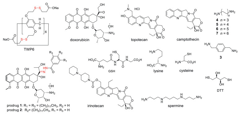
Figure 1.
Chemical structures of host TWP6, anti-tumor drugs, thiols, biomolecules, prodrugs 1, 2 and guests 3-7.
Smart supramolecular vesicles based on glutathione-reactive pillar[6]arene and acid-labile prodrug: Dual drug loading and sequential release
Yamin Liu , Siyang Jiang , Weipeng Mao , Pintao Li , Fang Zhou , Da Ma

"Smart" nanomaterials responsive to physiological or external stimuli are of great importance for biomedical applications [1-3]. When pH, reducing agent, reactive oxygen species, photo-irradiation or other type of stimuli is applied, smart nanomaterials undergo chemical or morphological transformation to enhance bioactivity [4-9].
Self-assembly nanomaterials based on host-guest interaction are of specific importance for the convenience to construct complex systems and introduce stimuli responsiveness [10-24]. Cellular environment contains high concentration of glutathione (GSH) and other biological thiols [25-27]. By developing thiol-reactive host molecules, smart supramolecular nanomaterials responsive to biological thiols may be designed, and used for the delivery and controlled release of cargo.
Here, we report GSH thiol-reactive pillar[6]arene (TWP6), which is the first example of reducing agent-reactive host molecule. Water-soluble TWP6 is based on the backbone of pillar[n]arene, a new type of macrocyclic hosts with interesting recognition property and applications [28-37]. TWP6 could encapsulate variable anti-tumor drugs, and react with biological or artificial thiols. TWP6 was used to assemble supramolecular vesicles with doxorubicin (DOX)-based acid-labile prodrug via host-guest interaction. Subsequently, two anti-tumor drugs were loaded, which could undergo sequential release responding to GSH and acidic condition. These smart nanomaterials were used for combination chemotherapy in vitro.
TWP6 was derived from pillar[6]arene with 12 disulfide-bonds introduced to side chains (Fig. 1). Carboxylic acid groups were installed on the end of side chains to improve aqueous solubility and enhance binding affinity with cationic guests. Synthetic procedure is described in supporting information. We studied recognition property of TWP6 toward anti-tumor drugs, biomolecules and common guests in sodium phosphate buffer (pH 7.4, 20 mmol/L) by direct titration or indicator displacement assay.
TWP6 was discovered to be a high affinity host molecule for suitable anti-tumor drugs. As shown in Table 1, the Ka value for TWP6·topotecan was (3.2 ± 0.1) × 104 L/mol. TWP6 had higher Ka value toward DOX ((2.6 ± 0.7) × 105 L/mol) and irinotecan ((1.9 ± 0.1) × 105 L/mol). Job plot confirmed the 1:1 binding stoichiometry for TWP6·DOX, TWP6·irinotecan and TWP6·topotecan (Figs. S23-S25 in Supporting information). Amino acid (lysine) and artificial or biological thiols (DTT, cysteine and GSH) demonstrated low binding affinity with TWP6 (< 103 L/mol). By contrast, the complex of TWP6·spermine had a high Ka value of (2.5 ± 0.4) × 105 L/mol, more than two-orders of magnitude higher than any of the above biomolecules. TWP6 showed spacer length-dependent binding with different diamino guests with the maximum binding strength at n = 5. Consequently, TWP6 is a high affinity and selective host molecule towards variable guests and biomolecules. Taking advantage of the high affinity binding between host and anti-tumor drug, we may construct supramolecular vesicles based on TWP6 and prodrug.
 DownLoad:
CSV
DownLoad:
CSV

|
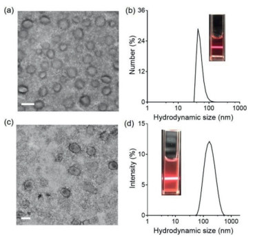
DOX-based prodrugs 1 and 2 were used to construct supramolecular vesicles, which contained an acid-labile hydrazone linker, and one or two hydrophobic alkyl chains (Fig. 1). Supramolecular vesicles were constructed by sonicating an aqueous solution of prodrug and TWP6 with a molar ratio of 1:1. Supramolecular vesicles were characterized by transmission electron microscopy (TEM) and dynamic light scattering (DLS). TEM images of TWP6·1 and TWP6·2 showed vesicle architectures (Fig. 2). The average vesicle size for TWP6·1 and TWP6·2 was 49 nm and 135 nm with a wall thickness of 12.1 nm and 9.3 nm, respectively. DLS observation showed a hydrodynamic size of 50 nm for TWP6·1 and 164 nm for TWP6·2. The critical aggregation concentration (CAC) for TWP6·1 and TWP6·2 was determined by conductivity to be 20.4 µmol/L and 22.7 µmol/L, respectively (Figs. S26 and S27 in Supporting information). ζ-Potential of supramolecular vesicles for TWP6·1 and TWP6·2 was determined to be −33.1 mV and −31.9 mV, respectively. Therefore, by using prodrug with different numbers of alkyl chains, supramolecular vesicles with variable sizes could be prepared.
Model hydrophobic dye nile red and hydrophobic drug CPT were loaded into supramolecular vesicles based on TWP6 and prodrug 1. We first studied thiol-induced release of cargo. As shown in Fig. 3a, for the three thiols, GSH was the sole reagent to trigger the release of nile red from supramolecular vesicles. By comparison, negligible nile red release was observed when cysteine, DTT or lysine was applied. Encapsulation of CPT was slightly less stable compared to that of nile red. Nevertheless, the use of GSH resulted in a significantly faster release of CPT (Fig. 3b). Based on these observations, supramolecular vesicles were selectively responsive to GSH. By contrast, cysteine and DTT were unable to disrupt supramolecular vesicles to release hydrophobic cargo. Next, we investigated acid-induced release of DOX. The fluorescence of DOX was quenched when conjugated to hydrazone, and recovered as hydrazone was cleaved to release DOX. As shown in Fig. 3c, while negligible DOX release was observed at neutral pH, DOX was rapidly released under an acidic pH of 4.0. The released DOX may be encapsulated by TWP6. A further dilution in vitro or in vivo will dissociate the complex of TWP6 and DOX to deploy its anti-tumor bioactivity.
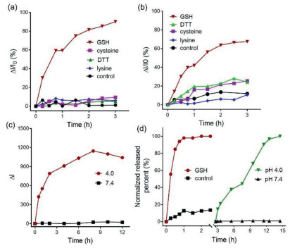
Using smart supramolecular vesicles, we were able to achieve sequential release of dual anti-tumor drugs. As shown in Fig. 3d, GSH was first applied to CPT-loaded supramolecular vesicles, and CPT release was observed. Subsequently, acidic condition of pH 4.0 was applied to release DOX. When using TWP6 and prodrug 2, similar selective responsive release towards GSH and sequential release was observed (Figs. S29-S32 in Supporting information). Consequently, thiol-reactiveness of TWP6 was the major reason for intelligent stimuli-responsiveness.
Mechanism study on molecular and nanoscale levels was carried out to illustrate the GSH-selective cargo release. First, thiol-reactivity of TWP6 was investigated by 1H NMR spectroscopy. As shown in Figs. S37 and S38 (Supporting information), after incubation with GSH or cysteine, there was resonance pattern change, indicating reaction occurred between TWP6 and biological thiols. By comparison, TWP6 alone was stable under the same condition (Fig. S36 in Supporting information). While 1H NMR spectra are relatively complex to interpret, thiol-disulfide exchange reaction is expected to occur between TWP6 and thiols. As shown in Scheme 1, we believe there are two thiol-disulfide exchange modes: (1) installation of thiol-moiety (m) on TWP6 product; (2) incorporation of new disulfide moiety (n) on TWP6 product. Next, TEM and DLS were used to study thiol-induced nanoparticle transformation with supramolecular vesicles based on prodrug 1. Supramolecular vesicles were allowed to react with GSH, cysteine or DTT for 5 h at 37 ℃. Hydrodynamic size and ζ-potential of supramolecular vesicles were monitored. Subsequently, nanoparticles were characterized by TEM. As shown in Fig. S39 (Supporting information), supramolecular vesicles retained spherical morphology after incubation with thiols. Nanoparticle size did not undergo significant change after treated with cysteine or DTT, while supramolecular vesicles grew in size when treated with GSH. ζ-Potential of supramolecular vesicles barely changed for the groups treated with cysteine or DTT (Fig. S34 in Supporting information). By contrast, when treated with GSH, ζ-potential increased, which indicated a significant change in nanoparticle surface property. Consequently, when treated with GSH, supramolecular vesicles undergo dramatic change, leading to a decreasing drug encapsulation ability.
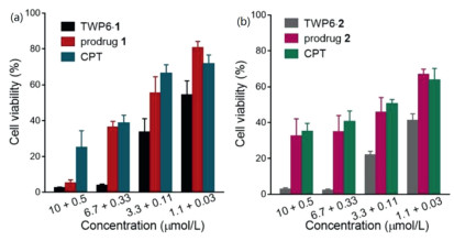
Lastly, these smart supramolecular vesicles were used for combination chemotherapy in vitro. In cellular environment, CPT was expected to be released in cytoplasm when exposed to cellular GSH. DOX could be released triggered by acidic environment in endosomal/lysosomal compartments. We first studied the biocompatibility of TWP6. TWP6 was confirmed to be nontoxic toward human cancer HeLa cells and human normal Chang liver cells (Figs. S40 and S41 in Supporting information). CPT-loaded supramolecular vesicles based on prodrug 1 or 2 were incubated with HeLa cells for 48 h, and viability was determined by MTT assay. As shown in Fig. 4, at high concentrations, CPT-loaded supramolecular vesicles showed a higher cytotoxicity compared to that of prodrug or CPT alone. At lower concentrations, supramolecular vesicles also demonstrated the higher cytotoxicity. Nevertheless, the difference of cytotoxicity was reduced compared to the high concentration groups, which may be due to dissociation of supramolecular vesicles. Consequently, CPT-loaded supramolecular vesicles showed combination therapeutic efficacy.
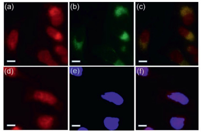
Internalization and intracellular distribution of supramolecular vesicles were also investigated. HeLa cells were incubated with supramolecular vesicles based on TWP6·1, and cells were thoroughly washed with PBS. Nuclei of cells were stained with 4, 6-diamidino-2-phenylindole (DAPI), and lysosomes were stained with Lysotracker Green. Fluorescence microscopy was used to image cells. As shown in Figs. 5a-c, there was significant overlap between DOX (red) and lysosome (green), which indicated DOX was internalized via endosome/lysosome-mediated endocytosis. We propose that nonfluorescent prodrug underwent acid-induced degradation in lysosome to release fluorescent DOX. In addition, an overlap between DOX (red) and nuclei (blue) indicated that DOX had been successfully delivered into cell nuclei (Figs. 5d-f).
In summary, thiol-reactive pillar[6]arene is designed and prepared to construct smart supramolecular vesicles with DOX-based acid-labile prodrug. Supramolecular vesicles are able to load DOX and CPT via prodrug and supramolecular encapsulation, respectively. Supramolecular vesicles are selectively responsive to GSH to release CPT, followed by a sequential release of DOX induced by acidic pH. Combination therapeutic efficacy is evaluated in vitro. By introducing two orthogonal stimuli-responsiveness types to host and prodrug molecules, supramolecular vesicles demonstrate intelligent drug delivery characteristics.
There are no conflicts to declare.
The authors are grateful to National Natural Science Foundation of China (Nos. 21921003 and 21672042) and Zhengzhou University for financial support.
Supplementary material associated with this article can be found, in the online version, at doi:10.1016/j.cclet.2021.07.024.
F. Zhang, G. Zhu, O. Jacobson, et al., ACS Nano 11(2017) 8838-8848. doi: 10.1021/acsnano.7b03003
K. Yang, Y. Chang, J. Wen, et al., Chem. Mater. 28(2016) 1990-1993. doi: 10.1021/acs.chemmater.6b00696
W. Cheng, H. Cheng, S. Wan, X. Zheng, M. Yin, Chem. Mater. 29(2017) 4218-4226. doi: 10.1021/acs.chemmater.7b00047
J. Yao, M. Yang, Y. Duan, Chem. Rev. 114(2014) 6130-6178. doi: 10.1021/cr200359p
S. Mura, J. Nicolas, P. Couvreur, Nat. Mater. 12(2013) 991-1003. doi: 10.1038/nmat3776
Y. Lu, A.A. Aimetti, R. Langer, Z. Gu, Nat. Rev. Mater. 2(2017) 16075. doi: 10.1038/natrevmats.2016.75
H. Wang, Y.Q. Yan, Y. Yi, et al., CCS Chem. 2(2020) 739-748. doi: 10.31635/ccschem.020.202000283
C. Ju, R. Mo, J. Xue, et al., Angew. Chem. Int. Ed. 53(2014) 6253-6258. doi: 10.1002/anie.201311227
L. Han, C. Tang, C. Yin, ACS Appl. Mater. Interfaces 8(2016) 23498-23508. doi: 10.1021/acsami.6b07173
Y. Kang, X. Tang, Z. Cai, X. Zhang, Adv. Funct. Mater. 26(2016) 8920-8931. doi: 10.1002/adfm.201602998
C. Wang, Z. Wang, X. Zhang, ACC Chem. Res. 45(2012) 608-618. doi: 10.1021/ar200226d
G. Yu, K. Jie, F. Huang, Chem. Rev. 115(2015) 7240-7303. doi: 10.1021/cr5005315
Y. Cao, Y. Chen, Z. Zhang, et al., Chin. Chem. Lett. 32(2021) 349-352. doi: 10.1016/j.cclet.2020.03.058
S. Fa, T. Kakuta, T.A. Yamagishi, T. Ogoshi, CCS Chem. 1(2019) 50-63. doi: 10.31635/ccschem.019.20180014
C. Kim, S.S. Agasti, Z. Zhu, L. Isaacs, V.M. Rotello, Nat. Chem. 2(2010) 962-966. doi: 10.1038/nchem.858
C. Hou, L. Liu, S. Meng, et al., Chin. Chem. Lett. 32(2021) 214-217. doi: 10.1016/j.cclet.2020.11.030
D. Mao, Y. Liang, Y. Liu, et al., Angew. Chem. Int. Ed. 56(2017) 12614-12618. doi: 10.1002/anie.201707164
W. Mao, D. Mao, F. Yang, D. Ma, Chem. Eur. J. 25(2019) 2272-2280. doi: 10.1002/chem.201804835
S. Jiang, S. Lan, D. Mao, et al., Chem. Commun. 54(2018) 9486-9489. doi: 10.1039/C8CC05552A
C. Sun, H. Zhang, S. Li, et al., ACS Appl. Mater. Interfaces 10(2018) 25090-25098. doi: 10.1021/acsami.8b06598
Q. Cheng, K.X. Teng, Y.F. Ding, et al., Chem. Commun. 55(2019) 2340-2343. doi: 10.1039/C8CC09432B
C. Sun, L. Yue, Q. Cheng, Z. Wang, R. Wang, ACS Mater. Lett. 2(2020) 266-271. doi: 10.1021/acsmaterialslett.0c00002
C. Gao, Q. Huang, Q. Lan, et al., Nat. Commun. 9(2018) 2967. doi: 10.1038/s41467-018-05437-5
Q. Cheng, S. Li, Y. Ma, H. Yin, R. Wang, Chin. Chem. Lett. 31(2020) 1235-1238. doi: 10.1016/j.cclet.2019.10.020
L.Y. Niu, Y.S. Guan, Y.Z. Chen, et al., J. Am. Chem. Soc. 134(2012) 18928-18931. doi: 10.1021/ja309079f
X. Liu, W. Shao, Y. Zheng, et al., Chem. Commun. 53(2017) 8596-8599. doi: 10.1039/C7CC04932C
C.W. Sathiyajith, R.R. Shaikh, Q. Han, et al., Chem. Commun. 53(2017) 677-696.
H. Zhang, Z. Liu, Y. Zhao, Chem. Soc. Rev. 47(2018) 5491-5528.
T. Ogoshi, T. Yamagishi, Y. Nakamoto, Chem. Rev. 116(2016) 7937-8002. doi: 10.1021/acs.chemrev.5b00765
G. Yu, X. Zhou, Z. Zhang, et al., J. Am. Chem. Soc. 134(2012) 19489-19497. doi: 10.1021/ja3099905
Q. Duan, Y. Cao, Y. Li, et al., J. Am. Chem. Soc. 135(2013) 10542-10549. doi: 10.1021/ja405014r
W. Feng, M. Jin, K. Yang, Y. Pei, Z. Pei, Chem. Commun. 54(2018) 13626-13640.
Y. Chang, K. Yang, P. Wei, et al., Angew. Chem. Int. Ed. 53(2014) 13126-13130. doi: 10.1002/anie.201407272
B. Li, Z. Meng, Q. Li, et al., Chem. Sci. 8(2017) 4458-4464. doi: 10.1039/c7sc01438d
L.L. Tan, H. Li, Y. Tao, et al., Adv. Mater. 26(2014) 7027-7031. doi: 10.1002/adma.201401672
Z.Y. Li, Y. Zhang, C.W. Zhang, et al., J. Am. Chem. Soc. 136(2014) 8577-8589. doi: 10.1002/chin.201431021
L. Luo, G. Nie, D. Tian, et al., Angew. Chem. Int. Ed. 55(2016) 12713-12716.

Figure 1 Chemical structures of host TWP6, anti-tumor drugs, thiols, biomolecules, prodrugs 1, 2 and guests 3-7.

Figure 2 TEM images of (a) TWP6·1 and (c) TWP6·2 (scale bar: 200 nm); DLS size distribution of supramolecular vesicles (b) TWP6·1 and (d) TWP6·2. [TWP6] = [1] = [2] = 10 µmol/L.

Figure 3 Time-dependent nile red release (a), CPT release (b) from supramolecular vesicles when incubating with GSH, cysteine, DTT, lysine or none of the above agents (control) at 37 ℃. (c) pH-dependent release of DOX from supramolecular vesicles at 37 ℃ (pH 7.4 or 4.0). (d) Drug release from CPT-loaded supramolecular vesicles at 37 ℃, when GSH and pH 4.0 were applied sequentially. Prodrug 1 was used to construct supramolecular vesicles ([TWP6] = [1] = 50 µmol/L). Thiols and biomolecules were used with a concentration of 1 mmol/L.

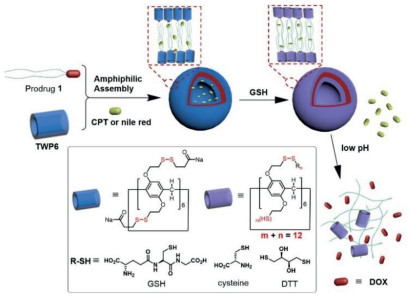
Scheme 1 Schematic illustration for the formation of supramolecular vesicles, and GSH/acid-induced sequential cargo release.

Figure 4 Viability of HeLa cells treated with (a) CPT-loaded supramolecular vesicles based on TWP6·1 or (b) CPT-loaded supramolecular vesicles based on TWP6·2. X axis represents concentration of TWP6/prodrug (front) and CPT (back) in µmol/L. Incubation time: 48 h. n = 6.

Figure 5 Fluorescence microscopic images of HeLa cells treated with supramolecular vesicles ([TWP6] = [1] = 10 µmol/L), and stained with Lysotracker Green (a-c) or DAPI (d-f): (a, d): DOX fluorescent channel; (b) Lysotracker Green fluorescence channel; (e) DAPI fluorescence channel; (c, f) merged. Scale bar: 10 µm.
Table 1. Ka value (L/mol) for the complex of TWP6 and drug/guest in sodium phosphate buffer (20 mmol/L, pH 7.4).
|
|
 下载: 导出CSV

 扫一扫看文章

扫一扫关注我们