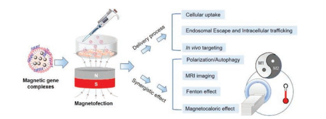
Figure 1.
Magic magnetic nanoparticles for efficient gene delivery and associated synergistic effects.
Magnetofection: Magic magnetic nanoparticles for efficient gene delivery
Qunjie Bi , Xu Song , Ao Hu , Tianying Luo , Rongrong Jin , Hua Ai , Yu Nie

Nowadays, magnetic nanoparticles (MNPs) are widely used in the biomedical field, such as drug delivery, bioimaging, diagnostic analysis [1-3]. MNPs mainly include pure metals such as Fe, Co, Ni nanoparticles, magnetic nano-metal alloys such as FePt and CoPt, nano-ferrites such as Fe3O4, γ-Fe2O3, and metal-doped iron oxides such as MnFe2O4, CoFe2O4 and ZnFe2O4 [4]. The essential feature of MNPs used in magnetofection must be ferromagnetic. CoFe2O4 and MnFe2O4 nanoparticles showed stronger magnetism than other magnetic materials, but their toxicity limits their use in biomedical applications [5-7]. While the most used Fe3O4 and γ-Fe2O3 nanoparticles not only have good superparamagnetic properties, but also show good biological safety [8]. The main preparation methods of MNPs are coprecipitation [9], high-temperature thermal decomposition [10], hydrothermal/solvent-thermal [11] and microemulsion methods [12]. The potential application of MNPs in biomedicine is attributed to the special physical properties, especially superparamagnetism and high magnetic responsiveness [13-16]. The MNPs with superparamagnetism only show magnetism in the presence of an applied magnetic field. The main applications are as follows. MNPs are used in magnetic separation, mainly for the extraction of DNA, proteins, and other biological molecules [17]. What is more, superparamagnetic nanoparticles can generate large amounts of heat originating from relaxation in an alternating magnetic field. Therefore, MNPs can be used as hyperthermia agents for treatment [18]. Meanwhile, MNPs can be used as magnetic resonance imaging (MRI) contrast agents by changing the relaxation time of tissues to affect the signal strength of tissues and improve the contrast of different tissues in MRI [19]. In addition, MNPs can be used as gene carriers to improve gene transfection efficiency [20], which is covered in more detail in this mini-review.
In recent years, gene therapy has made great progress featuring high efficacy and low side effects [21-23]. However, gene therapy is stymied by a lack of cell-specific, safe and efficient gene-delivery vectors, leading to low transfection efficiency [24]. The application of MNPs could promote the efficiency of gene transfection by holding the carriers at the targeting site via an external magnetic field [25, 26]. C. Mah [25] and C. Plank et al. [26] were the first to elucidate the magnetofection. This technique can enhance transfection efficiency and realize target control [27]. In this paper, we focus on the application and mechanism of MNPs in gene delivery, as well as recent synergistic effects together with magnetofection (Fig. 1).
Gene therapy is regarded as one of the most promising therapeutic strategies [28]. This therapeutic modality is generally based on the introduction of the exogenous gene to living cells, which encodes a specific therapeutic protein to correct or modulate many serious diseases, such as cancers and genetic disorders [27]. However, the success of gene therapy lies in the efficient transportation of large and fragile DNA molecules into the nucleus of targeted cells without significant degradation by nucleases. Extensive researches have been conducted to exploit efficient and safe nonviral vectors, which can protect and release the genetic cargos at the site of action [29-33]. Inspired by the strategy in magnetic drug delivery systems, which achieve target accumulation successfully under magnetic fields [34], Prof. Plank et al. has induce magnetic particles in gene delivery [27, 35]. They have defined magnetofection as nucleic acid delivery under the influence of a magnetic field, in which the magnetic nanoparticles act as associated vectors. Transfection with MNPs under the magnetic field shows unique advantages all through gene delivery [36].
It is well known that nucleic acids exhibit a negative surface charge [37]. Therefore, when applied to gene transfection, MNPs must be surface-modified to compact with genes or gene/carrier complexes, and meanwhile to enhance the stability and biocompatibility [38-41]. There are two ways for MNPs to load genes.
The first and the most straightforward approach is to coat cationic substances on the surface of MNPs, and then bind them with negatively charged genes through electrostatic absorption [42, 43]. Polyethylenimine (PEI), composed of many amino groups, can be commonly used for gene delivery [44]. For instance, PEI could be coated on the surface of 11-mercaptoundecanoic acid modified Au/Ni/Si nanospears by layer by layer approach [42]. The final modified MNPs could be positively charged for gene loading. Also, PEI could directly modify the surface of iron oxide nanoparticles to generate a core-shell structure, which could successfully bind with siRNA to form well-dispersed nanoparticles [43]. Besides PEI, other polycations like poly-arginine, poly-L-lysine (PLL) and chitosans could be modified on the surface of MNPs for successful gene binding [45, 46].
The second strategy is to compact with gene/carrier complexes, in which the surface charge is not the determining factor [38]. The study of several MNPs with different surface charge has confirmed that point [47]. In this strategy, genes generally bind to the polycation firstly. Then, MNPs with the opposite charge compacted with the gene/carrier complexes through electrostatic adsorption. Different charged MNPs, with the modification of citric acid (CA), carboxylmethyl-dextran (CMD), γ-aminopropyltriethoxy silane (APTES), betaine, and PEI, respectively, were confirmed the ability of the comparable gene compression and increased gene transfection efficiency by applying a magnetic field.
Cationic magnetic gene carriers could promote cell binding and absorption due to negative charges bringing from the sugar calyx and lipid bilayer on the cell membrane [48]. Meanwhile, it could also interact with the negatively charged serum proteins during circulation, which hampers cellular uptake [49]. Serum proteins could be adsorbed on the surface of nanoparticles by electrostatic interaction [50]. The particle size of MNPs increases and the surface charge goes from positive to negative [46, 51]. This fact may hinder MNPs binding to negatively charged membranes and thus MNPs internalization [52, 53]. Sometimes, the absorbed serum proteins even reduced the binding of MNPs to DNA [53]. Either way, there is no doubt that the application of the magnetic field can assist cationic gene carriers to resist the interference of serum [52-54]. Under the magnetic field, cellular uptake of MNPs/DNA complexes is almost free from the interference of serum [55]. Or with the external magnetic field, cellular uptake of PEI-PAAIO/pEGFP complexes even enhanced for more than 7-fold in serum incubation [54]. Further study also confirmed that pDNA/BPEISPION (conjugation of BPEI with superparamagnetic iron oxide nanoparticles for pDNA complexation) survived beyond 24 h [56]. While naked pDNA was degraded within 24 h in the DNase existence conditions [56]. All these studies reflected that MNPs could not only increase the cellular uptake, but also maintain the stability of genes in the presence of serum proteins.
It was also found that magnetofection could significantly accelerate the accumulation of MNPs/gene complexes on the cell surface and shorten the cellular uptake time. The maximum transfection level was achieved after 10 min incubation with a magnetic field, compared to the 2-4 h in the magnetic field absent group [26]. Y. Nie's group has proved magnetofection could achieve rapid enrichment of MNPs/gene complexes on the cell surface, and promote them to rapidly engraft into target cells without damage [53]. The speed of MNPs reaching the target cell is closely related to the intensity and duration of the magnetic field, and the range and distance of the magnetic field [57].
Generally, cellular uptake of nanoparticles is found through phargocytosis, macropinocytosis, clathrin- or caveolin-mediated endocytosis [58-62]. While cell uptake mechanism of magnetofection was fist explored by Prof. Plank's group [63]. They found that almost similarly to PEI polyplexes, the internalization pathway of MNPs involvsed clathrin-mediated endocytosis (in human lung epithelial cells and liver cancer cells) or caveolin-mediated endocytosis (in human cervical cancer cells) [64]. Followingly, M. Arsianti [46] and S. Ota et al. [65] confirmed that many different MNPs assemblies were all internalized by clathrin-mediated endocytosis (in baby hamster kidney cells and HeLa cells). All these studies indicate that magnetofection promotes cell uptake without affecting the entry pathway of nanoparticles [46, 53].
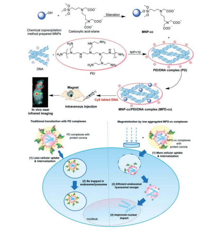
Beside cellular internalization, MNPs also facilitate endosomal escape and nuclear import. Y. Nie's group conducted an in-depth study on the gene magnetic transfection mechanism of magnetic polyethyleneimine/DNA (MPD-cc) complexes (Fig. 2) [53]. Magnetic field could help most MPD-cc complexes distribute in the non-lysosomal region of cytoplasm, and then entry into the nucleus within 24 h. However, without the magnetic field, most of the complexes in all groups were still located in lysosomes, and almost no Cy5 labeled DNA (red) was detected in the nuclear region [53]. Perhaps, the decreased interaction time between the gene carrier complex and the serum under the action of the magnetic field reduces the adsorption of proteins, which promotes the internalization of the complexes and the subsequent transfection process.
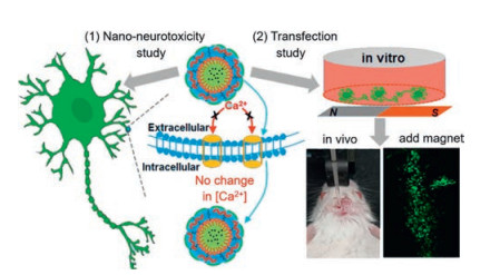
Under the external magnetic field, MNPs could be easy to aggregate, which is beneficial for realizing superior properties for targeted gene delivery [66]. Thus, MNPs have received great attention in targeted gene systems [66]. Under the magnetic field, interfering RNA (siRNA) loaded MMPs could be transferred to target tissues, realizing outstanding magnetism, biocompatibility and low toxicity [45, 67]. Neurons are notoriously difficult to transfect and sensitive to toxicity [68]. PEG modified MNP-PLGAPEI could achieve successful transfection of neurons and reduce nano neurotoxicity in the hippocampus of mice under the magnet field (Fig. 3) [68].
MRI is one of the most widely used imaging methods in clinical medicine [69, 70]. It detects the magnetic moment created by single protons in omnipresent hydrogen atoms. The intensity of the magnetic field will change after the hydrogen protons in the water bombarded by pulsed electromagnetic waves, while the reactions of hydrogen protons are different in different tissues. By collecting signals from the most abundant water in the human body, MRI can make high-resolution images of most tissues [71]. SPIONs, with a size of less than 20 nm, were one of the main nanostructures being studied as an MRI contrast agent [19, 72, 73]. SPIONs could significantly reduce proton transverse relaxation time T2 at a very low concentration [74]. Meanwhile, modified SPIONs could be used to deliver genes for diagnosis and treatment integration [75].
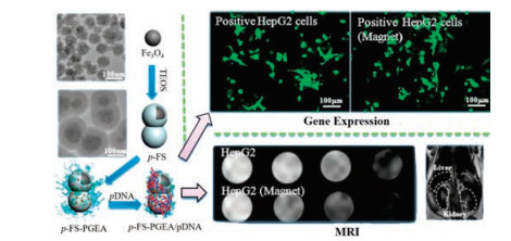
High gene transfection could simultaneously appear with MRI contrast, thus MRI guided visible gene transfection could be realized [75]. For instance, BUCT-PGEA (ethanolamine-functionalized poly(glycidyl methacrylate) grafted from the surface of Fe3O4@SiO2 nanoparticles) not only utilized the external magnetic field to further enhance the transfection efficiency, but also achieved the real-time magnetic resonance imaging (Fig. 4) [75]. The transfection efficiency was 3-fold higher under the magnetic field than under normal culture. Stearic acid-modified PEI-SPIO nanocrystals, which use low molecular weight PEI as the shell and SPIO nanocrystals as the core, could also possess ultrasensitive imaging capacity and effectively bind DNA for efficient gene transfection [76]. The MRI-visible MNPs system provides an effective platform for gene delivery [77, 78].
Under alternating magnetic field, SPIONs can generate a large amount of thermal energy through relaxation effect. This phenomenon could be used for antitumor treatment, which called magnetic mediated hyperthermia (MMH) [79]. R. Gilchrist et al. [80] first proposed the concept of MMH, when they found MNPs could accumulate in tumor site and bring with the temperature increase. Then, MMH of MNPs was found to be applied in tumor suppression [81]. However, MMH was also found to trigger a series of molecular effects, including a rapid increase in the synthesis of heat shock proteins (HSPs), which could protect cellular proteins from degradation at high temperature [82].
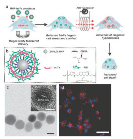
Combined therapy of MMHs and gene transfection could enhance antitumor efficacy. Q. Tang and coworkers [83] developed a novel heat-inducible gene expression system, by modifying Mn-Zn ferrite MNPs with PEI and then loading heterogonous genes. The expression of heterogonous genes could be elevated to 10 to 500- fold over background by moderating hyperthermia. In this study, heat promoted gene expression by facilitating the transcription and translation of heat-induced genes. In addition, downregulating HSP expression could improve the antitumor efficacy of MMH treatment (Fig. 5) [84]. Highly magnetic zinc-doped iron oxide nanoparticles (ZnFe2O4) were applied to deliver microRNA (let-7a), which targeted multiple downstream pathways modulated by HSPs. Compared with let-7a alone or magnetic hyperthermia alone, ZnFe2O4 combined with let-7a could significantly decrease insulin like growth factor 1 receptor, RAS and high mobility group AT-hook 2/c-MYC accompanied by MMH, thus promoting the apoptosis of brain cancer cells.
H.J.H Fenton discovered in 1894 that several metals have special oxygen transfer properties, which improve the use of hydrogen peroxide [85]. Some metals have a strong catalytic power to generate highly reactive hydroxyl radicals (·OH). Since this discovery, the iron catalyzed hydrogen peroxide has been called Fenton's reaction. The disproportionation of hydrogen peroxide (H2O2) with Fe2+ ions, can efficiently generates a specific kind of reactive oxygen species (ROS) [85, 86], including hydroxyl radical and superoxide anion [87, 88]. Apoptosis can be induced when the concentration of ROS exceeds a certain threshold [89, 90]. The Fenton reaction of MNPs has been demonstrated in the past decades [91]. Jing Zhu and coworkers [92] used Fe(III)-porphyrin nanosonosensitizers, which modified with bis(DPA-Zn)-RGD as the carrier of siRNA. This study used tumor targeting peptide-RGD guided tumor accumulation. siRNA was transferred to cancer cells to effectively down-regulate the expression of SOD2. The Fe(III) induced cascade not only reduced intracellular glutathione levels, but also produced cytotoxic Fenton reaction which enhanced the therapeutic effect. As aforementioned, combined targeted ability of MNPs and Fenton reaction may also improve tumor therapeutic effect.
Recent researches confirmed that MMH and Fenton reaction promoted apoptosis by inducing autophagy [93-95]. Autophagy is a key biological process for maintaining cell homeostasis [96]. Recent studies verified that autophagy induced by nanomaterials was a universal mechanism [96]. Iron oxide nanoparticles could also induce autophagy in a variety of cells, including macrophages, vascular endothelial cells, glioma cells, etc. [97, 98]. However, autophagy response plays distinct roles in different cells at different scenarios. Dextran coated SPIONs, tracking therapeutic cells in vivo, which could induce autophagy in dendritic cells, which protected them from apoptosis [99]. In addition, SPIONs could promote autophagy by inducing the production of ROS, thus killing tumor cells [93, 97]. Moreover, SPIONs could induce a temperature-dependent autophagy, rapidly destroying cell membranes and promoting tumor apoptosis [95]. However, the combined study of autophagy and magnetic transfection has not been reported. Whether autophagy can promote or inhibit magnetic transfection needs further studies in the future.
Based on the superparamagnetism and high magnetic responsiveness of MNPs, magnetofection has gradually explored, developed, and expanded in the past decades. The intrinsic magnetic saturation strength of MNPs, surface modification, and the parameters of the external magnetic field greatly affect the efficiency of gene transfection, especially the efficiency and targeting in vivo. Together with other effects of MNPs (such as MRI and MMH), magnetofection not only realized the integration of diagnosis and treatment but also significantly improved the synergistic treatment effect. With more deep understanding of Fenton reaction, autophagy phenomena and extra magnetic field, we believe it could also promote and board the application of magnetofection in further studies. Meanwhile, the stability of MNPs in vivo and technology for large-scale industrial production should be improved to upgrade the clinical application of magnetofection.
The authors declare that they have no known competing financial interests or personal relationships that could have appeared to influence the work reported in this paper.
This study was supported by National Key Research and Development Program of China (No. 2017YFC1104601), National Natural Science Foundation of China (NSFC, No. 81873921), SinoGerman cooperation group project (No. GZ1512) and Sichuan Science and Technology Program (No. 2019JDJQ0027).
Supplementary material related to this article canbefound, in the online version, at doi:https://doi.org/10.1016/j.cclet.2020.07.030.
J.P. Liu, E. Fullerton, O. Gutfleisch, D.J. Sellmyer, Nanoscale Magnetic Materials and Applications, Springer US, Boston, 2009.
Z. Chen, C. Wu, Z. Zhang, et al., Chin. Chem. Lett. 29(2018) 1601-1608. doi: 10.1016/j.cclet.2018.08.007
Q. Cheng, T. Peng, A. Liu, Chin. Chem. Lett. 16(2005) 1059-1062.
R. Hao, R. Xing, Z. Xu, et al., Adv. Mater. 22(2010) 2729-2742. doi: 10.1002/adma.201000260
M. Makarewicz, M. Podsiadly, M. Balanda, Acta Phys. Pol. A 115(2009) 568-571. doi: 10.12693/APhysPolA.115.568
S. George, T. Xia, R. Rallo, et al., ACS Nano 5(2011) 1805-1817. doi: 10.1021/nn102734s
W.S. Cho, R. Duffin, C.A. Poland, et al., Nanotoxicology 6(2012) 22-35. doi: 10.3109/17435390.2011.552810
A. Akbarzadeh, M. Samiei, S. Davaran, Nanoscale Res. Lett. 7(2012) 1-13. doi: 10.1186/1556-276X-7-1
S. Santra, C. Kaittanis, J. Grimm, J.M. Perez, Small 5(2009) 1862-1868. doi: 10.1002/smll.200900389
S. Sun, H. Zeng, D.B. Robinson, et al., J. Am. Chem. Soc. 126(2004) 273-279. doi: 10.1021/ja0380852
X. Wang, J. Zhuang, Q. Peng, Y. Li, Nature 437(2005) 121-124. doi: 10.1038/nature03968
J. Zhi, Y. Wang, Y. Lu, J. Ma, G. Luo, React. Funct. Polym. 66(2006) 1552-1558. doi: 10.1016/j.reactfunctpolym.2006.05.006
G. Cheng, J. Xing, Z. Pi, et al., Chin. Chem. Lett. 30(2019) 656-659. doi: 10.1016/j.cclet.2018.12.003
S. He, L. He, B. Liu, et al., Chin. Chem. Lett. 30(2019) 1031-1034. doi: 10.1016/j.cclet.2019.03.013
A. Lu, E.L. Salabas, F. Schüth, Angew. Chem. Int. Ed. 46(2007) 1222-1244. doi: 10.1002/anie.200602866
M. Tadic, S. Kralj, Y. Lalatonne, L. Motte, Appl. Surf. Sci. 476(2019) 641-646. doi: 10.1016/j.apsusc.2019.01.098
J. He, M. Huang, D. Wang, Z. Zhang, G. Li, J. Pharm. Biomed. Anal. 101(2014) 84-101. doi: 10.1016/j.jpba.2014.04.017
S. Dutz, R. Hergt, Int. J. Hyperth. 29(2013) 790-800. doi: 10.3109/02656736.2013.822993
G. Liu, J. Gao, H. Ai, X. Chen, Small 9(2013) 1533-1545. doi: 10.1002/smll.201201531
F. Scherer, M. Anton, U. Schillinger, et al., Gene Ther. 9(2002) 102-109. doi: 10.1038/sj.gt.3301624
K.A. High, M.G. Roncarolo, N. Engl. J. Med. 381(2019) 455-464. doi: 10.1056/NEJMra1706910
Y. He, Y. Nie, L. Xie, H. Song, Z. Gu, Biomaterials 35(2014) 1657-1666. doi: 10.1016/j.biomaterials.2013.10.073
Q. Jiang, Y. Nie, X. Chen, et al., Adv. Funct. Mater. 27(2017) 1701571. doi: 10.1002/adfm.201701571
D. Luo, W.M. Saltzman, Nat. Biotechnol. 18(2000) 893-895. doi: 10.1038/78523
C. Mah, I. Zolotukhin, T.J. Fraites, et al., Mol. Ther. 1(2000) S239. doi: 10.1006/mthe.2000.0174
C. Plank, U. Schillinger, F. Scherer, et al., Biol. Chem. 384(2003) 737-747.
O. Mykhaylyk, Y.S. Antequera, D. Vlaskou, C. Plank, Nat. Protoc. 2(2007) 2391-2411. doi: 10.1038/nprot.2007.352
T. Luo, H. Liang, R. Jin, Y. Nie, J. Gene Med. 21(2019) e3090.
X. Chen, Y. Chen, H. Xin, T. Wan, Y. Ping, Proc. Natl. Acad. Sci. U. S. A. 117(2020) 2395-2405. doi: 10.1073/pnas.1912220117
Y. Nie, M. Günther, Z. Gu, E. Wagner, Biomaterials 32(2011) 858-869. doi: 10.1016/j.biomaterials.2010.09.032
Y. He, Y. Nie, G. Cheng, et al., Adv. Mater. 26(2014) 1534-1540. doi: 10.1002/adma.201304592
H. Liang, X. Chen, R. Jin, et al., Small 16(2020) 1906538. doi: 10.1002/smll.201906538
Y. He, G. Cheng, L. Xie, et al., Biomaterials 34(2013) 1235-1245. doi: 10.1016/j.biomaterials.2012.09.049
A.R. Kuehnle, M.R. Kuehnle, Patent, US5516670A, 1996.
C. Plank, M. Anton, C. Rudolph, J. Rosenecker, F. Krotz, Expert Opin. Biol. Ther. 3(2003) 745-758. doi: 10.1517/14712598.3.5.745
L. Xie, W. Jiang, Y. Nie, et al., RSC Adv. 3(2013) 23571-23581. doi: 10.1039/c3ra43588a
J. Kim, A.C. Mirando, A.S. Popel, J.J. Green, Adv. Drug Deliv. Rev. 119(2017) 20-43. doi: 10.1016/j.addr.2016.11.003
Y. Ma, X. Wang, H. Gu, Chin. Sci. Bull. 57(2012) 4005-4011. doi: 10.1007/s11434-012-5184-1
X. Li, Q.R. Xie, J. Zhang, W. Xia, H. Gu, Biomaterials 32(2011) 9546-9556. doi: 10.1016/j.biomaterials.2011.08.068
W.S. Seo, J.H. Lee, X. Sun, et al., Nat. Mater. 5(2006) 971-976. doi: 10.1038/nmat1775
S.W. Kim, S. Kim, J.B. Tracy, A. Jasanoff, M.G. Bawendi, J. Am. Chem. Soc. 127(2005) 4556-4557. doi: 10.1021/ja043577f
X. Xu, S. Hou, N. Wattanatorn, et al., ACS Nano 12(2018) 4503-4511. doi: 10.1021/acsnano.8b00763
G. Liu, J. Xie, F. Zhang, et al., Small 7(2011) 2742-2749. doi: 10.1002/smll.201100825
A.P. Pandey, K.K. Sawant, Mater. Sci. Eng. C 68(2016) 904-918. doi: 10.1016/j.msec.2016.07.066
O. Veiseh, F.M. Kievit, H. Mok, et al., Biomaterials 32(2011) 5717-5725. doi: 10.1016/j.biomaterials.2011.04.039
M. Arsianti, M. Lim, C.P. Marquis, R. Amal, Biomacromolecules 11(2010) 2521-2531. doi: 10.1021/bm100748p
X. Wang, L. Zhou, Y. Ma, H. Gu, IEEE Trans. Nanotechnol. 8(2009) 142-147. doi: 10.1109/TNANO.2009.2013946
H.J. Vaughan, J.J. Green, S.Y. Tzeng, Adv. Mater. 32(2020) e1901081. doi: 10.1002/adma.201901081
K. Nagase, M. Hasegawa, E. Ayano, Y. Maitani, H. Kanazawa, Int. J. Mol. Sci. 20(2019) 430. doi: 10.3390/ijms20020430
A. Lesniak, F. Fenaroli, M.R. Monopoli, et al., ACS Nano 6(2012) 5845-5857. doi: 10.1021/nn300223w
M. Geppert, C. Petters, K. Thiel, R. Dringen, J. Nanopart. Res. 15(2013) 1349. doi: 10.1007/s11051-012-1349-8
M. Arsianti, M. Lim, C.P. Marquis, R. Amal, Langmuir 26(2010) 7314-7326. doi: 10.1021/la9041919
L. Xie, Q. Jiang, Y. He, et al., Biomater. Sci. 3(2015) 446-456. doi: 10.1039/C4BM00317A
S.L. Sun, Y.L. Lo, H.Y. Chen, L.F. Wang, Langmuir 28(2012) 3542-3552. doi: 10.1021/la204529u
T. Zhang, J. Wu, Q. Xu, et al., Int. J. Pharm. 520(2017) 1-13. doi: 10.1016/j.ijpharm.2017.01.041
R. Namgung, K. Singha, M.K. Yu, S. Jon, et al., Biomaterials 31(2010) 4204-4213. doi: 10.1016/j.biomaterials.2010.01.123
E.P. Furlani, X. Xue, Pharm. Res. 29(2012) 1366-1379. doi: 10.1007/s11095-012-0681-0
F. Zhao, Y. Zhao, Y. Liu, et al., Small 7(2011) 1322-1337. doi: 10.1002/smll.201100001
P. Foroozandeh, A.A. Aziz, Nanoscale Res. Lett. 13(2018) 339. doi: 10.1186/s11671-018-2728-6
B.D. Chithrani, W.C.W. Chan, Nano Lett. 7(2007) 1542-1550. doi: 10.1021/nl070363y
H. Gao, Y. Xiong, S. Zhang, et al., Mol. Pharm. 11(2014) 1042-1052. doi: 10.1021/mp400751g
H. Gao, Z. Yang, S. Zhang, Z. Pang, X. Jiang, J. Drug Targeting 22(2014) 450-459. doi: 10.3109/1061186X.2014.886038
S. Huth, J. Lausier, C. Rudolph, et al., Mol. Ther. 7(2003) 967.
S. Huth, J. Lausier, S.W. Gersting, et al., J. Gene Med. 6(2004) 923-936. doi: 10.1002/jgm.577
S. Ota, Y. Takahashi, A. Tomitaka, et al., J. Nanopart. Res. 15(2013) 1653. doi: 10.1007/s11051-013-1653-y
L. Shen, B. Li, Y. Qiao, Materials 11(2018) 324. doi: 10.3390/ma11020324
Y. Namiki, T. Namiki, H. Yoshida, et al., Nat. Nanotechnol. 4(2009) 598-606. doi: 10.1038/nnano.2009.202
Y. Cui, X. Li, K. Zeljic, et al., ACS Appl. Mater. Interfaces 11(2019) 38190-38204. doi: 10.1021/acsami.9b15014
S. Ray, Z. Li, C.H. Hsu, et al., Theranostics 8(2018) 6322-6349. doi: 10.7150/thno.27828
Y. Fang, J. Jia, J. Yang, J. Zheng, C. Yi, Chin. Chem. Lett. 29(2018) 1277-1280. doi: 10.1016/j.cclet.2017.10.023
Z. Zhou, L. Yang, J. Gao, X. Chen, Adv. Mater. 31(2019) 1804567. doi: 10.1002/adma.201804567
C. Wu, Y. Xu, L. Yang, et al., Adv. Funct. Mater. 25(2015) 3581-3591. doi: 10.1002/adfm.201501031
L.L. Israel, A. Galstyan, E. Holler, J.Y. Ljubimova, J. Control. Release 320(2020) 45-62. doi: 10.1016/j.jconrel.2020.01.009
C.W. Kessinger, O. Togao, C. Khemtong, et al., Theranostics 1(2011) 263-273. doi: 10.7150/thno/v01p0263
R. Wang, Y. Hu, N. Zhao, F.J. Xu, ACS Appl. Mater. Interfaces 8(2016) 11298-11308. doi: 10.1021/acsami.6b01697
Q. Wan, L. Xie, L. Gao, et al., Nanoscale 5(2013) 744-752. doi: 10.1039/C2NR32438E
C. Wu, J. Li, P. Pang, et al., Biomaterials 35(2014) 8249-8260. doi: 10.1016/j.biomaterials.2014.06.014
H. Gao, H. Feng, Y. Bai, et al., J. Biomed. Nanotechnol. 15(2019) 1764-1770. doi: 10.1166/jbn.2019.2805
J. Liu, N. Li, L. Li, et al., Oncol. Lett. 6(2013) 1550-1558. doi: 10.3892/ol.2013.1618
R. Gilchrist, R. Medal, W.D. Shorey, et al., Ann. Surg. 146(1957) 596-606. doi: 10.1097/00000658-195710000-00007
R. Gordon, J. Hines, D. Gordon, Med. Hypotheses 5(1979) 83-102. doi: 10.1016/0306-9877(79)90063-X
C.S.S.R. Kumar, F. Mohammad, Adv. Drug Deliv. Rev. 63(2011) 789-808. doi: 10.1016/j.addr.2011.03.008
Q. Tang, D. Zhang, X. Cong, M. Wan, L. Jin, Biomaterials 29(2008) 2673-2679. doi: 10.1016/j.biomaterials.2008.01.038
P.T. Yin, B.P. Shah, K.B. Lee, Small 10(2014) 4106-4112.
H.J.H. Fenton, J. Chem. Soc., Trans. 65(1894) 899-910.
H. Ranji-Burachaloo, P.A. Gurr, D.E. Dunstan, G.G. Qiao, ACS Nano 12(2018) 11819-11837. doi: 10.1021/acsnano.8b07635
T. Mai, J.Z. Hilt, Colloids Surf. A 576(2019) 9-14. doi: 10.1016/j.colsurfa.2019.05.003
S. Wang, H. Liao, F. Li, D. Ling, Chin. Chem. Lett. 30(2019) 847-852. doi: 10.1016/j.cclet.2019.03.025
M.R. Ramsey, N.E. Sharpless, Nat. Cell Biol. 8(2006) 1213-1215. doi: 10.1038/ncb1106-1213
S.S. Sabharwal, P.T. Schumacker, Nat. Rev. Cancer 14(2014) 709-721. doi: 10.1038/nrc3803
D. Zheng, Q. Lei, J. Zhu, et al., Nano Lett. 17(2017) 284-291. doi: 10.1021/acs.nanolett.6b04060
J. Zhu, C. Chu, D. Li, et al., Adv. Funct. Mater. 29(2019) 1904056. doi: 10.1002/adfm.201904056
S. Du, J. Li, C. Du, et al., Oncotarget 8(2017) 9410-9424. doi: 10.18632/oncotarget.14114
M.I. Khan, A. Mohammad, G. Patil, et al., Biomaterials 33(2012) 1477-1488. doi: 10.1016/j.biomaterials.2011.10.080
T. Sadhukha, T.S. Wiedmann, J. Panyam, Biomaterials 35(2014) 7860-7869. doi: 10.1016/j.biomaterials.2014.05.085
Y. Zhang, L. Zhang, J. Gao, L. Wen, Acc. Chem. Res. 52(2019) 3164-3176. doi: 10.1021/acs.accounts.9b00397
M.I. Khan, A. Mohammad, G. Patil, et al., Biomaterials 33(2012) 1477-1488. doi: 10.1016/j.biomaterials.2011.10.080
Q. Feng, Y. Liu, J. Huang, et al., Sci. Rep. 8(2018) 2082. doi: 10.1038/s41598-018-19628-z
Q. Wu, R. Jin, T. Feng, et al., Int. J. Nanomed. 12(2017) 3993-4005. doi: 10.2147/IJN.S135189

Figure 1 Magic magnetic nanoparticles for efficient gene delivery and associated synergistic effects.

Figure 2 Magnetic force accelerates lysosome escape and promotes nuclear import. Copied with permission [53]. Copyright 2015, Elsevier.

Figure 3 Under the magnetic field, gene transfection of primary hippocampal neurons with MNPs was realized. Reproduced with permission [68]. Copyright 2018, American Scientific Publishers.

Figure 4 Magnetic nanoparticle system utilized the external magnetic field to further enhance the transfection efficiency and achieve the real-time magnetic resonance imaging. Reproduced with permission [75]. Copyright 2016, American Chemical Society.

 扫一扫看文章

扫一扫关注我们
 DownLoad:
 下载:
下载:
