Scheme 1.
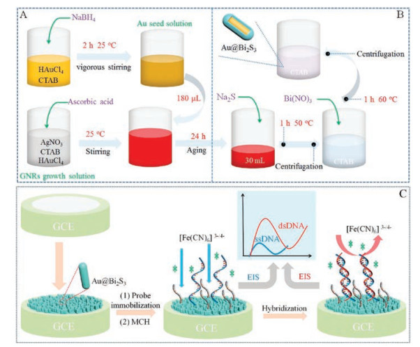
Schematic diagram for preparation of gold nanorods (A) and the core-shell structured Au@Bi2S3 (B). (C) The fabrication process of the Au@Bi2S3-based DNA electrochemical biosensor and its analytical application.
Synthesis of core-shell structured Au@Bi2S3 nanorod and its application as DNA immobilization matrix for electrochemical biosensor construction
Feng Gao , Juan Song , Bin Zhang , Hidekazu Tanaka , Fei Gao , Weiwei Qiu , Qingxiang Wang

Take advantages of its narrow band gap (1.2–1.7 eV), outstanding energy conversion efficiency and high absorption coefficient (104–105 cm-1), bismuth sulfide (Bi2S3) receives great attention in the application of electrochemical field [1, 2]. In addition, with the development of material science and nanotechnology, the nanosized Bi2S3with various morphologies have been synthesized and utilized as high-performance electrochemical sensing materials [3-5]. For example, Dong and coworkers [3] have constructed a nanorod Bi2S3-based electrochemical sensor for ascorbic acidanalysis, achieving high sensitivity and excellent reliability. Fu [4] synthesized three-dimensional Bi2S3 nanowire network, and utilized the prepared Bi2S3 nanowire network to construct an electrochemical ammonia sensor. The result showed that the sensor had an excellent specific response to ammonia due to their weak coordination adsorption action. Our group [5] also synthesized a unique broccoli-like Bi2S3 using an imidazoline derivative as the template and investigated its electrocatalysis toward the bovine hemoglobin. The results showed that the Bi2S3could effectively promote the direct electrochemistry of hemoglobin, and the prepared electrode displayed excellent electrocatalysis of hydrogen peroxide as an enzyme biosensor.
However, the pure Bi2S3 nanomaterial is easily aggregated during the synthesis process, which not only decreases its effective surface area, but also weakens the electrochemical properties [6]. On the purpose of getting around these shortcomings, the other functional materials such as carbon material [7, 8], polymers [9], and precious metals [10, 11] are usually introduced as supporting material or auxiliary material for the synthesis of the Bi2S3-based nanocomposites.
Gold nanoparticles (AuNPs) are an important and widely used precious metal nanomaterial bearing lots of excellent physicochemical properties such as good biocompatibility, high electronic conductivity, and outstanding adhesive ability with organic groups including —NH2, —SH, and so forth [12-15]. Based on these features, AuNPs are frequently utilized as a scaffold for bioprobe immobilization [16, 17] and electrochemical signal amplifier [18, 19]. In recent years, the AuNPs have also been utilized as functional material to prepare Bi2S3-based composite, and be extended to the application in the field of electrochemistry. For example, Zhang et al. [20] synthesized AuNPs/Bi2S3 nanorods and applied them as signal tags to catalyze silver deposition. Through such a strategy, an ultrasensitive electrochemical immunosensor for Escherichia Co1:O157:H7 was achieved using silver stripping signal as the analytical signal. In addition, Li et al. [21] have synthesized a new composite of KNbO3-AuNPs@Bi2S3, and used the composite as an antibody carrier for the analysis of prostate-specific antigen. The result showed that the AuNPs/Bi2S3 connected by Au-S covalent bond could effectively promote electron transfer kinetics and electrochemiluminescence signal intensity. However, we have not found the report concerning the use of AuNP/Bi2S3 composite as a matrix for probe DNA immobilization and electrochemical sensing analysis yet.
Herein, a rod-like core-shell structured Au@Bi2S3 composite was designed and prepared through direct growth of Bi2S3 shell on pre-synthesized gold nanorods. The sample was characterized by X-ray diffraction (XRD), transmission electron microscopy (TEM) and energy-dispersive X-ray spectroscopy (EDS). Electrochemical characterization experiment suggested that the gold core could dramatically decrease the electron transfer resistance of Bi2S3. Furthermore, the Au@Bi2S3coated glassy carbon electrode (GCE) was utilized as a scaffold for the self-assembly fixing of the amino-modified oligonucleotide probe to fabricate an electrochemical DNA biosensor. Under the optimized conditions, the target DNA could be well detected with a low detection limit of femtomolar level (2 fmol/L). This work broadens the application of Au and Bi2S3 nanocomposite in electrochemical sensing fields, and provides a new method for facile construction of a sensitive biosensor.
Firstly the gold nanorods were synthesized according to Ag+-assisted seed-mediated approach [22] with minor modification (Scheme 1A). Typically, an aliquot of 5.0 mL 0.02 mmol/L HAuCl4 solution was mixed first with 5.0 mL 200 mmol/L CTAB. Then 0.6 mL of ice-cold NaBH4 (10 mmol/L) was prepared and dropped into above mixture under vigorous stirring. During this process, the solution was transformed from dark-yellow to brown-yellow, which is a characteristic of formation of the gold seed in the solution. Followed by, put the gold seed solution into a water bath (25 ℃) for more than 2 h to decompose the unreacted NaBH4.
What is more, the mixture of 75.0 mL of 200 mmol/L cetyltrimethylammonium bromide (CTAB), 75.0 mL of 1 mmol/L HAuCl4, 1.25 mL of 4 mmol/L AgNO3 and 1.05 mL of 100 mmol/L l-ascorbic acid was prepared and continuously stirred, which was acted as the gold nanorods growth solution. Thereafter, 180 μL prepared gold seed solution was added into the freshly prepared growth solution, and kept the reaction temperature at 25 ℃. In 15 min, the color of the reaction solution became dark red, suggesting the reduction of Au3+ to Au0 species. After further grown for 24 h in the mixture solution, the gold nanorods (AuNR) were collected by centrifugation at 8000 rpm for 15 min, and finally, the gold nanorods dispersion was obtained by suspending the synthesized Au nanorods in 20.0 mmol/L CTAB.
Core-shell structured Au@Bi2S3 AuNR were prepared through direct growth of Bi2S3 on the surface of AuNR (Scheme 1B). In detail, 3.0 mL 0.1 mol/L Na2S was added into 30 mL of above-prepared AuNR dispersion in a vessel open to the air with vigorous stirring at room temperature. The mixture was aged for 1 h in water bath at 50 ℃. The particles were then collected by centrifugation and then dispersed in 20.0 mmol/L CTAB. Thereafter, 4.5 mL of 0.1 mol/L Bi(NO3)3 was added into the solution and reacted with S2- capped gold nanorods in a 60 ℃ water bath for 1 h to grow Bi2S3 on the surface of AuNR. Finally, the formed Au@Bi2S3 nanorods were obtained through centrifugation. The stock solution of the Au@Bi2S3 was prepared by dispersing 1 mg Au@Bi2S3 sample in 10 mL 20.0 mmol/L CTAB.
The biosensor was fabricated as following procedure: First, 10 μL prepared Au@Bi2S3 dispersion was dropped onto a GCE pretreated through physical polishing with alumina slurry, and electrochemically polishing via a CV process in 0.5 mol/L H2SO4, as described in the literature [23]. Then 10 μL of the prepared Au@Bi2S3 dispersion was dropped onto the surface of the pretreated GCE and dried under room temperature. Afterward, the loosely attached Au@Bi2S3 on the electrode was removed by gentle rinsing with DDW for three times, and the obtained electrode was signed as Au@Bi2S3/GCE. It is of note that, the bare GCE after electrochemical treatment in H2SO4, bears some functional oxygen-containing groups, which is beneficial for assembly of Au@Bi2S3 through tight coordination and electrostatic interaction with surface Bi3+ of the material. Thus, the stability of the modified electrode is ensured. To further assemble the amino group modified pDNA (5′-NH2-TCTTTGGGACCACTGTCG-3′) on electrode through coordination between - NH2 and Bi2S3 [24], the Au@Bi2S3/GCE was immersed in 0.1 μmol/L pDNA for 4 h at 30 ℃. Then the electrode was incubated in 1 mmol/L 6-mercapto-1-hexanol (MCH) for 2 h to passivate the residual sites between the DNA probes. Finally, the electrode was rinsed with phosphate buffer saline (PBS) to removed physically attached pDNA, and the biosensor (pDNA/Au@Bi2S3/GCE) was achieved. For comparison, the Bi2S3/GCE was also fabricated through a similar procedure only replacing single phase Bi2S3 with Au@Bi2S3. All the detailed reagents and apparatus were listed in Supporting information.
The composition and structure of the synthesized composite were characterized by XRD. As displayed in Fig. 1A, the diffraction peaks and the corresponding crystal planes for Au nanorod precursor, 2θ = 38.75° (111), 39.8° (200), 64.92° (220) and 77.50° (311) are well consistent with the results of nano gold PDF# 89–3697, proving the successful synthesis of the gold nanorods through the Ag+-assisted seed-mediated method. In addition, the XRD pattern of CTAB (PDF#48-2454) is appeared in AuNR, suggesting that CTAB is adsorbed on the surface of AuNR during the synthesis process. The presence of CTAB can well prevent the aggregation of AuNR. For Au@Bi2S3, it is found that all the characteristic diffraction peaks of Au are visible but their intensity is much weaker than the pure AuNR, which is likely caused by the coating of Bi2S3 shell. In addition, one can clearly observe the diffraction peaks ascribing to Bi2S3 at 2θ = 15.67° (002), 17.58° (012), 25.0° (111), 28.6° (121), 40.0° (034), 45.5° (200), 52.6° (213), 57.4° (241), confirming that Bi2S3 has successfully grown on AuNR.
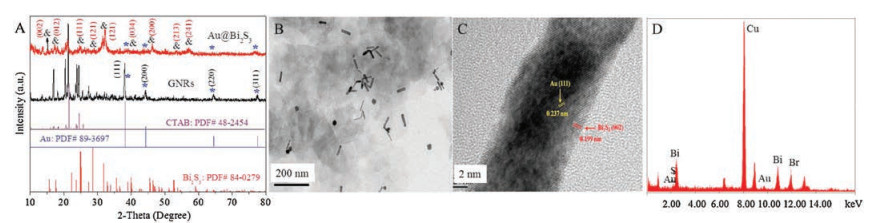
Then, the obtained products were characterized by TEM. As displayed in Fig. 1B, the Au@Bi2S3 particles show uniform rod shape with the average length of 58 nm, diameter of 20 nm, and draw ratio of 6:1. Fig. 1C is the HRTEM image of Au@Bi2S3, from which one can see that the distance between the crystal planes in the middle of the nanorod is 0.237 nm, corresponding to cubic phase of Au crystal (111) lattice face [25]. The distance between the crystal planes at the shell of the nanorod is 0.205 nm, which belongs to the orthorhombic phase of Bi2S3 crystal (002) lattice facet. This result further testifies that the Au@Bi2S3 composite has a core-shell structure. The EDS (Fig. 1D) analysis demonstrates that the particles are comprised by the elements of Au (9.71 keV), S (2.31 keV) and Bi (10.84 keV), further confirming the successful synthesis of Au@Bi2S3 by the proposed method.
The electrochemical property of Au@Bi2S3 and the fabrication of pDNA/Au@Bi2S3/GCE were characterized by cyclic voltammetry (CV) and electrochemical impedance spectra (EIS) (Fig. S1 in Supporting information). Both of the results suggest successful fabrication of the biosensor. Under the optimized conditions (Fig. S2 in Supporting information), the analytical performance of the biosensor is evaluated by EIS since it is a simple and effective approach for electrodecharacterization and biosensing analysis [26]. Fig. 2A shows the Ret value difference (ΔRet) on the biosensor before (curve a) and after hybridization with 0.1 nmol/L nDNA (curve b), tmDNA (curve c), smDNA (curve d) and cDNA (curve e) and the corresponding histogram (Inset). The result shows that the Ret value presents negligible variation after the pDNA/Au@Bi2S3/GCE is hybridized with nDNA, indicating that the pDNA on the constructed biosensor could not react with nDNA and also the non-specific absorption of nDNA on Au@Bi2S3 does not occur. Being different, the resistance on the biosensor increases markedly, when the biosensor is incubated in cDNA solution for hybridization reaction. The phenomenon indicates that hybridized duplex DNA has been formed, and the increased steric-hindranceand electrostatic repulsion block the diffusion of [Fe(CN)6]3-/4- from bulk solution to electrode surface. In addition, when the pDNA/Au@Bi2S3/GCE is hybridized with smDNA or tmDNA, the change of the Ret value is much less than the case of tDNA, but still larger than on pDNA/Au@Bi2S3/GCE, suggesting that only partial hybridization happens between pDNA and smDNA or tmDNA. Thus, it is concluded that the constructed biosensor has excellent hybridization selectivity to the complementary DNA from base mismatched and non-complementary DNA.
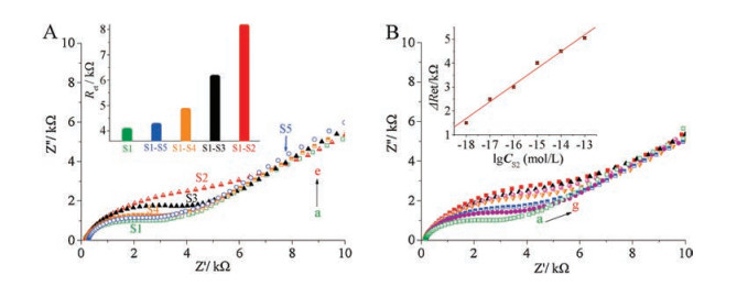
The sensitivity of the DNA biosensor is studied through incubation of the biosensor in cDNA solution with the different concentrations and comparing their EIS changes. Fig. 2B is the Nyquist plots recorded on pDNA/Au@Bi2S3/GCE before and after hybridization with cDNA in the concentration range from 10 fmol/L to 1 nmol/L. As shown in Fig. 2B, the Ret displays a steady increase with the increase of concentrations of cDNA. The Ret variations (ΔRet) between Ret at the hybridized electrode and that at pDNA/Au@Bi2S3/GCE are well correlated to the logarithm of cDNA concentration (lgCcDNA) in the range from 10 fmol/L to 1 nmol/L with a regression equation of ΔRet/(103 Ω) = 0.735 lg(CcDNA/M) + 14.777 (r = 0.9933) (inset of Fig. 2B). Then based on 3σ (σ was the standard deviation of a blank solution with 5 parallel measurements), the detection limit is estimated to be 2 fmol/L.
The reproducibility of the biosensor for DNA analysis was probed through hybridization of five parallel-made time pDNA/Au@Bi2S3/GCE with 0.1 nmol/L cDNA. When the ΔRet values are adopted as analytical signal, a relative standard deviation (RSD) of 5.73% is achieved, suggesting an acceptable reproducibility of the developed pDNA/Au@Bi2S3/GCE for DNA analysis. In addition, the chemical stability and durability of the fabricated pDNA/Au@Bi2S3/GCE were investigated by storing the prepared biosensor at 4 ℃ for one week and hybridized with target DNA fragment. The results showed that after storage, the Au@Bi2S3-based biosensor showed only 3% increase of Ret value, indicating the excellent stability of the biosensor. When the biosensor was hybridized with 1 pmol/L cDNA, the hybridization response (ΔRet) was very close to the case before storage, indicating that the developed biosensor remained the hybridization efficiency after storage, namely a good durability.
In summary, core-shell structured Au@Bi2S3 nanoparticles were synthesized via a facile seed-mediated method. Then the synthesized Au@Bi2S3 was further applied for fabrication of DNA biosensor. The electrochemical experimental results showed that the target DNA could be quantified in a wide concentration range from 10 fmol/L to 1 nmol/L and a low detection limit of 2 fmol/L. The high sensitivity can be explained by the synergetic effect of huge surface area and high electronic conductivity of Au@Bi2S3. Moreover, the sensor shows superior properties of good reproducibility, stability, and simple operation, showing that the Au@Bi2S3-based DNA biosensor promising to be applied in practical application.
The work is supported by the National Natural Science Foundation of China (Nos. 21802064, 21275127), Natural Science Foundation of Fujian Province, China (Nos. 2018J01435, 2017J01419), and Foundation of Key Laboratory of Sensor Analysis of Tumor Marker, Ministry of Education, Qingdao University of Science and Technology.
Supplementary material related to this article can befound, in the online version, at doi:https://doi.org/10.1016/j.cclet.2019.04.056.
M. Wang, H. Yin, N. Shen, et al., Biosens. Bioelectron. 53(2014) 232-237. doi: 10.1016/j.bios.2013.09.069
Q.X. Wang, F. Gao, S.X. Li, W. Weng, Z.S. Hu, Chin. Chem. Lett. 19(2008) 585-588. doi: 10.1016/j.cclet.2008.03.001
Y.P. Dong, L. Huang, J. Zhang, X.F. Chu, Q.F. Zhang, Electrochim. Acta 74(2012) 189-193. doi: 10.1016/j.electacta.2012.04.063
T.X. Fu, Mater. Res. Bull. 99(2018) 460-465. doi: 10.1016/j.materresbull.2017.11.038
X. Chen, Q. Wang, L. Wang, et al., Biosens. Bioelectron. 66(2015) 216-223. doi: 10.1016/j.bios.2014.11.020
X. Yan, Y. Gu, C. Li, et al., Sensor. Actuat. B-Chem. 257(2018) 936-943. doi: 10.1016/j.snb.2017.11.037
D.R. Kumar, S. Kesavan, M.L. Baynosa, V.Q. Nguyen, J.J. Shim, J. Colloid Interface Sci. 530(2018) 361-371. doi: 10.1016/j.jcis.2018.06.069
S. Vadivel, A.N. Naveen, V.P. Kamalakannan, P. Cao, N. Balasubramanian, Appl. Surf. Sci. 351(2015) 635-645. doi: 10.1016/j.apsusc.2015.04.101
Q. Zhu, F. Gao, Y. Yang, et al., Sensor. Actuat. B-Chem. 207(2015) 819-826. doi: 10.1016/j.snb.2014.10.120
B. Pandit, G.K. Sharma, B.R. Sankapal, J. Colloid Interface Sci. 505(2017) 1011-1017. doi: 10.1016/j.jcis.2017.06.092
S.V.P. Vattikuti, J. Shim, C. Byon, J. Solid State Chem. 258(2018) 526-535. doi: 10.1016/j.jssc.2017.11.017
M. Ma, T. Zhe, W. Song, et al., Sensor. Actuat. B-Chem. 253(2017) 818-829. doi: 10.1016/j.snb.2017.07.003
S.J. Li, Y.F. Shi, L. Liu, et al., Electrochim. Acta 85(2012) 628-635. doi: 10.1016/j.electacta.2012.08.118
W. Bai, H. Huang, Y. Li, et al., Electrochim. Acta 117(2014) 322-328. doi: 10.1016/j.electacta.2013.11.175
M. Lin, H. Han, D. Pan, H. Zhang, Z. Su, Microchim. Acta 182(2015) 805-813. doi: 10.1007/s00604-014-1391-6
Y. Guo, Y. Wang, G. Yang, J.J. Xu, H.Y. Chen, Biosens. Bioelectron. 85(2016) 897-902. doi: 10.1016/j.bios.2016.06.013
L. Tang, Z. Guo, X. Zheng, Sensor. Actuat. B-Chem. 279(2019) 1-6. doi: 10.1016/j.snb.2018.09.078
Y. Lai, C. Zhang, Y. Deng, et al., Chin. Chem. Lett. 30(2019) 160-162. doi: 10.1016/j.cclet.2018.07.011
N. Alizadeh, A. Salimi, R. Hallaj, J. Electroanal. Chem. 811(2018) 8-15. doi: 10.1016/j.jelechem.2017.12.080
J. Zhang, X. Song, Z. Xiong, et al., J. Electrochem. Soc. 164(2017) H572-H578. doi: 10.1149/2.1151706jes
J. Li, H. Ma, D. Wu, et al., Biosens. Bioelectron. 74(2015) 104-112. doi: 10.1016/j.bios.2015.06.027
H. Huang, S. Huang, X. Liu, et al., Biosens. Bioelectron. 24(2009) 3025-3029. doi: 10.1016/j.bios.2009.03.018
J. Zhou, X. Li, L. Yang, et al., Anal. Chim. Acta 899(2015) 57-65. doi: 10.1016/j.aca.2015.09.054
H.H. Huang, J. Chen, Y.Z. Meng, et al., Mater. Res. Bull. 48(2013) 3800-3804. doi: 10.1016/j.materresbull.2013.05.091
S. Paul, S. Ghosh, D. Barman, S.K. De, Appl. Catal. B-Environ. 219(2017) 287-300. doi: 10.1016/j.apcatb.2017.07.057
F. Gao, X. Cai, X. Wang, et al., Sensor. Actuat. B-Chem. 186(2013) 380-387. doi: 10.1016/j.snb.2013.06.020

Scheme 1 Schematic diagram for preparation of gold nanorods (A) and the core-shell structured Au@Bi2S3 (B). (C) The fabrication process of the Au@Bi2S3-based DNA electrochemical biosensor and its analytical application.

Figure 1 XRD pattern (A), TEM (B), HRTEM (C) and EDS (D) images of the synthesized Au@Bi2S3.

Figure 2 (A) Nyquist plots of the biosensor (a) upon hybridization with 10 fmol/L nDNA (b), mDNA (c), smDNA (d) and cDNA (e). The inset shows the corresponding histogram. (B) Nyquist plots of the biosensor before (a) and after hybridization with 10 fmol/L (b), 100 fmol/L (c), 1 pmol/L (d), 10 pmol/L (e), 0.1 nmol/L (f), 1 nmol/L (g) cDNA. Inset is the linear plot of ΔRet values versus the logarithm of cDNA concentrations (lgCcDNA).

 扫一扫看文章

扫一扫关注我们
 DownLoad:
 下载:
下载: