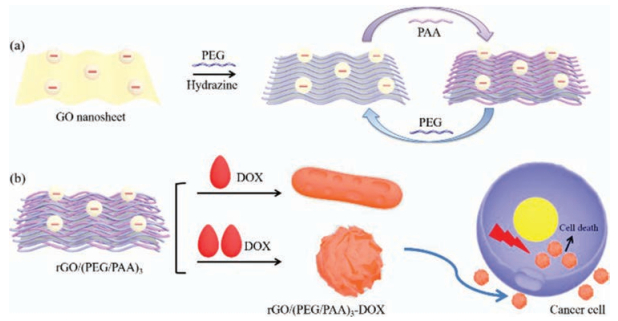
Scheme 1.
(a) Layer-by-layer assembly around the rGO template by PEG and PAA. (b) The construction of the rGO/(PEG/PAA)3-DOX clusters that enter cancer cells.
Designing cancer nanodrugs that are highly loaded, pH-responsive, photothermal, and possess a favored morphology: A hierarchical assembly of DOX and layer-by-layer modified rGO
Xiangming Li , Yihe Zhang , Zequn Ma , Chengjun He , Yaling Wu , Qi An

Cancer continues to threaten human health around the world. According to the World Health Organization, approximately 84 million people died of cancer between 2005 and 2015 [1]. Nanodrugs that simultaneously assume chemical therapy, photothermal effect, pH-responsive release profiles, and imaging functions are increasingly being researched because of their cancer-treating capabilities [2-6]. Recently, research into various two-dimensional (2D) inorganic nanosheets have especially gained attention because of their outstanding photothermal effect, drug loading capacity, and imaging capabilities [7-11]. Among these 2D nanosheets, reduced graphene oxide (rGO) has been the most widely studied nanodrug component [12-17]. rGO is able to efficiently load hydrophobic chemical drugs as well as present an advantageous photothermal effect [18, 19]. Many rGO-based nanodrugs also release their active chemical component in a pHresponsive way. Because rGO sheets present nonspecific interactions with serum proteins [20, 21], surface modification of rGO sheets in these nanodrugs are always necessary to avoid poor colloidal stability and acute toxicity [22-25]. Surface modification of poly(ethylene glycol) (PEG) using covalent or noncovalent interactions have been the most frequently applied modification strategies of rGO sheets. The choice of a surface modification method significantly influences the loading capacity of the chemical therapy drug, which is frequently Doxorubicin hydrochloride (DOX), and the efficacy of the composite nanodrug. The development of an effective surface modification method that promotes DOX-loading capabilities, permits a pH-responsive release profile, and obtains an effective cancer-killing capability remains highly desirable.
Besides the above-mentioned merits for the 2D nanosheets that compose part of the nanodrug, another important factor in tumor treatment is the enhanced permeation and retention (EPR) effect of solid tumor tissues. The neovasculature in solid tumors exhibits fenestrations ranging from 200 nm to 12, 000 nm in size, and this hyperpermeability, in combination with other tumor-associated fluid transport abnormalities, such as impaired lymphatic drainage, gives rise to the EPR phenomenon [3]. Morphological engineering that meets the morphological criteria for the favorable tumor-killing agents has rarely been realized for nanodrugs containing 2D nanosheets. To date, designing one nanodrug device that combines the advantages of the 2D inorganic nanosheets and the favored morphological traits remains an outstanding challenge.
In this article, we report on our design of a cancer-killing nanodrug that simultaneously assumes a chemical therapy effect, photothermal effect, and a favorable morphological trait. The nanodrug was a spherical cluster formed between DOX and surface-modified rGO (as shown in Scheme 1). A new rGO surface engineering method of layer-by-layer assembly of polymers PEG and PAA was employed to fulfill such a specific nanodrug device. The nanodrug features the high-loading capability of DOX, a pHresponsive release profile, a self-fluorescent capability, and a favored morphology by EPR effect. The nanodrug kills cancer cells effectively under near infrared (NIR) irradiation and permits fluorescent observation because of the self-fluorescence of DOX. The choice of the types of polymers—PEG and PAA as well as the three layer-by-layer assembly cycles around the rGO cores—were indispensable in achieving such a spherical clustered nanodrug.
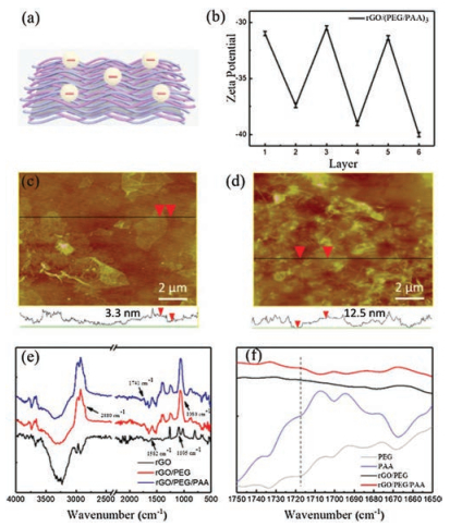
The LbL assembly of PAA and PEG was driven by hydrogen bonds as illustrated in Fig. 1a [24]. The zeta potential, AFM, TEM and ATR-FTIR characterizations of the rGO/(PEG/PAA)n (n = 0–3) were carried out to confirm the successful assembly of multilayered polyelectrolyte films around the rGO cores. The zeta potentials of the rGO/(PEG/PAA)n complex presented alternating negative potentials at around -30.0 mV and -40.0 mV during LbL assembly, corresponding to the outermost layer of PEG or PAA, as shown in Fig. 1b. AFM analysis indicated that the thickness of the rGO/PEG, the rGO/PEG/PAA and rGO/(PEG/PAA)3 sheets were 3.3 nm, 5.0 nm, and 12.5 nm respectively (Figs. 1c–d and Fig. S1 in Supporting information). The consecutive increase of the sheet thicknesses verified the assembly of the polymer layers around the rGO cores. TEM images in Fig. S1 showed the morphology of rGO/PEG and rGO/PEG/PAA. No signs of aggregations appeared in their images and the morphologies were soft fabric-like sheets which resembled the morphology of rGO. In order to further prove the successful assemblies of PEG and PAA around rGO, the ATR-FTIR of the samples was measured. The bands at 1582 cm-1 and 1105 cm-1 were ascribed to the aromatic C=C and epoxy C—O of rGO (Fig. 1e). A set of new peaks appeared after the LbL modification of PEG and PAA around the rGO template (Fig. 1e). The vibrational peaks at 1080 cm-1 and 2880 cm-1 were ascribed to the C—O band and hydroxyl of PEG. The peak corresponding to the COOH groups at 1741 cm-1verified that PAA was successfully assembled. After PEG or PAA was included within a rGO/(PEG/PAA)n multilayer, a new peak at 1719 cm-1 appeared as compared with pure PEG or PAA (Fig. S2 in Supporting information), indicating the formation of hydrogen bonds between PEG and PAA (Fig. 1f) [24]. The UV–vis spectra of rGO/(PEG/PAA)3 complex (Fig. S3 in Supporting information) showed the characteristic peak of rGO at around 270 nm.
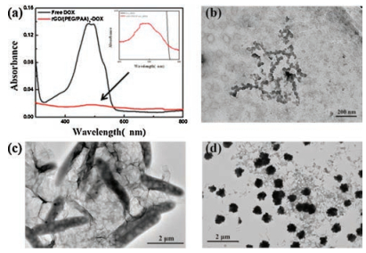
To form nanoclusters, the antitumor drug DOX was assembled with the rGO/(PEG/PAA)3. The particles were formed by mixing the rGO/(PEG/PAA)3 dispersion and DOX solutions. The morphologies of the assemblies were strongly influenced by both DOX concentrations and the number of polymer layers around the rGO cores. With the increase of the DOX concentration from 0.05 mg/mL, 0.15 mg/mL, to 0.25 mg/mL, assemblies with a variety of morphologies were formed. These morphologies included: ⅰ) irregular clusters assembled by nano-dots of around 50 nm, ⅱ) nanobars with dimensions of around 300 nm in width and 2–3 μm in length, and ⅲ) spherical clusters with rough surfaces with diameters of around 750 nm (Figs. 2b–d & Fig. S4 in Supporting information). Furthermore, the size of the drug nanoparticles can be controlled by controlling the concentration of DOX solution (Fig. S5 in Supporting information). When the concentrations are 0.01 mg/mL, 0.05 mg/mL and 0.1 mg/mL, the size of the drug nanoparticles was around 35 nm, 50 nm, 75 nm, respectively.
As shown in Fig. S6 in Supporting information, only the rGOPEG/PAA composite with 3.5 bilayers led to spherical particles, and the rGO-PEG/PAA composite with fewer polymer bilayers only led to irregular assemblies. The spherical clusters were selected to study in detail because previous literature indicated that nanoparticular clusters with sizes within the 200–1000 nm range would effectively accumulate in cancer tissues [3]. In addition, cell experiments (presented below) indicated that the spherical clusters contained more DOX and accumulated in cells more effectively than the nanobars.
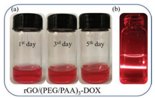
The spherical nanoparticles have an average size of 752.1 nm and a surface potential of -33.8 mV. The UV–vis spectra of rGO/ (PEG/PAA)3-DOX particles showed the characteristic absorption peaks of DOX at 480 nm (Fig. 2a), confirming that DOX was loaded into the assembly. The loading efficiency of the rGO/(PEG/PAA)3-DOX was 0.021 mg/mL. ATR-FTIR of the assembly displayed a much stronger vibrational peak at 1719 cm-1 than that of the rGO/(PEG/PAA)3 precursor, indicating that a substantial amount of hydrogen bonds between PAA and DOX formed with the formation of these assemblies (Fig. S7 in Supporting information). The rGO/(PEG/PAA)3 and rGO/(PEG/PAA)3-DOX complexes could stably suspend in PBS for up to 5 days (Fig. S8 in Supporting information, note that a longer period of time was not tested). In addition, the rGO/(PEG/PAA)3-DOX complex was also stable in the cell culturing media for at least 5 days (Fig. 3a), whereas typically the Tyndall effect (Fig. 3b) demonstrates the existence of distributed nanoparticles therein. Compared with the free drug DOX, as observed in the collagen hydrogel, the rGO/(PEG/PAA)3-DOX complex diffused more slowly in the mimetic gel-like environment of the extracellular matrix (Fig. S9 in Supporting information). This low diffusibility can decrease the toxicity towards healthy tissues of the rGO/(PEG/PAA)3-DOX complex away from its application position (e.g., an injection site) and increase its uptake by the tumor tissues via the EPR effect.
The rGO/(PEG/PAA)3-DOX assemblies released DOX in PBS over an extended period longer than 8 h and was responsive to solution pH values (Fig. 4a). An acidic environment remarkably facilitated the release of DOX. The amounts of DOX released in weakly acidic (pH 6.0) solution during 8 h were nearly 2.7 times of that in a neutral solution (pH 7.0) and 2.2 times of that in a basic (pH 8.0) solution. The cause of such a pH-responsive release profile was attributed to the pH-responsive functional groups in DOX and PAA. The acidic environment altered the PEG/PAA multilayer assembly states [24] and also partially destroyed the electrostatic interactions or hydrogen bonds between rGO/(PEG/PAA)3 and DOX (Fig. S10 in Supporting information). The absence of the vibrational peak at 1719 cm-1 after the clusters were dispersed in the acidic solution demonstrated that the hydrogen bond was damaged therein (Fig. S11 in Supporting information).
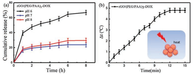
The photothermal effect of the rGO/(PEG/PAA)3-DOX complex is another property that facilitate its capability to kill cancer cells. Cell apoptosis could be induced by local temperature changes around the complex and the generation of active species [25]. The temperature of the rGO/(PEG/PAA)3-DOX dispersion increased from by 4.8 ℃ upon IR irradiation at 940 nm of 1 W/cm2 for 15 min (Fig. 4b).
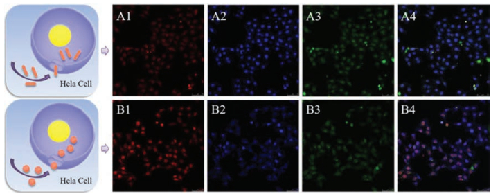
The autofluorescence of DOX allowed the observation of endocytosis of the rGO/(PEG/PAA)3-DOX complex through Hela cells. The spherical clusters could be endocytosed effectively after incubation for 24 h and still displayed clear fluorescence (Fig. 5). In comparison, the nanobars displayed very weak fluorescence and indicated a low DOX loading amount therein. The size of the clusters should favor EPR effect in tumor tissues, and is advantageous compared with the 2D rGO nanosheets [3]. Flow cytometry results (Fig. S12 in Supporting information) indicated that the nanoclusters presented FL signals with higher intensities than the nanobars, when the suspension of the nanodrugs were injected into the measurement. While after cultivation for 24 h in the existence of the nanodrugs, cells presented clear FL signal in the cytometry measuerments, indicating that both types of nanodrugs were internalized by cells effectively. Consistent the FL intensity presented by the nanodrugs, the cells incubated with nanoclusters presented higher FL intensities than cells incubated with nanobars.
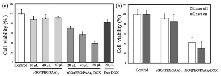
The cytotoxicity of the spherical complex towards Hela cells was evaluated by MTT assay. The rGO/(PEG/PAA)3 precursor without DOX presented a low cytotoxicity throughout the dose volume from 20 μL to 60 μL. In comparison, when incubated with the rGO/(PEG/PAA)3-DOX complex, the cell viability was significantly reduced to about 38.7% at a volume of 60 μL (0.25 mg/mL), as shown in Fig. 6a. When compared with pure DOX (20 μL, 0.25 mg/mL), the rGO/(PEG/PAA)3-DOX presented a superior effect of killing cancer cells at the same dosage, which is consistent with a previous report [3]. The effectiveness of the rGO/(PEG/PAA)3-DOX particle could be attributed to its successful cell internalization and pH-responsive release. Because these drug nanoparticles were formed by the hydrogen bonds between PAA and DOX. When the nanoparticles get into cells, the hydrogen bond can be easily broken in the acid environment of the cancer cells. The DOX can release from the nanoparticles and kill cancer cells. The photothermal effect of the complex further facilitated the effective killing of Hela cells as shown in Fig. 6b. The cell viability decreased to 100.0% and 84.7% after IR irradiation for 15 min when incubated without and with the rGO/(PEG/PAA)3 precursor, respectively. When incubated with the rGO/(PEG/PAA)3-DOX complex, a further decrease of cell viability to 30.7% was obtained after IR irradiation. These results confirmed that our complex acted as a combined chemical and photochemical agent against cancer cells.
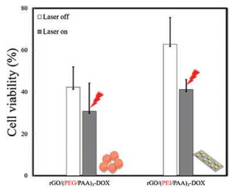
In the last step, we evaluated the influence of the choice of the polymers PEG and PAA in building the spherical complex and obtaining an effective cell-killing result. Our results indicated that the choice of the building polymers significantly influenced the cell-killing effect of the complex. The reference cell experiments conducted with the rGO/(PEI/PAA)3-DOX composite resulted in cell viability of 62.7% without IR irradiation and 41.1% with irradiation after parameter optimizations. In addition, the rGO/(PEI/PAA)3 and rGO/(PEI/PAA)3-DOX composite presented a performance remarkably inferior to the rGO/(PEG/PAA)3 and rGO/(PEG/PAA)3-DOX complex (Fig. 7 and Fig. S13 in Supporting information). The possible reason for the unique effectiveness of the rGO/(PEG/PAA)n-DOX composite might be that the precursor rGO/(PEG/PAA)n presented effective intermolecular interactions towards DOX that led to both a high drug-loading capacity and a favorable spherical morphology. In comparison, the rGO/(PEI/PAA)n precursor and DOX failed to form the spherical complex (Fig. S14 in Supporting information) and had inferior cancer cell-killing abilities (Fig. S15 in Supporting information). Both the number of the polymer layers assembled around the rGO cores and the assembly partner of PAA remarkably influenced the assembly with DOX, suggesting that a delicate influence of supramolecular interactions exists when designing functional materials.
In summary, we developed an effective strategy to build the spherical rGO/(PEG/PAA)3-DOX complex as an efficient and pHresponsive nanodrug to kill Hela cells. This rGO/(PEG/PAA)3-DOX complex combined chemotherapy, photothermal therapy effects, and a favorable morphological effect. The clusters successfully enter Hela cells as observed thanks to the self-fluorescence of the complex. The complex was prepared by first alternatively assembling PEG and PAA polymers around the rGO cores via hydrogen bond interactions, followed by DOX loading which led to a spherical complex with a diameter of around 750 nm. The nanospheres released DOX effectively in a weakly acidic environment, accumulated discriminatively in Hela cells but did not enter healthy cells, and killed Hela cells effectively upon IR irradiation. The choice of polymers played an important role in building the spherical and effective nanodrug. This report suggests an effective way of nanodrug engineering through the utilization of the supramolecular interactions between the loaded drug and the pseudo 2D precursors. Considering the wide research interests on various pseudo active 2D inorganic drug carrying materials and the wide availability of different types of LbL polymers, we hope our research provides an effective drug design method that is also potentially applicable to nanodrugs that involve other types of inorganic nanosheets.
This work was supported by the Fundamental Research Funds for the Central Universities (Nos. 2652015295, 2652016092), and the National Natural Science Foundation of China (NSFC), (Nos. 21673209, 21303169).
Supplementary data associated with this article can be found, in the online version, at https://doi.org/10.1016/j.cclet.2018.03.019.
F. Danhier, O. Feron, V. Préat, J. Controlled Release 148(2010) 135-146. doi: 10.1016/j.jconrel.2010.08.027
Y. Wen, W.S. Meng, J. Pharm. Innovation 9(2014) 158-173. doi: 10.1007/s12247-014-9183-4
B. Chertok, M.J. Webber, M.D. Succi, et al., Mol. Pharmaceutics 10(2013) 3531-3543. doi: 10.1021/mp4003283
S. Mohan, C.C. David, B. Harris, et al., J. Biomed. Nanotechnol. 12(2016) 481-490. doi: 10.1166/jbn.2016.2196
S. Supriya, B. Vinay, N. Abhignyan, et al., J. Biomed. Nanotechnol. 12(2016) 2202-2219. doi: 10.1166/jbn.2016.2312
Y. Qu, B.Y. Chu, J.R. Peng, et al., NPG Asia Mater. 7(2015) e207. doi: 10.1038/am.2015.83
C.X. Sun, L. Wen, J.F. Zeng, et al., Biomaterials 91(2016) 81-89. doi: 10.1016/j.biomaterials.2016.03.022
S.G. Wang, X. Li, Y. Chen, et al., Adv. Mater. 27(2015) 2775-2782. doi: 10.1002/adma.v27.17
Z.Y. Xiao, C.T. Xu, X.H. Jiang, et al., Nano Res. 9(2016) 1934-1947. doi: 10.1007/s12274-016-1085-y
Y. Chen, D.L. Ye, M.Y. Wu, et al., Adv. Mater. 26(2014) 7019-7026. doi: 10.1002/adma.201402572
R. Cai, D. Yang, J. Wu, et al., Nano Res. 9(2016) 2520-2530. doi: 10.1007/s12274-016-1138-2
H. Wen, C. Dong, H. Dong, et al., Small 8(2012) 760-769. doi: 10.1002/smll.v8.5
Y. Zhang, T.R. Nayak, H. Hong, et al., Nanoscale 4(2012) 3833-3842. doi: 10.1039/c2nr31040f
K. Yang, L. Feng, X. Shi, et al., Chem. Soc. Rev. 42(2013) 530-547. doi: 10.1039/C2CS35342C
T.Y. Jiang, W.J. Sun, Q.W. Zhu, et al., Adv. Mater. 27(2015) 1021-1028. doi: 10.1002/adma.201404498
R.R. Xing, T.F. Jiao, Y.M. Liu, et al., Polymers 8(2016) 181. doi: 10.3390/polym8050181
Y. Liu, J. Peng, S.H. Wang, et al., NPG Asia Mater. 10(2018) e458. doi: 10.1038/am.2017.225
Z. Liu, J.T. Robinson, X. Sun, et al., J. Am. Chem. Soc. 130(2008) 10876-10877. doi: 10.1021/ja803688x
J. Wu, Y. Wang, X. Yang, et al., Nanotechnology 23(2012) 355101. doi: 10.1088/0957-4484/23/35/355101
D. Li, M.B. Muller, S. Gilje, et al., Nat. Nanotechnol. 3(2008) 101-105. doi: 10.1038/nnano.2007.451
S.T. Yang, Y. Chang, H. Wang, et al., J. Colloid Interface Sci. 351(2010) 122-127. doi: 10.1016/j.jcis.2010.07.042
Y. Chang, S.T. Yang, J.H. Liu, et al., Toxicol. Lett. 200(2011) 201-210. doi: 10.1016/j.toxlet.2010.11.016
J.H. Liu, S.T. Yang, H. Wang, et al., Nanomedicine 7(2012) 1801-1812. doi: 10.2217/nnm.12.60
S. Sukhishvili, S. Granick, Macromolecules 35(2002) 301-310. doi: 10.1021/ma011346c
X.M. Li, Y.H. Zhang, Y.L. Wu, et al., ACS Appl. Mater. Interfaces 7(2015) 19353-19361. doi: 10.1021/acsami.5b05463

Scheme 1 (a) Layer-by-layer assembly around the rGO template by PEG and PAA. (b) The construction of the rGO/(PEG/PAA)3-DOX clusters that enter cancer cells.

Figure 1 (a) Schematic illustration of a rGO/(PEG/PAA)n (n = 1–3) composite nanosheet. (b) Zeta potential of rGO/(PEG/PAA)n during the assembly process. AFM images and height profiles of the (c) rGO/PEG and (d) rGO/(PEG/PAA)3 sheets. (e) the ATR-FTIR spectra of rGO, rGO/PEG and rGO/PEG/PAA, and (f) the enlarged view of the ATR-FTIR spectra of pure PEG, PAA, rGO/PEG and rGO/PEG/PAA within the range 1650–1750 cm-1.

Figure 2 (a) UV–vis spectra of free DOX and rGO/(PEG/PAA)3-DOX. The inset is the zoomed-in image of the spectra of the rGO/(PEG/PAA)3-DOX complex around 500 nm, characteristic of DOX absorbance. The TEM images of rGO/(PEI/PAA)3-DOX assemblies prepared with following concentrations of DOX: (b) 0.05 mg/mL, (c) 0.15 mg/mL, and (d) 0.25 mg/mL.

Figure 3 (a) The rGO/(PEG/PAA)3-DOX was stable in a cell-culturing media for at least 5 days without stirring. (b) The Tyndall effect of the rGO/(PEG/PAA)3-DOX dispersion.

Figure 4 (a) The release profile of rGO/(PEG/PAA)3-DOX in a PBS buffer with varying pH values of pH 6 (black), pH 7 (blue), and pH 8 (red). (b) The photothermal performance of the spherical rGO/(PEG/PAA)3-DOX complex.

Figure 5 Confocal fluorescence microscopic images of Hela cells coincubated with rGO/(PEG/PAA)3-DOX. (A1-A4) Hela cells incubated with rGO/(PEG/PAA)3-DOX nanobars for 24 h. (b) Hela cells incubated with rGO/(PEG/PAA)3-DOX spherical assemblies for 24 h. Chanel 1: Fluorescence signal of the rGO/(PEG/PAA)3-DOX nanodrugs; Chanel 2: Hoechst 33258 (cell nuclei) signal; Chanel 3: Fluorescence signal of living cells; Chanel 4: Overlapped field. Magnification: 40×.

Figure 6 (a) MTT assay of Hela cells coincubated with different volumes of rGO/(PEG/PAA)3, rGO/(PEG/PAA)3-DOX or free DOX dispersions. (b) MTT assay of Hela cells coincubated with rGO/(PEG/PAA)3 or spherical rGO/(PEG/PAA)3-DOX assemblies with and without NIR irradiation.

 扫一扫看文章

扫一扫关注我们
 DownLoad:
 下载:
下载:
