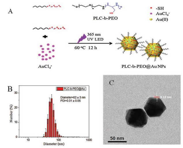
Figure 1.
(A) One-pot fabrication of the NIR-absorbing plasmonic PLC-b-PEO@Au NPs; DLS (B) and TEM data (C) for the plasmonic PLC-b-PEO@Au NPs.
One-pot photoreduction to prepare NIR-absorbing plasmonic gold nanoparticles tethered by amphiphilic polypeptide copolymer for synergistic photothermal-chemotherapy
Siqi Yang , Linzhu Zhou , Yue Su , Rong Zhang , Chang-Ming Dong

In the past decades, a myriad of gold nanoparticles (Au NPs) with excellent optical, catalytic and bio-physicochemical properties have attracted great attention in nanotechnology and nano-medicine as they intrinsically possess the localized surface plasmon resonance [1-8] Especially, some kinds of Au NPs with complicated architectures including gold nanoshells, nanorods, nanocages, and nanostars can absorb near-infrared light (NIR Ⅰ, 650 - 950 nm; NIR Ⅱ, 1000–1500 nm) and efficiently convert it into heat, making them great potentials for noninvasive photothermal therapy (PT) of cancers, bioimagings, and theranostics [3-8]. Importantly, the hyperthermia effect might enhance NPs accumulation and penetration in tumor site, the cellular permeability and uptake, and the antitumor immunity that would further enable the tumor ablation of single PT or the synergistic therapeutic efficacy of the combined photothermal-chemotherapy (PT-CT) [9-13]. On the other hand, synthetic polymers are generally utilized not only to tune the plasmonic wavelength, but to enhance the stability and the biocompatibility of Au NPs [14]. However, obvious drawbacks including complicated fabrication procedures, toxic reducing agents, and non-biodegradable polymeric templates are urgent to be overcome, which will otherwise hinder the plasmonic Au NPs from biomedical applications and clinical transitions [15-18]. Therefore we are interested in how to fabricate NIR-absorbing plasmonic Au NPs in a simple and green manner.
Owing to good biocompatibility and biodegradability, synthetic polypeptides (i.e., poly(amino acid)s) and their amphiphilic poly (ethylene oxide) (PEO) copolymers have been widely investigated for various drug delivery vesicles, also providing opportunities to construct biocompatible plasmonic Au NPs [19-22]. To address the above-mentioned problems, herein we develop one-pot photore-duction strategy for the preparation of plasmonic Au NPs with the NIR-absorption at 700–1100 nm by using the thiols-pendant poly (L-cysteine)-b-PEO (PLC-b-PEO) copolymer as a reducing and stabilizing agent (Fig. 1A). The as-prepared polypeptide copolymer-tethered Au NPs (i.e., PLC-b-PEO@Au NPs) have been thoroughly characterized by means of vis-NIR spectroscopy, XPS, DLS, TEM, and high resolution FE-TEM with two-dimensional element mapping, which convincingly demonstrated that the PLC-b-PEO@Au NPs has a thin layer of PLC-b-PEO copolymer dangled on the surface of Au NPs. The PLC-b-PEO@Au NPs possess strong NIR absorption at 700–1100 nm and excellent photothermal conver sion properties including ultrahigh photothermal conversion efficiency of 62.1% and good photostability, enabling them promising for both PT and PT-CT treatments of cancers.
Inspired by UV photoreduction of Au3+ to generate plasmonic Au NPs with visible light absorption [17], we tested the 365 nm UV irradiation (using a LED lamp) of HAuCl4 in the presence of amphiphilic thiols-pendant polypeptide copolymer (PLC-b-PEO) in DMF/H2O solution. When the mixed solution of PLC-b-PEO (5 mL, 1 mg/mL) with HAuCl4 (2.5 mL, 5 mg/mL, Au/S = 1 mol/mol) was irradiated at 60 ℃ and then stirred at room temperature for different times, the resulting solution became pink red and then turned purple-black. This phenomenon indicates the formation of plasmonic Au NPs that were probably tethered and stabilized by amphiphilic PLC-b-PEO [9-12, 14-15]. The vis-NIR spectra show increasing broad NIR absorption at 700–1100 nm besides a maximal one at about 565 nm (Fig. S1A in Supporting information). This evidences increasing amounts of plasmonic Au NPs was produced within 12 h UV irradiation in DMF/H2O solution and at 60 ℃, while black precipitate would occur for a longer time of irradiation. After 12 h UV irradiation and continuous stirring for different times at room temperature, the NIR absorbance ranging at 700–1100 nm gradually increased over the time, suggesting the continuous growth and/or aggregation of Au NPs in the presence of PLC-b-PEO (Fig. S1B) [12, 14-15]. As control, weak NIR absorption was observed without the polypeptide copolymer/UV irradiation or with the UV irradiation at room temperature (Figs. S2-S4 in Supporting information). As shown in Figs. 1B and C, both dynamic light scattering (DLS) and transmission electron microscopy (TEM) showed that the resultant PLC-b-PEO@Au NPs had an irregular polyhedron morphology with a hydrodynamic diameter (62 ± 3) nm (Fig. S5 in Supporting information), which was slightly bigger than (52 ± 2) nm for the self-assembled PLC-b-PEO micelles (Fig. S6 in Supporting information). Note that the morphology control of the PLC-b-PEO@Au NPs deserves to be further studied in the ongoing work. As can be discerned by TEM, the PLC-b-PEO@Au NPs have a thin copolymer layer of about 3 nm (also see the following high resolution FE-TEM analysis). Note that the TEM size of the PLC-b-PEO@Au NPs is basically in agreement with that determined by DLS. As the irregular polyhedron morphology and the refractive index difference between Au NPs and the thin polypeptide copolymer layer would enhance a localized surface plasmon resonance (LSPR) coupling, which greatly shifted the LSPR peak to the red region that become broader with increasing NPs diameter [10].
Then we optimized the NIR-absorbing property of the PLC-b-PEO@Au NPs by changing reaction time and various molar ratios of Au to S element (Au/S = 0.6–1.2, mol/mol). As found by vis-NIR spectroscopy (Fig. 2A), the plasmonic PLC-b-PEO@Au NPs that were prepared under optimal ratio of Au:S = 1:1 presented the strongest NIR absorption compared to the control. FT-IR spectra showed new vibrational peak at 1050 cm-1 for sulfinic or sulfonic acids, which were produced from the oxidization of thiols on PLC-b-PEO with concurrent gold reduction (Fig. S7 in Supporting information). The XPS of PLC-b-PEO@Au NPs gave the doublet peaks at 162.0 eV (S 2p3/2) and 163.5 eV (S 2p1/2), which were assigned to the coordinate bonding of sulfur-gold (S-Au). Moreover, the peak centered at 166.8 eV was assigned to oxidized sulfur species (Fig. 2B). In all, these results persuasively evidenced that the thiols-pendant polypeptide copolymer PLC-b-PEO could reduce HAuCl4 species under UV irradiation at 60 ℃ to produce the NIR-absorbing plasmonic PLC-b-PEO@Au NPs that were tethered by PLC-b-PEO via multivalent Au-S bondings [12, 14-15]. In addition, the gold weight percentage in PLC-b-PEO@Au NPs was determined to be 90.2 wt% by TGA analysis (Fig. S8 in Supporting information).
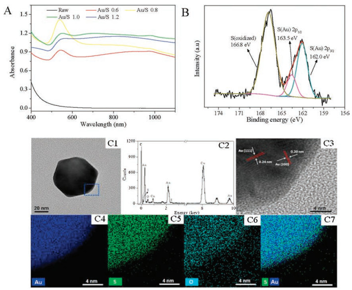
Both FE-TEM and energy-dispersive X-ray spectroscopy (EDX) were further used to investigate the microstructure and elemental composition of the PLC-b-PEO@Au NPs. Fig. 2-C1 is a typical FE-TEM image of a representative sample, which clearly showed irregular polyhedron morphology and the hybrid NPs surface was coated by the PLC-b-PEO copolymer layer. The EDX spectra in Fig. 2-C2 confirmed that the elements Au, S, C and O were distributed in whole NPs (note that the detected Cu element originates from the copper grid). The lattice fringes are clearly observed from Fig. 2-C3 and the d-spacing value is 0.24 nm and 0.20 nm, corresponding to the {111} and {200} lattice planes, respectively [23]. The two-dimensional (2D) elemental mapping analyses verify that PLC-b-PEO was homogeneously tethered on the gold nanocrystal surface (Fig. 2C4-C7). Taken together, these experimental findings convincingly demonstrated that the thiols-pendant polypeptide copolymer PLC-b-PEO acted not only as a reducing agent for the oxidized gold species of HAuCl4, but as a stabilizer for the reduced gold nanocrystal via forming multivalent S-Au bonds.
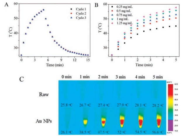
As the PLC-b-PEO@Au NPs have intrinsic NIR-absorbing capacity, we irradiated their solution using a continuous-wave 808 nm laser to test the photothermal effect. As shown in Fig. 3A, the solution temperature of the PLC-b-PEO@Au NPs (1 mg/mL) increased sharply over the irradiation time and a maximum of 30.8 ℃ was attained upon 5 min laser irradiation at a power intensity of 2 W/cm2, however, the control was elevated only by 2.4 ℃. Moreover, both laser-on and laser-off cycle curves were repeated three times without any change, demonstrating good photostability. The magnitude of temperature elevation increased with the concentration of PLC-b-PEO@Au NPs, also representing intrinsic NIR-absorbing characteristics (Fig. 3B). Similarly the temperature rise can be tuned by the laser power intensity (Fig. S9 in Supporting information). Furthermore, this temperature-elevating process was recorded by a thermo-imaging camera, during which the NPs solution temperature doubled from 26.1 ℃ to 52.0 ℃ within initial 3 min irradiation and then slowly increased to 56.6 ℃ at 5 min (Fig. 3C). Notably, the PLC-b-PEO@Au NPs gave higher photothermal conversion efficiency (η) of 62.1% that is largely more than that (13% ~ 22%) of the well-known gold nanorods and nanoshells [3-8]. This is attributed to the irregular polyhedral gold nanocrystals core stabilized by a thin copolymer layer, which enhanced the LSPR coupling and induced a "lightning rod" effect upon an NIR laser irradiation [10]. Meanwhile, there is a linear relationship between η and the concentration of PLC-b-PEO@Au NPs or with the laser power extinction (Figs. S10 and S11 in Supporting information), which is consistent with the previous report on the hyperthermia effect of the Au NPs [16]. Collectively, these experimental results evidence that the PLC-b-PEO@ Au NPs can act as an excellent photothermal nanoagent with strong NIR absorption, ultrahigh photothermal conversion efficiency, and good photostability. In addition, because the plasmonic PLC-b PEO@Au NPs has strong NIR-Ⅱ absorbance, its NIR-Ⅱ mediated PTT deserves to be further investigated in the future work.
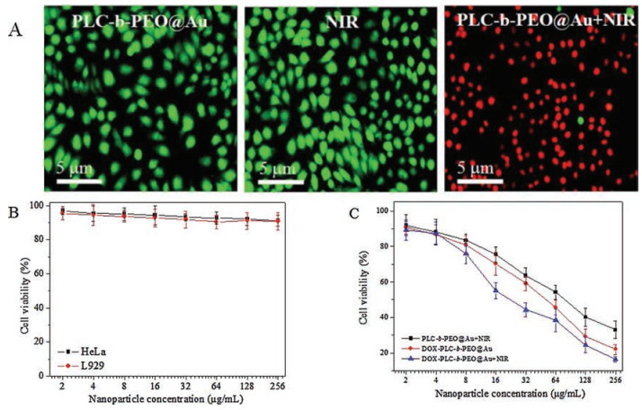
The hyperthermia effect for killing cancer cells was directly observed by using a double fluorescent staining technique. After the NIR irradiation (5 min, 808 nm, 2 W/cm2) and incubation for 12 h, the HeLa cells were co-stained with AO and EB to differentiate live (green) and dead (red) cells, respectively. The HeLa cells vividly kept alive upon with the NIR irradiation or PLC-b-PEO@Au NPs (256 mg/mL); however, they underwent nearly full death upon with same dose of PLC-b-PEO@Au NPs plus the NIR irradiation (Fig. 4A). These experiments further verified that the hyperthermia effect mediated by PLC-b-PEO@Au NPs plus the NIR irradiation could efficiently kill the cancer cells in vitro, enabling them promising for the PT treatment of cancers [3-8].
The anticancer drug doxorubicin (DOX) was stirred with the PLC-b-PEO@ Au NPs solution to produce the drug-loaded counterparts (i.e., DOX-PLC-b-PEO@ Au NPs), which gave a high drug-loading capacity of about 27 wt% and a bigger hydrodynamic diameter of (85 ± 3) nm than that ((62 ± 3) nm) of the blank ones (Fig. S12 in Supporting information). Owing to the complex interactions including hydrogen-bonding, van der Walls, and hydrophobic interactions among DOX, the tethered polypeptide copolymer chains, and the Au NPs surfaces, the loaded DOX was probably absorbed onto the interface layer between the copolymer and Au NPs during the self-assembly process [9-12].
With same dose of the NIR irradiation to the blank ones, the DOX-PLC-b-PEO@Au NPs solution (1 mg/mL) increased to 51.0 ℃ from 25.0 ℃ (Fig. S13 in Supporting information). Taking account of the DOX fraction in the DOX-PLC-b-PEO@Au NPs, the drug hardly had effect on the photothermal conversion ability of the drugloaded nanoparticles. The NIR-triggered cumulative DOX release was greatly enhanced to 44.5 wt% at 12 h compared to 26.0 wt% for non-triggered sample as the photo-converted heat accelerated the DOX diffusion in aqueous solution (Fig. S14 in Supporting information). This result illustrates that the built-in hyperthermia function of the drug-loaded DOX-PLC-b-PEO@Au NPs might improve the DOX-induced chemotherapy efficacy [12]. The cytotoxicity of PT, CT, and PT-CT of the PLC-b-PEO@Au NPs and/ or the drug-loaded counterparts was evaluated by a standard MTT assay, respectively. As control, the same dose of NIR irradiation and the PLC-b-PEO@Au NPs themselves induced less cytotoxicity with cell viability > 90% (Fig. 4B). However, the cells viabilities sharply decreased over the drug-loaded NPs concentration (with or without NIR irradiation) and/or the blank ones plus NIR irradiation, and the half-maximal inhibitory concentrations (IC50) of cells could be calculated from Fig. 4C. Based on the blank nanoparticles or DOX concentration, an IC50 value of 54.78 μg/mL or 13.53 μg/mL can be calculated for single PT. and single CT, respectively. Based on the concentration of the DOX-loaded PLC-b-PEO@Au NPs, a combined IC50 of 27.02 μg/mL was given under PT-CT treatment, which consisted of 7.29 μg/mL for the combination CT and 19.73 μg/mL for the combination PT, respectively. As for the combination therapy, the combination index (CI) is generally used to evaluate the synergistic effect between different treatments, and the CI values of < 1, 1, and > 1 indicate synergism, additive effect, and antagonism, respectively [24]. Herein, the DOX-loaded PLC-b-PEO@Au NPs gave a CI of 0.9, demonstrating a synergistic effect of the combination PT-CT on the HeLa cell line.
In summary, we developed one-pot photoreduction strategy to fabricate the NIR-absorbing plasmonic PLC-b-PEO@Au NPs, during which the thiols-pendant polypeptide copolymer PLC-b-PEO acted not only as a reducing agent for HAuCl4, but as a stabilizer for the reduced gold nanocrystal via forming multivalent S-Au bonds. The PLC-b-PEO@Au NPs possessed strong NIR absorption at 700–1100 nm, an ultrahigh photothermal conversion efficiency of 62.1%, and good photostability. The PLC-b-PEO@Au NPs mediated hyperthermia effect could efficiently kill the HeLa cells and the PT-CT treatment produced a synergistic effect in vitro. Importantly, this work establishes a versatile platform for one-pot fabrication of the NIR-absorbing plasmonic Au NPs in a simple and green manner.
The National Natural Science Foundation of China (No. 21474061) and The Innovation Fund (No. IFPM2016B004) of Shanghai Jiao Tong University & Affiliated Sixth People's Hospital South Campus are appreciated.
Supplementary data associated with this article can be found, in the online version, at https://doi.org/10.1016/j.cclet.2018.02.015.
X. Yang, M. Yang, B. Pang, et al., Chem. Rev. 115(2015) 10410-10488. doi: 10.1021/acs.chemrev.5b00193
W. Zhou, X. Gao, D. Liu, et al., Chem. Rev. 115(2015) 10575-10636. doi: 10.1021/acs.chemrev.5b00100
S. Lal, S.E. Clare, N.J. Halas, Acc. Chem. Res. 41(2008) 1842-1851. doi: 10.1021/ar800150g
A. Liu, G. Wang, F. Wang, et al., Coord. Chem. Rev. 336(2017) 28-42. doi: 10.1016/j.ccr.2016.12.019
L. Cheng, C. Wang, L. Feng, et al., Chem. Rev. 114(2014) 10869-10939. doi: 10.1021/cr400532z
Z. Zhang, J. Wang, C.Y. Chen, Adv. Mater. 25(2013) 3869-3880. doi: 10.1002/adma.v25.28
R.F. Zhao, X.X. Han, Y.Y. Li, et al., ACS Nano 11(2017) 8103-8113. doi: 10.1021/acsnano.7b02918
N. Zhang, D.X. Zhu, L. Feng, et al., J. Biomed. Nanotechnol. 13(2017) 134-143. doi: 10.1166/jbn.2017.2330
H.F. Deng, Y. Dai, G.H. Ma, et al., Adv. Mater. 27(2015) 3645-3653. doi: 10.1002/adma.201501420
S. Chen, Q. Lei, W.X. Qiu, et al., Biomaterials 117(2017) 92-104. doi: 10.1016/j.biomaterials.2016.11.056
L. Wang, Y.Y. Yuan, S.D. Lin, et al., Biomaterials 78(2016) 40-49. doi: 10.1016/j.biomaterials.2015.11.024
X. Wu, L. Zhou, Y. Su, et al., Biomacromolecules 17(2016) 2489-2501. doi: 10.1021/acs.biomac.6b00721
Q. Yang, J.R. Peng, Y. Xiao, et al., ACS Appl. Mater. Interfaces. 10(2018) 150-164. doi: 10.1021/acsami.7b14705
J.B. Song, P. Huang, H.W. Duan, et al., Acc. Chem. Res. 48(2015) 2506-2515. doi: 10.1021/acs.accounts.5b00059
Y. Liu, Y. Liu, J. Yin, et al., Macromol. Rapid. Commun. 36(2015) 711-725. doi: 10.1002/marc.v36.8
K. Jiang, D.A. Smith, A. Pinchuk, J. Phys. Chem. 117(2013) 27073-27080.
S.E. And, M.A. Elsayed, J. Phys. Chem. 110(2006) 14014-14019. doi: 10.1021/jp062972k
Y.F. Huang, S.C. Lu, Y.C. Huang, et al., Small 10(2014) 1939-1944. doi: 10.1002/smll.201303462
H. Cabral, N. Nishiyama, K. Kataoka, Acc. Chem. Res. 44(2011) 999-1008. doi: 10.1021/ar200094a
C. Deng, J.T. Wu, R. Cheng, et al., Prog. Polym. Sci. 39(2014) 330-364. doi: 10.1016/j.progpolymsci.2013.10.008
X. Wu, L. Zhou, Y. Su, et al., Polym. Chem. 7(2016) 5552-5562. doi: 10.1039/C6PY01189F
B.W. Zhao, Z.X. Zhou, Y.Q. Shen, Chin. J. Polym. Sci. 34(2016) 94-103. doi: 10.1007/s10118-016-1735-2
Y. Bustami, M. Moo-Young, W.A. Anderson, Sensor. Actuat. B-Chem. 245(2017) 753-764.
L. Ma, M. Kohli, A. Smith, ACS Nano 7(2013) 9518-9525. doi: 10.1021/nn405674m

Figure 1 (A) One-pot fabrication of the NIR-absorbing plasmonic PLC-b-PEO@Au NPs; DLS (B) and TEM data (C) for the plasmonic PLC-b-PEO@Au NPs.

Figure 2 (A) Vis-NIR spectra of the PLC-b-PEO micelles (1 mg/mL) and the plasmonic PLC-b-PEO@Au NPs with various ratios of Au/S; XPS spectra (B); (C1) FE-TEM photograph of PLC-b-PEO@Au NPs; (C2) Corresponding TEM–EDX profile recorded from one position (blue square) in the NPs; (C3) The magnified FE-TEM image taken from the blue square; (C4- C7) The two-dimensional (2D) element mappings for the detected three elements Au, S, O, and Au + S.

Figure 3 (A) The temperature change curves of PLC-b-PEO@Au NPs solution (1 mg/mL) for three on-off laser irradiation cycles (808 nm, 2 W/cm2); (B) The concentration dependent heating curves of the PLC-b-PEO@Au NPs solution upon 5 min NIR irradiation; (C)The IR thermal images of the PLC-b-PEO@Au NPs solution (1 mg/mL) upon NIR irradiation at different times.

Figure 4 (A) Fluorescence microscope images of HeLa cells treated with PLC-b-PEO@Au NPs (256 mg/mL) or the NIR irradiation (5 min, 808 nm, 2 W/cm2); (B) Cytotoxicity of the PLC-b-PEO@Au NPs incubated with L929 or HeLa cells; (C) Cytotoxicity of the PLC-b-PEO@Au NPs + NIR irradiation or the DOX-PLC-b-PEO@Au NPs or the DOX-PLC-b-PEO@Au NPs + NIR irradiation after incubated with HeLa cells. The data are presented as average ± standard error (n = 6).

 扫一扫看文章

扫一扫关注我们
 DownLoad:
 下载:
下载: