Scheme 1.
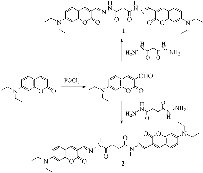
探针1和2的合成路线
Scheme 1.
Synthesis routes for probes 1 and 2

Cu2+ is an essential transition metal ion in human body, which plays critical roles in various physiological processes and normal development of tissues[1, 2]because of its redox pro-perties[3]. However, the imbalance of Cu2+ in cellular homeostasis, both excess and deficiency, can cause serious neurodegenerative diseases. For example, epileptic seizures[4], Parkinson's, Alzheimer's[5], Wilson's and Menke's disease[6, 7] are induced by the excess intake of Cu2+ ion[8], whereas Men-kessyndrome is induced by Cu2+ deficiency. Further-more, Cu2+ is an important environmental pollutant because of its widespread application[9, 10]. Therefore, it is very important to detect Cu2+ both in biological and environmental systems.
Unlike traditional analytical assay, fluorescent probes of specific ions have many advantages such as high sensitivity, excellent selectivity, instantaneous response, easy operation, low cost, low toxicity in vivo, and cumulative signaling behavior[11~14]. Recent studies have shown that acylhydrazone group is a fascinating metal ion recognition site in chemical sensors because they are rich in nitrogen and oxygen atoms[15, 16] and can selectively bind metal cations. Additionally, some acylamide group will change to the enol-form when forming a complex with metal ions, which could conjugate small π systems to a bigger one[17].
In this paper, probes 1 and 2 with acylhy-drazones as the copper ion detection unit and coumarins as the fluorescent signal group were synthesized (Scheme 1). They could selectively detect Cu2+ among various ions through a turn-off fluorescence signaling mechanism, and the detection process is accompanied by a significant color change, which can be directly observed by the naked eyes.
Commercially available analytical grade solvents and reagents were used directly in all reactions unless otherwise stated. 1, 2-Dichloroethane was used after purification by standard methods.
1H NMR spectra were obtained on Varian Mercury Plus 400MHz and Agilent Technologies DDZ 600MHz Spectrometer with tetramethylsilane as the internal standard. Absorption spectra and photoluminescence spectra were recorded by a Shimadzu UV-2550 spectrometer and a Perkin-Elmer LS-55 spectrometer, respectively. MS was obtained from a Thermo Scientific Orbitrap Elite mass spectrometer.
Under N2, anhydrous DMF (13.30 mL, 172.5 mmol) was dropped into POCl3(3.16 mL, 34.5 mmol) with stirring for 6 h in an ice bath. The solution of 7-N, N-diethylaminocoumarin (3.00 g, 13.8 mmol) in anhydrous 1, 2-dichloroethane was added to the above solution and the mixture was stirred at 60 ℃ for 12 h. Subsequently, the mixture was poured into ice water and neutralized with NaOH solution (20%) to pH 7~8. The formed precipitate was filtered off and washed three times with water. The residue was chromatographed on silica, eluting with petroleum ether /CH2Cl2 (2:1, v/v) to give the product as an orange solid. Yield: 76%.1H NMR (400 MHz, CDCl3) δ: 10.10 (s, 1H, CHO), 8.25(s, 1H, coumarin H), 7.40~7.42 (s, 1H, coumarin H), 6.63~6.65(d, 1H, coumarin H), 6.49 (s, 1H, coumarin H), 3.45~3.51(q, J=21.3Hz, 4H, CH2), 0.96~0.97(t, J=3.2Hz, 6H, CH3).
Under N2, a solution of malonohydrazide (0.2 g, 1.5 mmol) in 30 mL DMSO was added to a solution of synthesized FDC (1.1 g, 4.5 mmol) in 50 mL ethanol, and then the mixture was heated to 80℃ and stirred for 4 h. After cooling to room temperature, the mixture was filtered, and the solid was washed with methanol (3×10 mL) to obtain an orange solid with a yield of 79%. m.p.: >250 ℃. MS (ESI) m/z: 587.26 [M+H]+. 1H NMR (400 MHz, DMSO-d6) δ: 11.54(s, 2H, N-H), 8.36~8.24 (m, 2H, C-H), 8.15 (s, 2H, N=C-H), 8.06(s, 2H, coumarin H), 7.40 (d, 2H, coumarin H), 6.70 (d, 2H, coumarin H), 6.55~6.50 (m, 2H, coumarin H), 3.43 (m, 8H, CH2), 1.13~1.10 (t, 12 H, CH3). 13C NMR(101MHz, DMSO-d6) δ: 169.73, 161.15, 156.85, 151.64, 138.35, 137.77, 130.465, 113.10, 110.09, 108.05, 96.96, 44.67, 29.55, 12.90.
Under N2, a solution of succinohydrazide (0.3 g, 2.0 mmol) in 20 mL DMSO was added to a solution of FDC (1.0 g, 4.0 mmol) in 50 mL methanol, and then the mixture was heated to 70 ℃ and stirred for 24 h. After cooling to room temperature, the mixture was filtered, and the solid was washed with methanol (3×10 mL) to obtain an orange solid with a yield of 79%. m.p.: >250 ℃. MS (ESI) m/z: 601.28 [M+H]+. 1H NMR (400 MHz, DMSO-d6) δ: 11.3(s, 2H, N-H), 8.31~8.22 (m, 4H, CH2), 8.21~8.17 (m, 2H, N=C-H), 8.04~8.03 (d, 2H, coumarin H), 7.64~7.57 (m, 2H, coumarin H), 7.56~7.52 (m, 2H, coumarin H), 6.75~6.71 (d, 2H, coumarin H), 3.47~3.44 (m, 8H, CH2), 1.14~1.12 (t, 12 H, CH3). 13C NMR(101MHz, DMSO-d6)δ: 173.79, 169.23, 161.19, 156.79, 151.58, 140.60, 138.77, 138.101, 137.77, 130.77, 113.28, 110.08, 108.50, 96.72, 44.58, 29.25, 27.98, 27.19, 12.83.
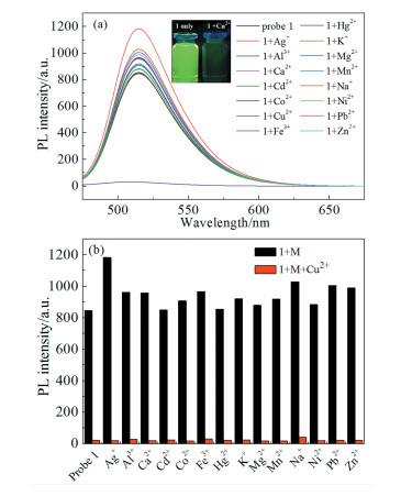
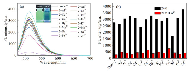
To detect the selectivity of probes for metal ions, the fluorescence spectra of probes after addition of various metal species, such as Ag+, Na+, K+, Ca2+, Mg2+, Cd2+, Hg2+, Mn2+, Co2+, Ni2+, Pb2+, Zn2+, Al3+ and Fe3+, were recorded under the same conditions. Probes 1 and 2 in DMSO/H2O (v/v, 9/1) solution showed yellow-green fluorescence under a UV-lamp. After adding Cu2+(1.0 eq) to the solution, the fluorescence of probe 1 is quenched immediately, while other metal ions can slightly increase the fluorescence of probe 1 (Fig. 1a). Both Ni2+ and Cu2+ can quench the fluorescence emission of probe 2, but the quenching effect of Cu2+ is much stronger than that of Ni2+ (Fig. 2a). In addition, the remarkable change in the color of the probe solution could be observed with the naked-eye under UV lamp (inset of Fig. 1a and 2a).
(溶剂:DMSO/H2O (v/v, 9/1),浓度:1.0×10-6 mol·L-1,激发波长:445nm, 激发狭缝宽度:5.0nm, 发射狭缝宽度:10.0nm, 室温)
(Solvent: DMSO/H2O (v/v, 9/1), c(probe 1):1.0×10⁻⁶ mol·L-1, λEx: 445nm, Ex-slit: 5.0nm, Em-slit:10.0nm, T=room temperature)
(溶剂:DMSO/H2O (v/v, 9/1),浓度:1.0×10-6mol·L-1,激发波长:445nm, 激发狭缝宽度:10.0nm, 发射狭缝宽度:20.0nm, 室温)
(Solvent: DMSO/H2O (v/v, 9/1), c(probe 1):1.0×10⁻⁶ mol·L-1, λex:445nm, Ex-slit:10.0nm, Em-slit: 20.0nm, T=room temperature)
Furthermore, fluorescence experiments of probes toward Cu2+ in the presence of other competitive metal ions were performed to study the interference immunity of probes in the sensing process. From Fig. 2a and Fig. 2b, it can be seen that the coexistence of equal amount of other cations did not bring out any notable interference on the Cu2+ recognition.
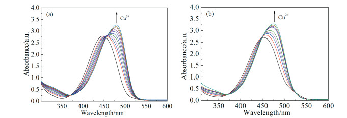
To determine the interaction mode of Cu2+ with probes, absorption titration was employed to monitor the changes in absorbance upon increasing the Cu2+ concentration. Upon the addition of Cu2+, the absorption band of each probe showed obvious bathochromic shift and an increase in absorption intensities, which indicates the interactions between probes and Cu2+ (Fig. 3). The λmax of probes 1 and 2 exhibited a bathochromic shift from 445 to 479 nm and 451 to 475 nm, respectively (Fig. 3a and 3b).
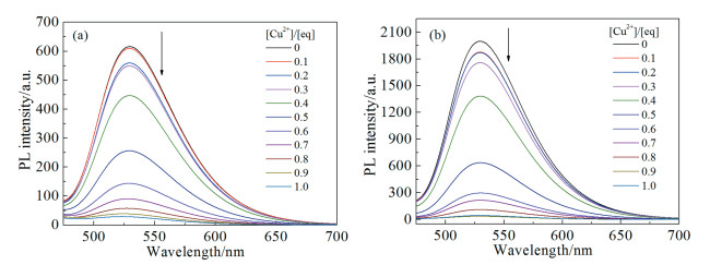
The quantitative nature of the sensing of Cu2+ by probes was elucidated by fluorescence titration as described in Fig. 4. With the increase of Cu2+ concentration, the fluorescence emission peaks at 529 nm of probes 1 (Fig. 4a) and 2 (Fig. 4b) decreased significantly, and the fluorescence was completely quenched when 1.0 eq Cu2+ was added.
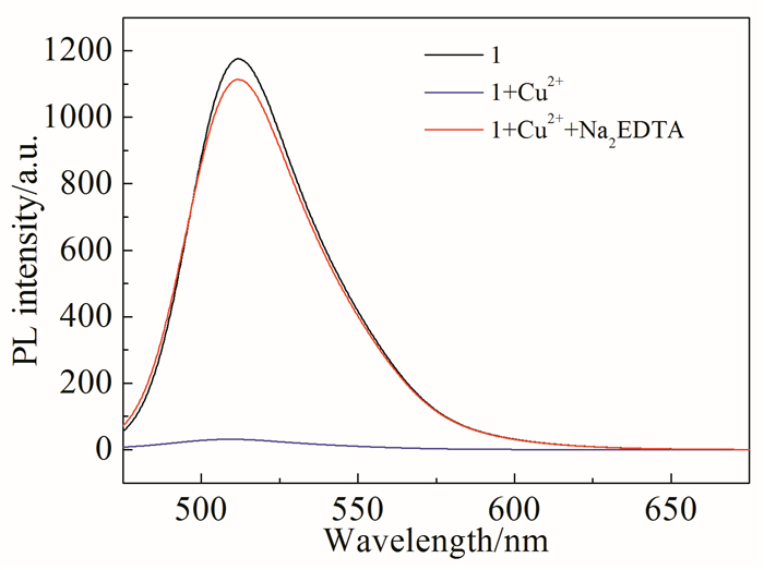
To examine the reversibility of the binding of the probe to Cu2+ in aqueous solution, EDTA was added to the probe 1-Cu2+ system. As shown in Fig. 5, the fluorescence signal upon the addition of Cu2+ was entirely reversed with the addition of EDTA. These results indicated that probe 1 was a reversible sensor for Cu2+. Probe 2 has the similar reversibility.
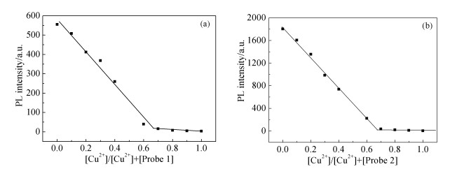
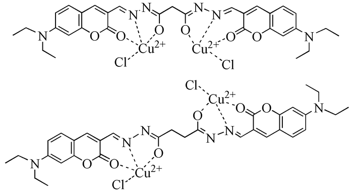
To gain insight into the sensing mechanism of probe towards Cu2+, Job's plot experiments were conducted by using fluorescence emission intensity at 529 nm, and the molar ratio of Cu2+ in probe 1 + Cu2+ complex varied from 0 to 1.0. As illustrated in Fig. 6a, the fluorescence intensity at 529 nm decreased to the minimum when the molar ratio of Cu2+ in complex was 0.668, confirming that the stoichiometric ratio of the binding of probe 1 to Cu2+ in DMSO/H2O (v/v, 9/1) solution is 1:2. The stoichiometric ratio of the binding of probe 2 to Cu2+ is also 1:2 (see Fig. 6b).
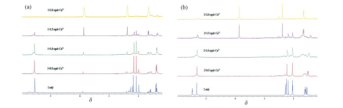
The 1H NMR titration spectra (Fig. 7) of probes 1 and 2 in the absence and presence of varying stoichiometry of CuCl2·2H2O in DMSO-d6 solution were carried out to further validate the binding behavior of probe with Cu2+. Upon addition of 2.0 eq Cu2+, the signal at δ 11.5 (N-H) in probe 1 disappeared, the signal (C-H) in acylhydrazone group shifted from δ 8.15 to δ 8.30 and the hydrogen atoms in coumarins showed a significant downfield shift (Fig. 7a), which confirmed that probe 1 may bind to Cu2+ in the enol-form, and in this way the H atom moves from the N atom to the O atom. The NMR titration test of probe 2 has similar results (Fig. 7b).
From the above data, the binding mode of probes 1 and 2 with Cu2+ in Fig. 8 is deduced.
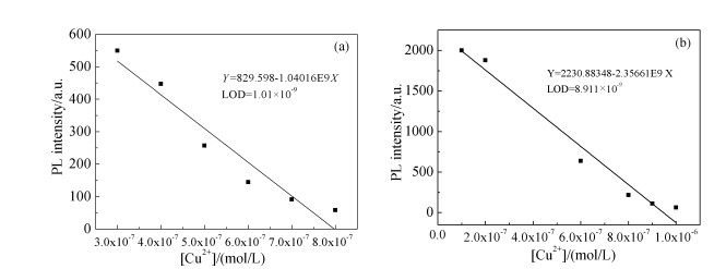
The LOD is very important to detect the analyte at low concentrations for practical purposes, which was calculated using the equation 3σ/κ[18, 19]. Fig. 9 showed that probes 1 and 2 can be applied to detect Cu2+ at an extremely low concentration level of 10-9 mol/L, which may fully meet the detection needs.
In this work, we have developed two acylhy-drazone based fluorescent probes for Cu2+. These probes display highly selective and sensitive recog-nition for Cu2+ over other metal ions through the "turn-off" effect of fluorescence intensity in DMSO/H2O (v/v, 9/1) solution. The fluorogenic response is based on cation-mediated reductive photo induced electron transfer (PET). These probes interact with Cu2+ through 1:2 binding ratio with detection limit of 10-9 mol/L. Therefore, the probes should potentially be used to monitor Cu2+ in environmental and biological systems.
Choi M G, Kim J, Hong J M, et al.. Tetrahed. Lett., 2016, 57:975-978. doi: 10.1016/j.tetlet.2016.01.052
Ding A X, Shi Y D, Zhang K X, et al.. Sens. Actuat. B, 2018, 255:440-447. doi: 10.1016/j.snb.2017.08.037
Lv T, Xu Y Q, Li H J, et al.. Bioorg. Med. Chem., 2018, 26:1448-1452. doi: 10.1016/j.bmc.2017.09.026
Zhang X, Sun P, Li F, et al.. Sens. Actuat. B, 2018, 255:366-373. doi: 10.1016/j.snb.2017.07.196
Nguyen K H, Hao Y Q, Zeng K, et al.. J. Photochem. Photobiol. A, 2018, 358:201-206. doi: 10.1016/j.jphotochem.2018.03.023
Chen S, Kuang Y F, Zhang P P, et al.. Sens. Actuat. B, 2017, 253:283-291. doi: 10.1016/j.snb.2017.06.140
Sil A, Islam S N, Patra S K.. Sens. Actuat. B, 2018, 254:618-628. doi: 10.1016/j.snb.2017.07.067
Ren A S, Zhu D J, Xie W, et al.. Inorg. Chim. Acta, 2018, 476:136-141. doi: 10.1016/j.ica.2018.02.015
Wang Y P, Liu S Z, Chen H B, et al.. Dyes Pigm., 2017, 142:293-299. doi: 10.1016/j.dyepig.2017.03.051
Jiang H, Lia Z J, Kang Y F, et al.. Sens. Actuat. B, 2017, 242:112-117. doi: 10.1016/j.snb.2016.11.033
Dai Y P, Wang P, Fu J X, et al.. Spectrochim. Acta A, 2017, 183:30-36. doi: 10.1016/j.saa.2017.04.015
Liao Z, Liu Y, Han S F, et al.. Sens. Actuat. B, 2017, 244:914-921. doi: 10.1016/j.snb.2017.01.074
Zheng X L, Ji R X, Cao X Q, et al.. Anal. Chim. Acta, 2017, 978:48-54. doi: 10.1016/j.aca.2017.04.048
Ryu H, Choi M G, Cho E J, et al.. Dyes Pigm., 2018, 149:620-625. doi: 10.1016/j.dyepig.2017.11.022
Wang Y, Ma Z Y, Zhang D L, et al.. Spectrochim. Acta A, 2018, 195:157-164. doi: 10.1016/j.saa.2018.01.049
Wang R J, Wang N S, Tu Y Y, et al.. J. Photochem. Photobiol. A, 2018, 364:32-39. doi: 10.1016/j.jphotochem.2018.05.035
Liu Y J, Tian F F, Fan X Y, et al.. Sens. Actuat. B, 2017, 240:916-925. doi: 10.1016/j.snb.2016.09.051
Liang H, Li Z C, Wu D Q, et al.. Sens. Actuat. B, 2018, 269:62-69.
Qu W J, Wei T B, Lin Q, et al.. Sens. Actuat. B, 2016, 232:115-124. doi: 10.1016/j.snb.2016.03.120

Figure 1 (a) Fluorescence spectra of 1 in the absence and presence of common metal ions. Inset: Photograph of 1 in the absence and presence of Cu2+ (1 eq); (b) Fluorescence response of 1 towards Cu2+ in the presence of other co-existed metal ions (1 eq)
(溶剂:DMSO/H2O (v/v, 9/1),浓度:1.0×10-6 mol·L-1,激发波长:445nm, 激发狭缝宽度:5.0nm, 发射狭缝宽度:10.0nm, 室温)
(Solvent: DMSO/H2O (v/v, 9/1), c(probe 1):1.0×10⁻⁶ mol·L-1, λEx: 445nm, Ex-slit: 5.0nm, Em-slit:10.0nm, T=room temperature)

Figure 2 (a) Fluorescence spectra of 2 in the absence and presence of common metal ions. Inset: Photograph of 2 in the absence and presence of Cu2+ (1 eq); (b) Fluorescence response of 2 towards Cu2+ in the presence of other co-existed metal ions (1 eq)
(溶剂:DMSO/H2O (v/v, 9/1),浓度:1.0×10-6mol·L-1,激发波长:445nm, 激发狭缝宽度:10.0nm, 发射狭缝宽度:20.0nm, 室温)
(Solvent: DMSO/H2O (v/v, 9/1), c(probe 1):1.0×10⁻⁶ mol·L-1, λex:445nm, Ex-slit:10.0nm, Em-slit: 20.0nm, T=room temperature)

Figure 3 UV-Vis spectra of (a) probe 1 and (b) probe 2 in DMSO/H2O (v/v, 9/1, c:1.0×10⁻⁶ mol·L-1) upon addition increasing amount of Cu2+

Figure 4 Fluorescence spectra of (a) probe 1 and (b) probe 2 in DMSO/H2O (v/v, 9/1, c: 1.0×10⁻⁶ mol·L-1) responding to various concentrations of Cu2+ (0~1.0 eq)

Figure 5 Fluorescence intensity of probe 1 before and after adding Cu2+ and that after treatment with EDTA (EDTA: Cu2+ 1 :1 M ratio)

Figure 6 Job's for the determination of the stoichiometry of (a) probe 1 and (b) probe 2 with Cu2+ in the complexes

Figure 7 The 1H NMR titration spectra of (a) probe 1 and (b) probe 2 in the absence and presence of varying stoichiometry Cu2+ in DMSO-d6 solution

 扫一扫看文章

扫一扫关注我们
 下载:
下载:

 下载:
下载:

