Received Date:
12 January 2022 Accepted Date:
08 April 2022 Revised Date:
20 February 2022 Available Online:
15 February 2023
Abstract:
MicroRNAs (miRNAs) have attracted significant attention in biomedical research and clinical diagnosis. However, due to their inherent characteristics of low abundance and the high complexity of corresponding biological matrices, simultaneous detection of multiple miRNAs at low abundance is still a challenge. In this work, a method coupling exponential amplification reaction (EXPAR) with matrix-assisted laser desorption/ionization time-of-flight mass spectrometry (MALDI-TOF MS) is developed for label-free and simultaneous detection of multiple miRNAs. The assay can be performed under isothermal conditions in a single reaction tube, and finished in less than 30 min. It exhibits good quantification ability and with attomolar-level sensitivity for miRNAs detection. It also shows high specificity to distinguish miRNAs at single-nucleotide resolution. We used the method to detect the miRNA-21, let-7a, miRNA-100, and miRNA-125b in samples of spiked human serum and breast cancer cells (i.e., MCF-7, MDA-MB-231 and SK-BR-3). The quantification results were well consistent with the standard real-time fluorescence EXPAR. Consequently, the label-free mass-spectrometric platform could be a potential tool for miRNAs analysis in complex biological samples, and may be used for clinical diagnosis.
MicroRNAs (miRNAs) are short noncoding single-stranded RNA molecules (18-25 nucleotides), possessing the property of diversity and high level of homology [1, 2]. MiRNAs are involved in many important cellular processes and are essential for cell proliferation, migration and apoptosis by regulating gene expression through specific miRNA-mRNA interactions [3, 4]. Emerging evidences have demonstrated that the abnormal expression of miRNAs is associated with many human diseases, making miRNAs as potential biomarkers for clinical diagnosis [5, 6]. Moreover, the pathogenesis of diseases is typically related to a simultaneous change of multiple miRNAs, such as the expression alteration of multiple miRNAs in obesity and lipodystrophy [7-9]. Consequently, the development of sensitive and rapid approaches for simultaneous analysis of multiple miRNAs is extremely desirable in the view of biomedical research and clinical diagnosis.
To date, the methods for miRNA profiling include the traditional Northern-blotting analysis, microarray assay, and real-time quantitative polymerase chain reaction (qRT-PCR) [10-12]. Northern-blotting analysis is sample-consuming and can hardly achieve accurate quantification. The microarray assay is limited by a restricted linear range of quantification and high complexity in sample labeling. qRT-PCR has the advantages of high sensitivity and accuracy, but is restricted by primer design wherein miRNA is normally too short to act as a PCR template [13]. During the past years, a number of new methods have been developed for sensitive, accurate and high throughput detection of miRNAs, based on techniques such as fluorescence, electrochemistry, electro-chemiluminescent and surface-enhanced Raman scattering (SERS) [14-17]. However, the techniques based on optical spectrum and electrochemistry normally suffer from limitations, such as the unstable electrochemical signals, the potential spectral overlap, the short bioluminescence time and the tedious synthesis procedure of molecular beacons or materials, which restrict the methods from practical applications [18].
Mass spectrometry has the advantages of high specificity, sensitivity, accuracy, throughput and resolving power, and has been used for various biomolecules analysis. Inductively coupled plasma mass spectrometry (ICP-MS) was used for the detection of multiple miRNAs by labeling target miRNAs with transition metal element or nanoparticles [19, 20]. These techniques are based on indirect analysis of target molecules, and suffer from the problems, such as cross-reaction related false positive result, limited labelling efficiency and high cost of labelling reagents. Shi and coworkers designed a label-free and multiplexed miRNAs assay based on a duplex-specific-nuclease (DSN)-enzyme-assisted signal-amplification technique coupled with electrospray ionization (ESI) MS [21]. Although ESI-MS has the advantage of high specificity and resolution, it requires a series of tedious sample preparation, such as dialysis and freeze concentration, to avoid the suppression effect from alkali metal ions or surfactants during sample ionization, which restrict the overall analysis throughput. As consequence, there is an urgent demand of facile and label-free methods with high sensitivity and specificity for the simultaneous analysis of multiple miRNAs molecules.
Matrix-assisted laser desorption/ionization time-of-flight mass spectrometry (MALDI-TOF MS), as a powerful analytical technique, shows superior performance in the detection of oligonucleotides and metabolic patterns [22-25]. The technology possesses the characteristic of salt tolerance and requires only simple sample preparation [26-28]. Coupling with PCR, MALDI-TOF MS has been used in various genetics and epigenetics research, and in clinical diagnosis of genetic disorders, including the quantitative analysis of gene expression, analysis of gene copy number variation (CNV), single nucleotide polymorphism (SNP) genotyping, DNA methylation identification, etc. [29-32]. However, PCR requires sophisticated operation under different temperatures and long turnaround time.
Isothermal recycling amplification can be performed under simple conditions without complex thermocycling [33, 34]. In this work, we present an exponential amplification reaction (EXPAR) coupled MALDI-TOF MS assay for label-free and simultaneous detection of multiple miRNAs. EXPAR employs short miRNAs as the trigger to initiate signal-amplification reactions, which displays high amplification efficiency and rapid amplification kinetics under isothermal conditions [35]. The EXPAR template contains two repeat regions that are complementary to miRNAs, thereby generating short products that can be easily discriminated and analyzed by MS. We chose miRNA-21, let-7a, miRNA-100 and miRNA-125b as model miRNAs to demonstrate the principle of the method due to their significant biological roles in breast cancer. For instance, let-7a has been reported to regulate tumorigenicity of breast cancer by targeting HMGA1; and the expression of the other three miRNAs are different between tumor and normal adjacent tissues, acting as biomarkers to monitoring the prognosis of breast cancer [36, 37].
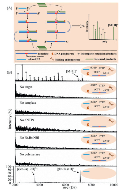
As illustrated in Fig. 1A, the EXPAR reaction is initiated through the hybridization of complementary sequence of the template with the target miRNA. Then, the DNA-RNA hybrid duplexes can be extended in the presence of Vent (exo−) DNA polymerase and deoxyribonucleotide triphosphates (dNTPs). After extension, the double-stranded DNA (dsDNA) with recognition site for the nicking enzyme Nt.BstNBI is cleaved to generate a downstream nick. The cleaved dsDNA strand is extended again from the 3′-end of the nicked site, resulting in simultaneous displacement of the synthesized strand. Since the template contains two repeat regions, the displaced strand has the same sequence as the target miRNA except for the change of uracil (U) to thymine (T) and the change of ribonucleotides to deoxyribonucleotides. The displaced strand can then hybridize with other templates, initiating new extension, cleavage, and strand displacement. Therefore, an exponential amplification of trace miRNA can be achieved. Finally, the extension products are detected by MALDI-TOF MS. Due to the sequence differences of different miRNAs, the sequences of amplicons are different as well, and hence their molecular weight. Therefore, multiplexed detection of miRNAs can be achieved by MALDI-TOF MS.
Figure 1
Figure 1.
(A) Schematic illustration of the EXPAR coupled MALDI-TOF MS for miRNAs detection. (B) EXPAR coupled MALDI-TOF MS assay for let-7a detection, and the control experiments with one reaction component missing or with only the let-7a. The concentration of let-7a was 100 nmol/L. The reaction time was 11 min. *: extension products. [M + H]+: molecular ion of the complete extension product.
To demonstrate the feasibility of the proposed method for the detection of miRNAs, let-7a was firstly selected as a mode target. The sequences of the oligonucleotides used in this work are listed in Table S 1 (Supporting information). As shown in Fig. 1B, when any one of the reaction components, i.e., target, template, dNTP, Nt.BstNBI and polymerase, was absent in the EXPAR reaction system, no signal could be detected by MALDI-TOF MS. On the contrary, when the complete EXPAR system was triggered by the target miRNA, obvious signal could be detected by MS. The molecular weights of extended oligonucleotides matched well with the peaks on MALDI-TOF MS (Table S 2 in Supporting information). The individual let-7a was also analyzed by MALDI-TOF MS, and there were only peaks of the singly and doubly charged molecular ions, which were different from the extension products. The results demonstrated that the asterisk labeled peaks on the mass spectrum in Fig. 1B were from incomplete extension instead of fragmentation of the target miRNA. To avoid the fragment peaks, we inactivated the cleavage enzyme by heating at 90 ℃ for 5 min, while the polymerase was still functional to consume the incomplete extension products. As shown in Fig. S 1 (Supporting information), after the additional step of cleavage enzyme inactivation, the MALDI-TOF mass spectrum was clean, where only the complete extension product was observed. However, the intensity of the complete extension product was decreased because it can also be consumed by the EXPAR. Furthermore, without the fragment peaks, the detection specificity would be restricted. Therefore, we performed the experiments without deactivating cleavage enzyme for better assay sensitivity and specificity. All the above results demonstrated the practicality of the proposed method for miRNA detection. Similar results were obtained for the other miRNAs, i.e., miRNA-21, miRNA-100 and miRNA-125b (Figs. S2-S 4 in Supporting information). The detailed sequences and corresponding molecular weights of extended oligonucleotides triggered by miRNA-21, miRNA-100 and miRNA-125b, respectively, are listed in Tables S3-S 5 (Supporting information).
Background amplification can normally be observed in EXPAR assays, and can lead to false positive results. Therefore, the respective EXPAR reaction time of background amplification was investigated for different target miRNAs, i.e., miRNA-21, let-7a, miRNA-100 and miRNA-125b, as shown in Fig. S 5 (Supporting information). In the absence of target miRNAs, when the reaction time was long enough, extension products can still be observed due to the background amplification. As a result, to avoid such background amplification while to achieve the best sensitivity, the optimal reaction times for the detection of miRNA-21, let-7a, miRNA-100 and miRNA-125b were 9 min, 11 min, 13 min and 13 min, respectively. Subsequent experiments were conducted with the optimized reaction times. Together with the MALDI-TOF MS analysis, the total assay time was less than 30 min.
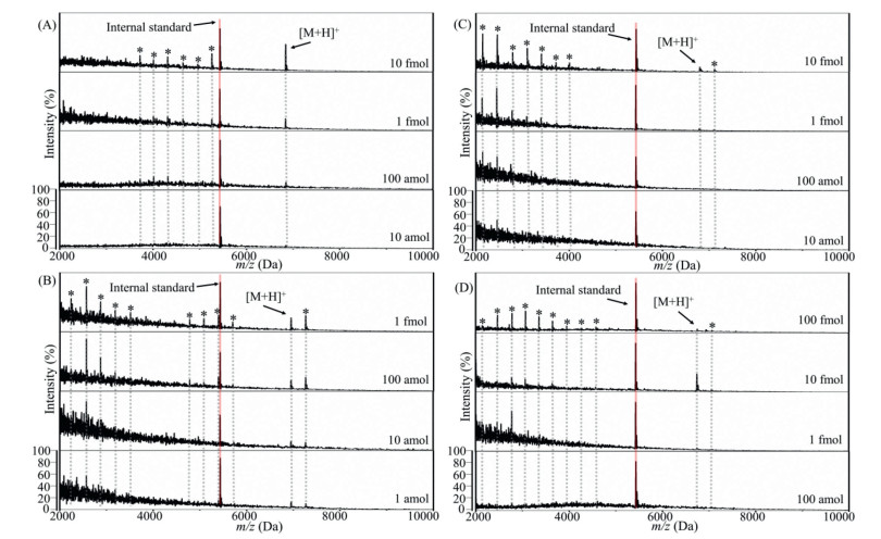
With the optimal reaction time, the detection sensitivity of the method was evaluated using 10-fold serial dilutions of different miRNAs. The mass spectra are shown in Fig. 2. A gradual increase in the peak intensity of the extension products was clearly observed with the increasing amounts of different target miRNAs. Especially for the detection of let-7a, the method achieved a high sensitivity with the detection limit as low as 1 amol sample amount (Fig. 2B). The correlation between mass spectrum peak area and the concentrations of miRNA was investigated by adding internal standard oligonucleotide. The peak area ratio between Areaex-oligo (the sum area of all detectable peaks of extension products) and Areais (the peak area of the internal standard) was well correlated with the logarithm (log) concentration of the target miRNA, as shown in Fig. S 6 (Supporting information). It was observed that the complete extension products with 100 fmol miRNA-125b as target showed a weaker peak compared to that with 10 fmol miRNA-125b as target. However, the fragment peaks with 100 fmol miRNA-125b as target were much stronger compared to those with 10 fmol miRNA-125b as target. Similar results were also observed for let-7a and miRNA-100. The results could be related to the ionization competition of samples in MALDI [38]. At high concentration, oligonucleotides with small molecular weight are more likely to interact with matrix, thereby inhibiting the ionization of the complete extension product with large molecular weight, while at low concentration both the incomplete and complete extension products could be efficiently ionized.
Figure 2
Figure 2.
The MALDI-TOF mass spectra of the EXPAR assay triggered by different amounts of (A) miRNA-21, (B) let-7a, (C) miRNA-100 and (D) miRNA-125b in 10 µL reaction mixture. The concentration of internal standard oligonucleotide was 20 nmol/L. *: extension products. [M + H]+: molecular ion of the complete extension product. The reaction was 9 min, 11 min, 13 min and 13 min for miRNA-21, let-7a, miRNA-100 and miRNA-125b, respectively.
To demonstrate the specificity of the method, the mixture of four miRNAs, including miRNA-21, let-7a, miRNA-100 and miRNA-125b, was used to initiate the EXPAR. As shown in Fig. 3A, MS signals from target miRNA extension products were obtained without any interference from other miRNAs, demonstrating the good specificity of the assay. One of the challenges in oligonucleotides analysis is to discriminate single-nucleotide differences. Accordingly, to further demonstrate the specificity of the method, members of the let-7 family (let-7a, -7b, -7c, -7e and -7i) were analyzed with the let-7a-specific template. We chose the let-7 family miRNAs based on their sequence similarity against let-7a. The EXPAR reaction is started from the 3′-end of the miRNA in the DNA-RNA duplex. As a result, the base mismatches closer to 3′-end would have more significant impact on the initiation of EXPAR. Among all the let-7 family miRNAs, it is most difficult to differentiate let-7a from let-7e, wherein the mismatched base (G) is far from the 3′-end [39]. As shown in Fig. 3B, no extension products could be detected for let-7b, -7c, -7i. In the presence of let-7e, there was indeed some extension products observed but much less intensive compared to let-7a. It is clear that the method possesses good specificity for the detection of target miRNAs.
Figure 3
Figure 3.
Specificity of the EXPAR coupled MALDI-TOF MS assay for miRNA detection. (A) MALDI-TOF mass spectra of the EXPAR assay with specific templates triggered by miRNAs mixture (miRNA-21, let-7a, miRNA-125b and miRNA-100, 100 nmol/L each). TCCCTGA, AACCCGT, TAGCTTATCAGA, TGAGGTAG. (B) MALDI-TOF mass spectra of the EXPAR assay with let-7a-specific template triggered by 1 nmol/L of let-7a, let-7b, let-7c, let-7e and let-7i, respectively. The red bases show the position(s) different from let-7a. The reaction time was 9 min, 11 min, 13 min and 13 min for miRNA-21, let-7 family miRNAs, miRNA-100 and miRNA-125b, respectively. *: extension products. [M + H]+: molecular ion of the complete extension product.
Since particular diseases can be closely related to multiple miRNAs [2, 5], it is important to detect multiple miRNAs simultaneously for applications, like clinical diagnosis. Mass spectrometry, compared to optical spectrometry and other analytical methods, has much higher resolving power and much larger peak capacity, thereby is an ideal technique for simultaneous detection of multiple targets. We further applied the proposed method to multiple miRNAs detection. Firstly, hybrid templates (miRNA-21-, let-7a-, miRNA-100- and miRNA-125b-template) were triggered by a single miRNA target. As shown in Fig. S7A (Supporting information), in the presence of target miRNA, corresponding signals of the extension products can be observed with the optimized reaction time of each target miRNA. Non-specific signals from background amplification could be observed when the reaction time was prolonged, for instance, the signals for miRNA-21 in the presence of let-7a as the target miRNA, which however did not influence the detection of let-7a at its optimal reaction time. Furthermore, the hybrid templates were triggered by a mixture of miRNAs (miRNA-21-, let-7a-, miRNA-100- and miRNA-125b), and the MS analysis was performed at 9 min, 11 min and 13 min of amplification reaction, respectively, to detect the corresponding miRNAs. As shown in Fig. S7B (Supporting information), the extension products generated from the corresponding miRNA target were also well observed. Fig. S7C (Supporting information) shows that there was no background amplification at the corresponding reaction time for the target miRNA, i.e., 9 min for miRNA-21, 11 min for let-7a, and 13 min for miRNA-100 and miRNA125b, even with the hybrid templates. All these results clearly demonstrated the feasibility of the proposed method for the simultaneous detection of multiple miRNAs.
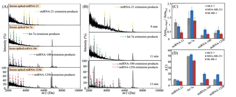
In order to assess the actual performance of the method with complex biological samples, a human serum sample was spiked with target miRNAs and used as simulated real samples. As shown in Fig. 4A, with the presence of only miRNA-21 in the spiked human serum, only MS signals of miRNA-21 extension products were observed. Similarly, in the presence of let-7a, miRNA-100 or miRNA-125b, the MS signals of the extension products triggered by the corresponding miRNA were observed, respectively. Furthermore, when all the four targets were spiked into the serum simultaneously, the amplification products could also be detected under the optimized reaction time for each target miRNA (Fig. 4B). In contrast, no signal was observed from the serum without any spiked miRNA (Fig. S 8 in Supporting information). The spiked serum sample was also analyzed by EXPAR with fluorescence detection, and did not show any nonspecific amplification of the 4 miRNAs (data not shown).
Figure 4
Figure 4.
MALDI-TOF spectra of the EXPAR assay with hybrid templates (miRNA-21-, let-7a-, miRNA-100-, and miRNA-125b-template) triggered by miRNAs extracted from (A) single miRNA-spiked serum and (B) four miRNAs-spiked serum. Relative quantification of target miRNAs extracted from breast cancer cells measured by (C) EXPAR-MALDI-TOF and (D) real-time fluorescence EXPAR. The initial concentration of miRNA in spiked serum was 1 µmol/L each, before miRNA extraction. The reaction time was 9 min, 11 min, 13 min and 13 min for miRNA-21, let-7a, miRNA-100 and miRNA-125b, respectively. Error bars represent the standard deviation of three measurements.
It is essential to detect miRNA expression levels for the early diagnosis of cancer [8, 37, 40]. To assess the performance of the method in profiling miRNA expression levels, we detected the endogenous miRNAs extracted from human breast cancer cells (MCF-7, MDA-MB-231, SK-BR-3). The detection results of miRNA-21, let-7a, miRNA-100 and miRNA-125b are shown in Fig. 4C. Relative quantification was performed using the relative peak area of the extension products. The abundances of let-7a, miRNA-100, and miRNA-125b in MDA-MB-231 were higher than those in the other two cancer cells. Meanwhile, we utilized the standard real-time fluorescence EXPAR with SYBR Green I to quantify the level of the miRNAs from the same batch of extracted samples (Fig. 4D). The results obtained by the EXPAR-MALDI-TOF MS were consistent with those by the real-time fluorescence EXPAR, demonstrating the applicability of the proposed method for profiling the expression levels of multiple miRNAs, which might be used for the differentiation of various cancer stages and cancer types. Nevertheless, it was observed that the standard deviations of three measurement by MALDI-TOF MS were larger than those by real-time fluorescence EXPAR. In MALDI-TOF MS, analytes are mixed with matrices and dried on a target plate. Due to the inherent heterogeneity of sample distribution in solid state, the ionization suffers from a stochastic element, restricting the quantification reproducibility and resulting in large standard deviation. In order to improve reproducibility and decrease standard deviation, some efforts have already been made in our work, such as homogenization of the sample and utilization of hydrophobic target plate.
In summary, we have developed an exponential amplification reaction coupled MALDI-TOF MS with the capability of detecting multiple miRNAs in one analysis. The method possesses good quantification ability, high sensitivity, with LOD at attomole level, and excellent specificity for discriminating different miRNAs with single nucleotide resolution. The assay is simple in sample preparation, and can be performed under isothermal conditions in a single reaction tube. The total assay time is less than 30 min. The multiplexed assay was successfully performed to determine miRNAs extracted from spiked serum samples and cancer cells, exhibiting good agreement with the real-time fluorescence EXPAR. Furthermore, as EXPAR employs a single primer to initiate signal-amplification reactions, and exponentially synthesizes short oligonucleotides to act as primers, it is particularly suited for the amplification of miRNAs. The synthetic oligonucleotides are typically less than 25 nucleotides, which is suitable for MALDI-TOF MS analysis. Consequently, the EXPAR coupled MALDI-TOF MS assay can be expanded to detected different miRNAs, and holds great promise in clinical diagnosis.
Declaration of competing interest
The authors declare that they have no known competing financial interests or personal relationships that could have appeared to influence the work reported in this paper
Acknowledgments
This work was supported by the National Natural Science Foundation of China (NSFC, Nos. 22022401, 22074022 and 21934001), and the Ministry of Science and Technology of China (Nos. 2020YFF0426500, 2020YFF0304502).
Supplementary materials
Supplementary material associated with this article can be found, in the online version, at doi:10.1016/j.cclet.2022.04.019.
Figure 1
(A) Schematic illustration of the EXPAR coupled MALDI-TOF MS for miRNAs detection. (B) EXPAR coupled MALDI-TOF MS assay for let-7a detection, and the control experiments with one reaction component missing or with only the let-7a. The concentration of let-7a was 100 nmol/L. The reaction time was 11 min. *: extension products. [M + H]+: molecular ion of the complete extension product.
Figure 2
The MALDI-TOF mass spectra of the EXPAR assay triggered by different amounts of (A) miRNA-21, (B) let-7a, (C) miRNA-100 and (D) miRNA-125b in 10 µL reaction mixture. The concentration of internal standard oligonucleotide was 20 nmol/L. *: extension products. [M + H]+: molecular ion of the complete extension product. The reaction was 9 min, 11 min, 13 min and 13 min for miRNA-21, let-7a, miRNA-100 and miRNA-125b, respectively.
Figure 3
Specificity of the EXPAR coupled MALDI-TOF MS assay for miRNA detection. (A) MALDI-TOF mass spectra of the EXPAR assay with specific templates triggered by miRNAs mixture (miRNA-21, let-7a, miRNA-125b and miRNA-100, 100 nmol/L each). TCCCTGA, AACCCGT, TAGCTTATCAGA, TGAGGTAG. (B) MALDI-TOF mass spectra of the EXPAR assay with let-7a-specific template triggered by 1 nmol/L of let-7a, let-7b, let-7c, let-7e and let-7i, respectively. The red bases show the position(s) different from let-7a. The reaction time was 9 min, 11 min, 13 min and 13 min for miRNA-21, let-7 family miRNAs, miRNA-100 and miRNA-125b, respectively. *: extension products. [M + H]+: molecular ion of the complete extension product.
Figure 4
MALDI-TOF spectra of the EXPAR assay with hybrid templates (miRNA-21-, let-7a-, miRNA-100-, and miRNA-125b-template) triggered by miRNAs extracted from (A) single miRNA-spiked serum and (B) four miRNAs-spiked serum. Relative quantification of target miRNAs extracted from breast cancer cells measured by (C) EXPAR-MALDI-TOF and (D) real-time fluorescence EXPAR. The initial concentration of miRNA in spiked serum was 1 µmol/L each, before miRNA extraction. The reaction time was 9 min, 11 min, 13 min and 13 min for miRNA-21, let-7a, miRNA-100 and miRNA-125b, respectively. Error bars represent the standard deviation of three measurements.

 DownLoad:


 下载:
下载:


