Figure 1.
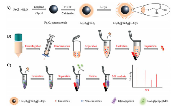
Illustration of (A) the synthesis procedure of Fe3O4@TiO2@L-Cys; (B) the urinary exosome purification process with Fe3O4@TiO2@L-Cys; and (C) the glycopeptide enrichment procedure.
Specific enrichment of urinary exosomes and exosomal glycopeptides by coefficient affinity of integrated L-cysteine and titania
Yijie Chen , Haolin Chen , Chenjie Yang , Yonglei Wu , Chunhui Deng , Nianrong Sun

It is well known that protein glycosylation is a kind of most vital post-translational modification (PTM) process [1, 2], which plays an important role in numerous biological processes, such as cell signaling [3, 4], proliferation [5], protein secretion [6] and tumor immunology [3, 7]. Abnormal glycosylation is closely related to occurrence and development of diseases, especially evolution and progression in tumor [2, 3, 7-9]. Therefore, glycosylation research is significant that it can provide much information on differences of disease-associated glycoforms, or obtain a better understanding of the disease mechanism for early diagnosis [10-13]. In addition, body fluids such as blood, serum, saliva, breast milk and urine are the easiest access for diagnostic and therapeutic purpose [6, 14-16]. Exosomes, as an important liquid biopsy target, were widely presented in various body fluids and tissues and contained many cargoes including glycoproteins [16-21]. Therefore, studies on exosomes and inclusive glycoproteins will be an effective way to monitor and analyze the physiological and pathological conditions.
However, challenges such as the complexity of biological samples and the relative low abundance of exosomes make it difficult to directly analyze exosomes and exosomal glycoproteins [22-25]. Therefore, it's necessary to conduct efficient enrichment process on a larger scale [17]. On account of the interaction between phospholipid bilayer membrane and titanium oxide, the excellent separation performance of titanium oxide towards exosomes in human urine was reported in a high-efficient and non-disruptive way [17, 26, 27]. Up to now, various strategies (hydrazide chemistry [6, 28], boronic acid chemistry [29-31], lectin affinity chromatography [32], hydrophilic interaction chromatography (HILIC) [25, 33, 34], etc.) have been developed for enrichment of glycopeptides prior to MS identification. Therein, boronic acid methods are not favorable in enriching low-abundance glycopeptides because of the weak interactions [35, 36]. Lectins have inherent specificity so that each kind of lectin can only recognize a specific glycan structure, which makes it hard to find a single lectin or lection-combination to enrich all glycopeptides [23]. Among the aforementioned methods, HILIC methods exhibit an outstanding ability in universal glycopeptide enrichment and have been the most common use [37, 38]. In this work, a novel strategy was proposed to solve the difficulties in exosome separation and exosomal glycoprotein analysis.
L-cysteine (L-Cys) is a hydrophilic amino acid with superior biocompatibility and abundant polar groups such as amino, carboxyl and thiol. According to early reports, interaction between titanium and the thiol group remains L-Cys immobilized on TiO2 [38, 39]. Considering titanium oxide was an ideal functional unit with ability to separate exosomes by binding the phosphate group of the exosomal phospholipid bilayer [40, 41], hence, we successfully integrated L-Cys and titania onto the surface of magnetic nanoparticles in a simple procedure (denoted as Fe3O4@TiO2@L-Cys), by which the coefficient affinity towards exosomes and exosomal glycoproteins was achieved. In addition to the great enrichment performance for exosomes and glycoproteins, the Fe3O4@TiO2@L-Cys displayed strongly magnetic responsiveness, which made the separation process rapid and effortless under external magnetic field. Notably, the exosome lysis was directly implemented after the exosomes were captured by Fe3O4@TiO2@L-Cys from human urine samples without extra elution process. Afterwards, the difunctional material was employed to enrich glycopeptides from these urinary exosomes based on hydrophilic interactions followed by LC-MS/MS analysis.
The synthetic process of Fe3O4@TiO2@L-Cys is manifested in Fig. 1A. Transmission electron microscopy (TEM) was applied to characterize this dual-functional material. It is clear in Fig. 2A that the boundaries of Fe3O4 were surrounded by agminated nanoparticles after modified by TiO2 and L-Cys. The diameter of Fe3O4 core was about 200 nm, and increased to approximately 300 nm after modification, which reveals the diameter of TiO2 and L-Cys shell was around 100 nm. TiO2 was utilized to form a protective shell, making this magnetic material a favorable substrate for exosome capture and further modification of hydrophilic groups. The ζ potential parameters under neutral condition were measured to monitor the synthetic process as demonstrated in Fig. 2B. It's clear that the ζ potential changed along with the progress of modification procedure. In detail, the ζ potential of Fe3O4 was recognized as 4.03 mV. After coated with TiO2 and L-Cys layer upon layer, the value gradually decreases to minus.
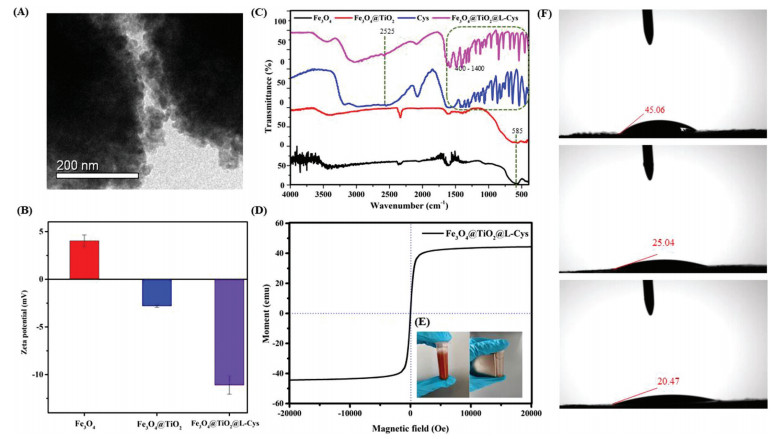
The energy dispersive X-ray (EDX) spectrum for element detection was shown in Supporting information (Fig. S1 in Supporting information), distinctly displaying that C, O, N, Fe, S and Ti elements are homogeneously distributed, implying the successful modification of TiO2 and L-cysteine. Furthermore, Fourier transform infrared (FTIR) spectroscopy was applied to characterize function groups on Fe3O4@TiO2@L-Cys. As shown in Fig. 2C, typical adsorption peak at 585 cm−1 was ascribed to Fe–O stretching vibration in FTIR spectra [42, 43]. Various peaks between 400 cm−1 and 1400 cm−1 and 2525 cm−1 vibration in fingerprint region were all attributed to the characteristic peaks in L-Cys [44]. Other broad absorption bands at 1667, 1585, 1340 cm−1 were assigned to the stretching modes of COOH, NH2 and CH groups, respectively [45]. This information further confirmed the successful modification of L-Cys. Moreover, the amount of L-Cys modified on Fe3O4@TiO2 nanomaterials was also compared as illustrated in Fig. S2 (Supporting information). The material performed best when the ratio of Fe3O4@TiO2 to L-Cys was 1:3.
The magnetic response, dispersity in aqueous solution and hydrophilicity of Fe3O4@TiO2@L-Cys were tested respectively. The results showed the saturated magnetic value of Fe3O4@TiO2@L-Cys was 44.31 emu/g (Fig. 2D), which was capable of conducting magnetic separation [18, 41]. As observed in Fig. 2E, the dispersed Fe3O4@TiO2@L-Cys can be separated from aqueous solution within 5 s using a magnet. Water contact angle test was conducted to prove the hydrophilicity of Fe3O4@TiO2@L-Cys. The water contact angel gradually decreased from 45.06° to 25.04°, and then to 20.47° after successive modification by TiO2 and L-Cys (Fig. 2F), indicating the introduction of titania shell and L-Cys can improve the hydrophilicity of Fe3O4, which will contribute to glycopeptide capture based on hydrophilic interaction. All above characterizations proved the successful synthesis and excellent properties of Fe3O4@TiO2@L-Cys. The TiO2 functionalized surface and hydrophilic exterior surface would contribute to intact exosome capture and glycopeptide enrichment.
As a proof of coefficient enrichment strategy based on Fe3O4@TiO2@L-Cys, the ability for exosome capture was demonstrated by using urine concentrate of human volunteers as sample. The urinary exosome purification process was manifested in Fig. 1B. Western blotting results confirmed the presence of three typical antigen markers of exosomes (TSG101, CD63, CD9) which were commonly expressed on the exosome surface (Fig. S3 in Supporting information), revealing the selective exosome capture ability of this difunctional material. These material-captured exosomes were lysed with the aid of ultrasound. After lysis, CD9 and TSG101 were invisible in images and the color of CD63 was also lighten, which indicated that the majority of exosomes were released from the materials after ultrasonication. These results confirmed the good capture performance of Fe3O4@TiO2@L-Cys and high release effect of direct lysis.
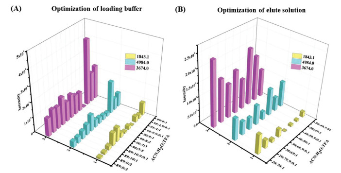
Another design of Fe3O4@TiO2@L-Cys was to analyze glycopeptides of exosomes further, and the glycopeptide enrichment procedure was shown in Fig. 1C. The capacity and specificity were primarily investigated by employing HRP as a standard glycoprotein. As shown in Fig. S2, the signals of glycopeptides were severely suppressed by abundant non-glycopeptides before enrichment when 100 fmol/µL HRP digest was used as a model sample. However, after enrichment with Fe3O4@TiO2@L-Cys, the interference was most eliminated. A total of 32 significant peaks corresponding to N-glycopeptides were identified (Figs. S4, S5 and Table S1 in Supporting information), demonstrating the excellent performance of Fe3O4@TiO2@L-Cys for enrichment of N-linked glycopeptides. Then the conditions of Fe3O4@TiO2@L-Cys for enrichment process were explored, mainly including incubation and elution condition, since it is well-known that the vital factor of HILIC-based materials in glycopeptides enrichment is the ratio of acetonitrile (ACN) to trifluoroacetic acid (TFA). Herein, different ratios of ACN and TFA in both loading buffer and eluting solution were explored for achieving the optimal performance [46-48]. After overall considering the number and intensity of identified glycopeptides, and background of interference of artifactual bands, the optimized enrichment parameters of ACN: H2O: TFA were chosen as 95:4.9:0.1 and 50:49:1 as incubating and eluting solvent, respectively (Fig. 3 and Fig. S6 in Supporting information). Furthermore, the sensitivity and selectivity of Fe3O4@TiO2@L-Cys were assessed in view of the low abundance of glycopeptides in biological samples. The results indicated the sensitivity could be as low as 0.1 fmol/µL (Fig. S7 in Supporting information), and the selectivity reached 1:100 (HRP/BSA, wt/wt, Fig. S8 in Supporting information), revealing high sensitivity and good anti-interference ability.
Next, to verify the successive effectiveness of Fe3O4@TiO2@L-Cys towards exosome capture and glycopeptide enrichment, we further applied this material to enrich glycopeptides from the aforementioned urine exosomes. A total of 146 glycopeptides corresponding to 77 glycoproteins were identified by nano-LC-MS/MS. Compared with the extraction of exosomes in other studies, separating procedure of exosomes in this work shortened the analysis time by effective binding of phosphate group on exosomes with titanium oxygen on material [18, 49]. The detailed information of identified glycopeptides was listed in Table S2 (Supporting information). To combine the glycopeptides analysis with the correlated biological function, gene ontology (GO) analysis was conducted through the database for annotation visualization, integrated discovery database (DAVID) and a web platform for scientific data visualization (Hiplot,
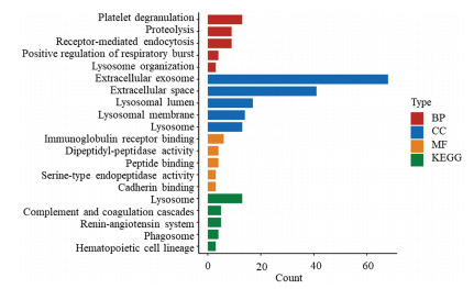
In summary, a novel dual-functional magnetic nanomaterial Fe3O4@TiO2@L-Cys constructed in a facile and convenient one-pot approach was proposed. With the utilization of titanium oxide, good capture performance towards exosomes and high release effect after direct lysis were realized. This novel material with abundant hydrophilic groups manifested high sensitivity and outstanding practical applicability in glycopeptide enrichment. A total of 146 glycopeptides corresponding to 77 glycoproteins were captured from human urine exosomes. Gene ontology and Kyoto encyclopedia of genes and genomes analysis all revealed that the identified exosomal glycoproteins exerted influence on diverse biological processes. These studies on exosome separation and glycoprotein analysis prove Fe3O4@TiO2@L-Cys to be a potent tool for exosomal glycoproteomics. However, only a small number of samples were used in this work, more samples should be conducted to provide a comprehensive understanding towards exosomes and the corresponding glycoproteome. More importantly, disease samples should be explored in future given the emerging of precise medicine area.
The authors declare no conflict of interest.
This work was supported by National Key R & D Program of China (No. 2018YFA0507501), the National Science Foundation for Distinguished Young Scholars of China (No. 21425518), the National Natural Science Foundation of China (Nos. 22074019, 22004017), and Shanghai Sailing Program (No. 20YF1405300).
Supplementary material associated with this article can be found, in the online version, at doi:
R.G. Spiro, Glycobiology 12 (2002) 43R–56R. doi: 10.1093/glycob/12.4.43R
D.F. Zielinska, F. Gnad, J.R. Wisniewski, M. Mann, Cell 141 (2010) 897–907. doi: 10.1016/j.cell.2010.04.012
K. Ohtsubo, J.D. Marth, Cell 126 (2006) 855–867. doi: 10.1016/j.cell.2006.08.019
Y. Li, J. Wang, N. Sun, C.H. Deng, Anal. Chem. 89 (2017) 11151–11158. doi: 10.1021/acs.analchem.7b03708
G. Qing, Q. Lu, Y. Xiong, et al., Adv. Mater. 29 (2017) 1604670. doi: 10.1002/adma.201604670
H Zhang, X.J. Li, D.B. Martin, R Aebersold, Nat. Biotechnol. 21 (2003) 660–666. doi: 10.1038/nbt827
M.M. Fuster, J.D. Esko, Nat. Rev. Cancer 5 (2005) 526–542. doi: 10.1038/nrc1649
L. Zhu, J. Zhao, Z. Guo, et al., Biosensors 11 (2021) 344. doi: 10.3390/bios11090344
M. Liu, L. Xi, T. Tan, et al., Chin. Chem. Lett. 32 (2021) 1726–1730. doi: 10.1016/j.cclet.2020.11.072
H.H. Freeze, E.A. Eklund, B.G. Ng, M.C. Patterson, Lancet. Neurol. 11 (2012) 453–466. doi: 10.1016/S1474-4422(12)70040-6
T. Hennet, Biochim. Biophys. Acta 1820 (2012) 1306–1317. doi: 10.1016/j.bbagen.2012.02.001
J. Yao, J. Wang, N. Sun, C. Deng, Nanoscale 9 (2017) 16024–16029. doi: 10.1039/C7NR04206J
Y. Tian, H. Zhang, Proteom. Clin. Appl. 4 (2010) 124–132. doi: 10.1002/prca.200900161
J. Roth, Chem. Rev. 102 (2002) 285–303. doi: 10.1021/cr000423j
Z. Li, C. Hu, J. Jia, et al., J. Biomed. Nanotechnol. 15 (2019) 1090–1096. doi: 10.1166/jbn.2019.2768
X. Sun, Y. Wang, K. Gu, et al., Mater. Express 11 (2021) 46–53. doi: 10.1166/mex.2021.1887
Y. Wu, N. Zhang, H. Wu, N. Sun, C. Deng, Microchim. Acta 188 (2021) 66. doi: 10.1007/s00604-021-04728-x
S. Guan, H. Yu, G. Yan, et al., J. Proteome Res. 19 (2020) 2217–2225. doi: 10.1021/acs.jproteome.9b00693
S.A. Melo, H. Sugimoto, J.T. O'Connell, et al., Cancer Cell 26 (2014) 707–721. doi: 10.1016/j.ccell.2014.09.005
S.A. Melo, L.B. Luecke, C. Kahlert, et al., Nature 523 (2015) 177–182. doi: 10.1038/nature14581
H. Zhang, Y. Lv, J. Du, et al., Anal. Chim. Acta 1098 (2020) 181–189. doi: 10.1016/j.aca.2019.11.012
J. Wang, J. Yao, N. Sun, C. Deng, J. Chromatogr. A 1512 (2017) 1–8. doi: 10.1016/j.chroma.2017.07.020
H. Xiao, W. Chen, J.M. Smeekens, R. Wu, Nat. Commun. 9 (2018) 1692. doi: 10.1038/s41467-018-04081-3
A. Chernykh, R. Kawahara, M. Thaysen-Andersen, Biochem. Soc. Trans. 49 (2021) 161–186. doi: 10.1042/BST20200222
G. Qing, J. Yan, X. He, X. Li, X. Liang, Trends Analyt. Chem. 124 (2020) 115570. doi: 10.1016/j.trac.2019.06.020
H. Dong, C. Tang, Z. He, et al., Chin. Chem. Lett. 31 (2020) 1812–1816. doi: 10.1016/j.cclet.2020.03.002
Z. Li, C. Hu, J. Jia, et al., J. Biomed. Nanotechnol. 17 (2021) 407–415. doi: 10.1166/jbn.2021.3028
J. Wohlgemuth, M. Karas, T. Eichhorn, R. Hendriks, S. Andrecht, Anal. Biochem. 395 (2009) 178–188. doi: 10.1016/j.ab.2009.08.023
S. Kong, Q. Zhang, L. Yang, et al., Anal. Chem. 93 (2021) 6682–6691. doi: 10.1021/acs.analchem.0c05482
X.M. Wang, Z.J. Hu, P.F. Guo, M.L. Chen, J.H. Wang, ACS Appl. Mater. Interfaces 12 (2020) 43273–43280. doi: 10.1021/acsami.0c12171
C. Zhang, X. Jin, L. Wang, et al., ACS Appl. Mater. Interfaces 13 (2021) 9714–9728. doi: 10.1021/acsami.0c22221
J.C. Trinidad, R. Schoepfer, A.L. Burlingame, K.F. Medzihradszky, Mol. Cell Proteomics 12 (2013) 3474–3488. doi: 10.1074/mcp.M113.030007
S. Mysling, G. Palmisano, P. Hojrup, M.T. Andersen, Anal. Chem. 82 (2010) 5598–5609. doi: 10.1021/ac100530w
P. Hagglund, J. Bunkenborg, F. Elortza, O.N. Jensen, P. Roepstorff, J. Proteome Res. 3 (2004) 556–566. doi: 10.1021/pr034112b
L. Zhang, Y. Xu, H. Yao, et al., Chemistry 15 (2009) 10158–10166. doi: 10.1002/chem.200901347
W. Chen, J.M. Smeekens, R. Wu, Mol. Cell Proteomics 13 (2014) 1563–1572. doi: 10.1074/mcp.M113.036251
Y. Wu, H. Lin, Z. Xu, et al., Anal. Chim. Acta 1096 (2020) 1–8. doi: 10.1016/j.aca.2019.11.032
N. Sun, Z. Wang, J. Wang, et al., J. Chromatogr. A 1595 (2019) 1–10. doi: 10.1016/j.chroma.2019.02.039
N. Zhang, N. Sun, C. Deng, Chem. Commun. 56 (2020) 13999–14002. doi: 10.1039/D0CC06147F
N. Zhang, X. Hu, H. Chen, C. Deng, N. Sun, Chem. Commun. 57 (2021) 6249–6252. doi: 10.1039/D1CC01530C
H. Zheng, S. Guan, X. Wang, et al., Anal. Chem. 92 (2020) 9239–9246. doi: 10.1021/acs.analchem.0c01572
H. Chen, Y. Li, H. Wu, N. Sun, C. Deng, ACS Sustain, Chem. Eng. 7 (2019) 2844–2851.
H. Chu, X. Hu, J. Yao, et al., Talanta 206 (2020) 120178. doi: 10.1016/j.talanta.2019.120178
Y. Wu, Q. Liu, C. Deng, Anal. Chim. Acta 1061 (2019) 110–121. doi: 10.1016/j.aca.2019.01.052
X. Feng, C. Deng, M. Gao, X. Zhang, Anal. Bioanal. Chem. 410 (2018) 989–998. doi: 10.1007/s00216-017-0602-5
W. Ma, L. Xu, X. Li, et al., ACS Appl. Mater. Interfaces 9 (2017) 19562–19568. doi: 10.1021/acsami.7b02853
N. Sun, J. Wang, J. Yao, C. Deng, Anal. Chem. 89 (2017) 1764–1771. doi: 10.1021/acs.analchem.6b04054
Q. Liu, Y. Xie, C. Deng, Y. Li, Talanta 175 (2017) 477–482. doi: 10.1016/j.talanta.2017.07.067
N. Zhang, N. Sun, C. Deng, Talanta 221 (2021) 121571. doi: 10.1016/j.talanta.2020.121571
Z. Xu, Y. Wu, Z. Deng, et al., Talanta 234 (2021) 122713. doi: 10.1016/j.talanta.2021.122713

Figure 1 Illustration of (A) the synthesis procedure of Fe3O4@TiO2@L-Cys; (B) the urinary exosome purification process with Fe3O4@TiO2@L-Cys; and (C) the glycopeptide enrichment procedure.

Figure 2 Characteristic of Fe3O4@TiO2@L-Cys. (A) TEM images, (B) ζ potential of Fe3O4, Fe3O4@TiO2 and Fe3O4@TiO2@L-Cys, (C) FTIR spectroscopy of Fe3O4, Fe3O4@TiO2, L-Cys and Fe3O4@TiO2@L-Cys. (D) Magnetic hysteresis loop Fe3O4@TiO2@L-Cys, (E) Fe3O4@TiO2@L-Cys dispersed in aqueous solution and separated in 5 s via a magnet and (F) contact angles of Fe3O4, Fe3O4@TiO2 and Fe3O4@TiO2@L-Cys.

Figure 3 Glycopeptide enrichment performance of Fe3O4@TiO2@L-Cys. The intensity of three typical glycopeptides enriched from HRP tryptic digests using different ratios of ACN: H2O: TFA in (A) loading buffer and (B) eluting solution.

 扫一扫看文章

扫一扫关注我们
 DownLoad:
 下载:
下载:
