Login In
Evolution of coke microcrystalline structure during calcination process of coal-based needle coke
- Corresponding author: ZHAO Xue-fei, zhao_xuefei@sohu.com
Figures(5)
Citation:
CHENG Jun-xia, ZHU Ya-ming, GAO Li-juan, ZHAO Xue-fei. Evolution of coke microcrystalline structure during calcination process of coal-based needle coke[J]. Journal of Fuel Chemistry and Technology,
;2020, 48(9): 1071-1078.

Figures(5)


TAMAS U, GUBICZA J, GABOR R, JENÖ G, CRISTIAN P. Microstructure of carbon blacks determined by X-ray diffraction profile analysis[J]. Carbon, 2002,40(6):929-937. doi: 10.1016/S0008-6223(01)00224-X
MARTINS M A, OLIVEIRA L S, FRANCE A S. Modeling and simulation of petroleum coke calcination in rotary kilns[J]. Fuel, 2001,80(11):1611-1622. doi: 10.1016/S0016-2361(01)00032-1
XIAO J, HUANG J, ZhONG Q, ZHANG H L, LI J. Modeling and simulation of petroleum coke calcination in pot calciner using two-fluid model[J]. J Metals, 2016,68(2):643-655.
KAKUTA M, TSUCHIYA N, TANAKA H, NOGUCHI K. Structural changes during graphitization of petroleum coke[J]. Carbon, 1984,22(2)237.
WALLOUCH R W, FAIR F V. Kinetics of the coke shrinkage process during calcinations[J]. Carbon, 1980,13:147-153. doi: 10.1016/0008-6223(80)90023-8
RHEDEY P J, NADKARNI S K. Coker feedstock characteristics and calcined coke properties[J]. J Metals, 2013,36(5):22-25. doi: 10.1007/BF03338449
HEINTA E A. Effect of calcination rate on petroleum coke properties[J]. Carbon, 1995,33(6):817-820. doi: 10.1016/0008-6223(95)00002-U
MITSUNAO K, YOSHIHARU O, CAI Xue-min. Structural changes during calcination of petroleum coke (Part 1)-Research on the heat treatment changes of coke's structure, organization and specific gravity[J]. Carbon Technol, 1983(2):16-20.
SACHSSE H. Encyclopaedia of Chemical Technolog[M]. NewYork:Ohn Wiley and Sons, 1990.
RAGAN S, MATSH H. Effects of calcination upon properties of needle-cokes[J]. J Mater Sci, 1983,12(18):3695-3705. doi: 10.1007/BF00540742
HU Jian-hong, Fundamental study on coal tar pitch refining and preparation of needle coke[D]. Beijing: China University of Minning & Tachnology, 2019.
ISMAGILOV Z R, SOZINOV S A, POPOVA A N, ZAPOTIN V P. Structural analysis of needle coke[J]. Coke Chem, 2019,62(4):135-142. doi: 10.3103/S1068364X19040021
SAOWADEE N, AGERSTED K, BOWEN J R. Lattice constant measurement from electron backscatter diffraction patterns[J]. J Microsc, 2017,266(2):200-210. doi: 10.1111/jmi.12529
ZENOU V Y, SNEJANA B. Microstructural analysis of undoped and moderately Sc-doped TiO2, anatase nanoparticles using Scherrer equation and Debye function analysis[J]. Mater Charact, 2018,144:287-296. doi: 10.1016/j.matchar.2018.07.022
SARKAR A, DASGUPTA K, BARAT P, MUKHERJEE P, SATHIYAMOORTHY D. Studies on neon irradiated amorphous carbon using X-ray diffraction technique[J]. Int J Mod Phys B, 2008,22(7):865-875. doi: 10.1142/S0217979208038119
BALACHANDRAN M. Study of stacking structure of amorphous carbon by X-ray diffraction technique[J]. Int J Electrochem Sci, 2012,7(4):3127-3134. doi: 10.1016/j.jpowsour.2012.01.007
MANOJ B. Investigation of nanocrystalline structure in selected carbonaceous materials[J]. Int J Min Met Mater, 2014,21(9):940-946. doi: 10.1007/s12613-014-0993-7
LIU Dong-dong, GAO Ji-hui, WU Shao-hua, QIN Yu-kun. XRD and Raman characterization of microstructure changes of char during pyrolysis[J]. J Harbin Inst Technol, 2016,48(7):39-45.
HAN W H, CAI Y X, LI X H, WANG J, WANG J, LI K, WEI X. Raman spectroscopy analysis of carbon structural evolution of diesel particulate matters with the treatment of nonthermal plasma[J]. Spectrosc Spect Anal, 2012,32(32):2152-2156.
DEWA K, ONO K, MATSUKAWA Y, TAKAHASHI K, SAITO Y, MATSUSHITA Y AOKI H, ERA K, AOKI T, YAMAGUCHI T. Determining the structure of carbon black using Raman spectroscopy and X-ray diffraction[J]. Carbon, 2017,114(4):132-138.
XIA Song-xun. Research on petroleum coke calcining process[D]. Changsha: Central South University, 2012.
CAO Jia-hui, SHEN Jun, WANG Yu-gao, LIU Gang, LI Rui-feng, XU Qing-bai. Changes in leachable content of polycyclic aromatic hydrocarbons before and after mixing petroleum pitch with coal tar pitch[J]. Petrochem Technol, 2019,48(7):702-708.
LIN D, QIU P, XIE X, ZHAO Y. Chemical structure and pyrolysis characteristics of demineralized Zhundong coal[J]. Energy Source, 2017,27:1-6. doi: 10.1080/15567036.2017.1403504
LIU Wan-yue, LIU Qin-pu, LIU Lin-song, LIU Di. Study on FT-IR features of middle and high rank coal structure in north part of Qinshui Basin[J]. Coal Sci Technol, 2019,47(2):186-192.
SAIKIA B K, BORUAH R K, GOGOI P K. FT-IR and XRD analysis of coal from Makum coalfield of Assam[J]. J Earth Syst Sci, 2007,116(6):575-579. doi: 10.1007/s12040-007-0052-0
POLITIS T G, NAZEM F F, FAN You-zhi. The calcination mechanism of needle coke[J]. Carbon Technol, 1992(3):23-24.
WHITTAKER M P, MILLER F C, FRITZ H C. Structural changes accompanying coke calcinations[J]. Ind Eng Chem Prod Res Dev, 1970,9(2):187-190. doi: 10.1021/i360034a014
KIM J D, ROH J S, KIM M S. Effect of carbonization temperature on crystalline structure and properties of isotropic pitch-based carbon fiber[J]. Carbon Lett, 2017,21:51-60. doi: 10.5714/CL.2017.21.051
SHI H, REMIMERS N, DAHN J R. Structure-refinement program for disordered carbons[J]. J Appl Crystallogr, 1993,26:827-836. doi: 10.1107/S0021889893003784
SHI H. Disordered carbons and battery applications[D]. Burnaby: Simon Frasier University, 1993.
IWASHITA N, INAGAKI M. Relations between structural parameters obtained by X-Ray powder diffraction of various carbon materials[J]. Carbon, 1993,31(7):1107-1113. doi: 10.1016/0008-6223(93)90063-G
NAKAMIZO M, KAMMERECK R, WALKER P L. Laser raman studies on carbons[J]. Carbon, 1974,12(3):259-267. doi: 10.1016/0008-6223(74)90068-2
GUPTA A K, RUISSIN T J, GUTIE H R, EKLUND P C. Probing grapheneedgesr, viar, Raman scattering[J]. ACS Nano, 2009,3(1):45-52.
CHEN Shi, SHI Yan-ping, WU Qi-lin. Microstructual evolution during preoxidation, carbonization and graphitization of PAN fiber[J]. New Chem Mater, 2016,44(5):121-123.
GRAF D, MOLITOR F, ENSSLIN K. Spatially resolved Raman spectroscopy of single -and Few-Layer Graphene[J]. Nano Lett, 2007,7(2):238-242. doi: 10.1021/nl061702a
FITZER E. Some remarks on Raman spectroscopy of carbon structures[J]. High Temp High Press, 1988,20:449-454.
HUA Zhong, WANG Yue-mei, XIAO Li, QIN Zheng-kun, FAN Wen-ji. Relations beltween hybrid C-C bond length and structure parameters of PAN-based carbon fibers[J]. New Chem Mater, 2005,20(3):274-277.
SHI Yan-ping. Raman spectroscopy to study the microstructure and properties of carbon fiber[D]. Shanghai: Donghua University, 2011.
KO T H. Raman spectrum of modified PAN-based carbon fibers during graphitization[J]. J Appl Polym Sci, 1996,59(4):577-580. doi: 10.1002/(SICI)1097-4628(19960124)59:4<577::AID-APP2>3.0.CO;2-Q
BU H, ZHAO M, WANG A. WANG X First-principles prediction of the transition from graphdiyne to a superlattice of carbon nanotubes and graphenenanoribbons[J]. Carbon, 2013,65:341-348. doi: 10.1016/j.carbon.2013.08.035
Yongchao ZHU , Wenjie LIANG , Hai XU . Raman spectroscopic layer-dependent of Bi2SeO5 nanosheets and their encapsulation performance for two-dimensional materials. Chinese Journal of Inorganic Chemistry, 2026, 42(3): 584-592. doi: 10.11862/CJIC.20250217
Jing Wang , Xue Qi , Yan Li , Zhihui Wen , Zhanheng Feng , Xiaohang Qiu . 散射光中的隐秘指纹——拉曼光谱. University Chemistry, 2026, 41(4): 280-288. doi: 10.12461/PKU.DXHX202507031
Danqing Wu , Jiajun Liu , Tianyu Li , Dazhen Xu , Zhiwei Miao . Research Progress on the Simultaneous Construction of C—O and C—X Bonds via 1,2-Difunctionalization of Olefins through Radical Pathways. University Chemistry, 2024, 39(11): 146-157. doi: 10.12461/PKU.DXHX202403087
Lu Zhuoran , Li Shengkai , Lu Yuxuan , Wang Shuangyin , Zou Yuqin . Cleavage of C―C Bonds for Biomass Upgrading on Transition Metal Electrocatalysts. Acta Physico-Chimica Sinica, 2024, 40(4): 2306003-0. doi: 10.3866/PKU.WHXB202306003
Ce Liang , Qiuhui Sun , Adel Al-Salihy , Mengxin Chen , Ping Xu . Recent advances in crystal phase induced surface-enhanced Raman scattering. Chinese Chemical Letters, 2024, 35(9): 109306-. doi: 10.1016/j.cclet.2023.109306
Haixia Wu , Kailu Guo . Sulfur reduction reaction mechanism elucidated with in situ Raman spectroscopy. Chinese Chemical Letters, 2025, 36(6): 110654-. doi: 10.1016/j.cclet.2024.110654
Ying Wang , Mingcheng Yang , Zhu Yin , Yingqi Wang , Jiajia Cheng . Transition metal-free poly(heptazine imide) photocatalyst for C-X bond construction from katritzky salts. Acta Physico-Chimica Sinica, 2026, 42(7): 100212-. doi: 10.1016/j.actphy.2025.100212
Junyou Ding , Xiaotong Li , Hongmin Lin , Bochao Ye , Xing Zhou , Feihu Cui , Yingming Pan , Haitao Tang . Highly regioselective hydrogermylation of unsaturated C-C bonds over ligand-control single atom palladium catalysts. Chinese Chemical Letters, 2025, 36(11): 111286-. doi: 10.1016/j.cclet.2025.111286
Shihao Tan , Caiyun Cui , Shuwei Ma , Liangsen Zhu , Xianguo Liu . Introducing nanocrystalline/amorphous heterostructures on laminated FeSiBCr to synchronously enhance absorption, expand absorption bandwidth and reduce matching thickness. Acta Physico-Chimica Sinica, 2026, 42(7): 100283-0. doi: 10.1016/j.actphy.2026.100283
Chengde Wang , Liping Huang , Shanshan Wang , Lihao Wu , Yi Wang , Jun Dong . A distinction of gliomas at cellular and tissue level by surface-enhanced Raman scattering spectroscopy. Chinese Chemical Letters, 2024, 35(5): 109383-. doi: 10.1016/j.cclet.2023.109383
Huihui LIU , Baichuan ZHAO , Chuanhui WANG , Zhi WANG , Congyun ZHANG . Green synthesis of MIL-101/Au composite particles and their sensitivity to Raman detection of thiram. Chinese Journal of Inorganic Chemistry, 2024, 40(10): 2021-2030. doi: 10.11862/CJIC.20240059
Shu Tian , Wenxin Huang , Junrui Hu , Huiling Wang , Zhipeng Zhang , Liying Xu , Junrong Li , Yao Sun . Exploring the frontiers of plant health: Harnessing NIR fluorescence and surface-enhanced Raman scattering modalities for innovative detection. Chinese Chemical Letters, 2025, 36(3): 110336-. doi: 10.1016/j.cclet.2024.110336
Hao Li , Hanzhi Lu , Linlin Hu , Xueli Zhang , Hua Shao , Fulun Li , Yanfei Shen . Dynamic surface-enhanced Raman spectroscopy-based metabolic profiling: A novel pathway to overcoming antifungal resistance. Chinese Chemical Letters, 2025, 36(7): 110342-. doi: 10.1016/j.cclet.2024.110342
Jiayin Li , Linan Yang , Feng Shi , Yan Long , Yuru Wang , Daniel D. Stuart , Hongbo Li , Quan Cheng , Lingfeng Min , Zhanjun Yang , Juan Li . Versatile Au nanozyme Raman probe strategy for ultrasensitive encoded photonic crystal-based SERS multiplex immunosensing. Chinese Chemical Letters, 2025, 36(11): 110883-. doi: 10.1016/j.cclet.2025.110883
Feiyan Ma , Haomiao Dou , Danni Luo , Ying Yan , Jie Zhou , Guangda Xu , Yue Wang , Longshan Zhao . Recent advances in chiral recognition based on surface-enhanced Raman scattering spectroscopy. Chinese Chemical Letters, 2026, 37(4): 111632-. doi: 10.1016/j.cclet.2025.111632
Ying-Qian Zhou , Chan-Ying Yao , Ze-Yu Ruan , Bang-Heng Lyu , Shan-Nan Du , Si-Guo Wu , Yan-Cong Chen , Wei Deng , Jun-Liang Liu , Ming-Liang Tong . Proton control of Raman relaxation in bis-hydrazone single-molecule magnets. Chinese Chemical Letters, 2026, 37(8): 111179-. doi: 10.1016/j.cclet.2025.111179
Kexin Yan , Zhaoqi Ye , Lingtao Kong , He Li , Xue Yang , Yahong Zhang , Hongbin Zhang , Yi Tang . Seed-Induced Synthesis of Disc-Cluster Zeolite L Mesocrystals with Ultrashort c-Axis: Morphology Control, Decoupled Mechanism, and Enhanced Adsorption. Acta Physico-Chimica Sinica, 2024, 40(9): 2308019-0. doi: 10.3866/PKU.WHXB202308019
Chunhui Zhang , Jie Wang , Jieyang Zhan , Runmin Yang , Guanggang Gao , Jiayuan Zhang , Linlin Fan , Mengqi Wang , Hong Liu . Highly sensitive hydrazine detection through a novel Raman scattering quenching mechanism enabled by a crystalline and noble metal–free polyoxometalate substrate. Chinese Chemical Letters, 2025, 36(3): 109719-. doi: 10.1016/j.cclet.2024.109719
Huihui LIU , Baichuan ZHAO , Tingting ZHANG , Chuanhui WANG , Zhi WANG , Congyun ZHANG . High-sensitivity surface-enhanced Raman scattering detection and self-cleaning performance of organic pollutants based on a filter membrane sandwich structure. Chinese Journal of Inorganic Chemistry, 2026, 42(6): 1299-1311. doi: 10.11862/CJIC.20260362
Jing Yan , Zenan Zhang , Dongwei Ma , Xinyi Zhang , Zhuodong Ye , Xuefang Chen . Melamine-assisted topotactic transformation of MOFs into needle-like α-MoC/β-Mo2C for high-performance electromagnetic wave absorption and corrosion resistance. Acta Physico-Chimica Sinica, 2026, 42(9): 100328-0. doi: 10.1016/j.actphy.2026.100328
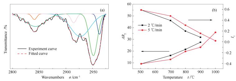
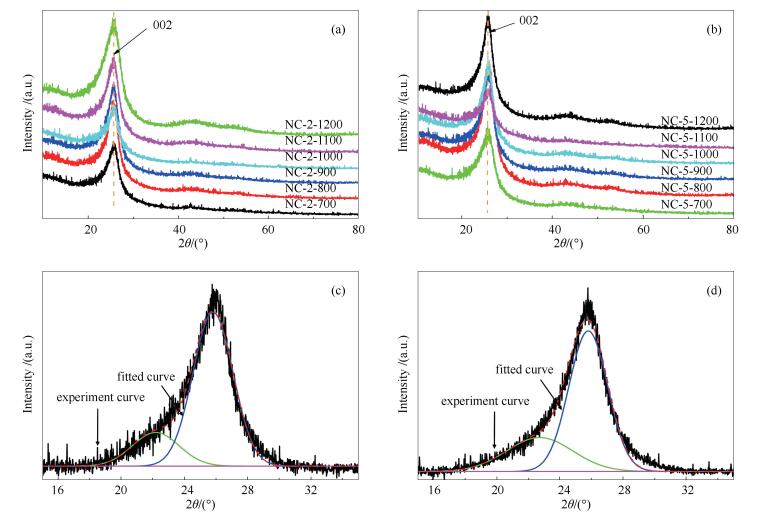
(a): 2 ℃/min; (b): 5 ℃/min; (c): NC-2-700 002 peak-fitting spectra; (d): NC-5-700 002 peak-fitting spectra
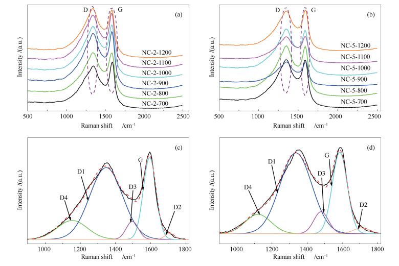
(a): 2 ℃/min; (b): 5 ℃/min; (c): curve-fitted Raman spectra of NC-2-700; (d): curve-fitted Raman spectra of NC-5-700
 DownLoad: