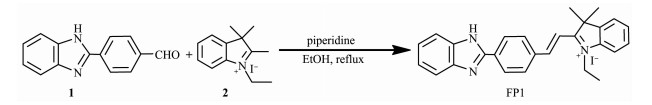
Scheme 1.
Synthesis of probe FP1

As an effective preservative, bisulfite has been widely added to foods, beverages and pharmaceutical products to prevent oxidation, browning, and microbial growth during production and storage[1-2]. It's also applied in the field of paper industry and water treatment[3]. However, bisulfite can be toxic in high doses, and extensive intake of it would cause asthmatic attacks and allergic reactions[4-5]. Due to the potential health issues of people, many countries have strictly controlled the threshold levels of bisulfite. For example, the total content of bisulfite (calculated by SO2) in granulated sugar is rigorously regulated as < 30 mg·kg-1 in China[6]. Thus, the development of conve-nient and rapid methods for sensitive and selective detection of bisulfite is of vital importance in the environment and in vivo.
At present, various traditional approaches for detecting bisulfite include flow injection analysis, electrochemistry, chromatography and titrimetric analysis[7-8]. But most of these methods are time-consuming and require expensive instruments, multiple reagents and complicated procedures. In contrast, fluorescent probe techniques have drawn much attention because of their simplicity, visualization, fast response, real-time monitoring and high sensitivity and selectivity[9-10]. On the basis of the nucleophilic reaction with aldehyde, selective deprotection of levulinate, coordinative interactions and Michael-type additions, a number of fluorescent probes for the detection of HSO3- have been reported[11-17]. Neverthel-ess, some of them have deficiencies such as incom-plete autofluorescence of the probes, slow response time and interference from cysteine, homocysteine, and hydrogen sulfide, which restrict their utility[18-19]. Hence, it's urgently needed to construct new practical fluorescent probes for the rapid detection of HSO3-.
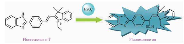
The benzimidazole group has been used as a platform for the design of fluorescent probe to detect metal ions, anions and small inorganic molecules[20]. Besides, the fluorophores linked to aromatic hetero-cycle by C=C double bond was often requisitioned as Michael addition receptor[21-22]. With this in mind, a novel turn-on benzimidazole-derived fluorescent probe FP1 for detecting HSO3- was reported based on a nucleophilic addition reaction. The benzimidazole moiety in probe FP1 was used as a fluorophore, and an unsaturated C=C double bond was incorporated as the binding site for HSO3-. Upon the addition of HSO3-, probe FP1 exhibited high selectivity and sensitivity as well as fast response. Moreover, it can be applied to detect HSO3- in sugar samples and living cells.
All reagents and solvents were purchased from commercial sources and directly used unless stated otherwise. The melting point was determined with an XT4A micromelting point apparatus and was uncorrected. All pH measurements were carried out with a PHS-3C digital pH-meter. Elemental analyses were carried out on a Perkin-Elmer 2400 instrument. Fluorescence spectra were performed on a FluoroMax-P spectrofluorimeter. 1H NMR and 13C NMR spectra were recorded on a Bruker Avance 400 spectrometer with TMS as an internal standard. Electrospray ionization mass spectra (ESI-MS) were acquired on a LCQ Fleet ion trap mass spectrometer. A Leica DMI 3000B fluorescent inverted microscope and HeLa cells were used in the live-cell imaging experiments.
Compounds 1 and 2 were prepared according to the literature methods[23-24]. As demonstrated in Scheme 1, probe FP1 was synthesized through a single step.
Compounds 1 (0.22 g, 1 mmol), 2 (0.30 g, 1 mmol) and catalytic amount piperidine (2~3 drops) were dissolved in ethanol (30 mL), then the reaction mixture was stirred under reflux for 6 h. After reaction completion (by TLC monitoring), the solvent was removed under reduced pressure, and the residue was purified by silica gel column chromatography using dichloromethane as eluent to obtain a red-brown solid. Yield:65%; m.p. 232~234℃. 1H NMR (400 MHz, DMSO-d6):δ 12.15 (s, 1H), 8.49 (d, J=8.4 Hz, 1H), 8.22 (d, J=15.8Hz, 1H), 8.05 (t, J=7.8 Hz, 1H), 7.73~7.69 (m, 4H), 7.54~7.46 (m, 3H), 7.38~7.32 (m, 3H), 7.12 (d, J=15.8Hz, 1H), 3.84 (q, J=7.2 Hz, 2H), 1.73(s, 6H), 1.58 (t, J=7.2 Hz, 3H). 13C NMR (100 MHz, DMSO-d6):δ 171.23, 153.45, 142.86, 142.35, 141.91, 138.44, 136.78, 133.57, 128.96, 127.42, 127.08, 126.82, 124.53, 211.49, 117.74, 116.61, 114.32, 55.63, 41.52, 33.87, 26.72. ESI-MS m/z:392.24[M]+. Anal. Calcd. for C27H26N3I(%):C, 62.43; H, 5.05; N, 8.09. Found(%):C, 62.14; H, 4.96; N, 8.32.
A stock solution of probe FP1 (1 mmol·L-1) was dissolved in ethanol. Deionized water was applied to prepare the solution of different analytes (F-, Cl-, Br-, I-, S2-, HS-, SO42-, HSO4-, HSO3-, SO32-, SCN-, CO32-, HCO3-, ClO-, NO3-, NO2-, Ac-, Cys, Hcy, glutathione(GSH)) with a concentration of 1 mmol·L-1. Test solutions were prepared by placing 30 μL of FP1 stock solution and an appropriate aliquot of analyte in a quartz cuvette (path length=1 cm), the resulting solution was diluted to 3 mL using the EtOH/Tris-HCl buffer solution (1:9, V/V, pH 7.4). After 2 min of mixing, the fluorescence spectra were recorded at room temp-erature (λex=292 nm, λem=484 nm, slit:10 nm/10 nm).
Crystal sugar, granulated sugar, and soft sugar were purchased from a supermarket and used in the sample test. Sample solution was prepared by dissolving 5 g of each sugar in deionized water and diluting to 10 mL. Aliquots of the sugar solution were added directly to the EtOH/Tris-HCl buffer solution (1:9, V/V, pH 7.4) containing probe FP1 (10 μmol·L-1), and the emission intensity at 484 nm was measured after 2 min. The sugar samples were then spiked with various concentrations of NaHSO3 (2.00, 4.00 and 6.00 μmol·L-1) that had been accurately prepared. The resulting samples were further treated with probe FP1 (10 μmol·L-1) for 2 min, and the emission intensities at 484 nm was recorded.
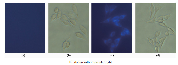
HeLa cells were cultured in Dulbecco's Modified Eagle's Medium (DMEM) supplemented with 10% Fetal Bovine Serum (FBS), 1% penicillin and streptomycin in a humid atmosphere of 5%(V/V) CO2 and 95%(V/V) air at 37℃, and then were seeded in a 24-well plate for 24 h. For living cells imaging experiments, cells were incubated with probe FP1 (5 μmol·L-1) for 30 min, and washed three times with pre-warmed PBS buffer. In a further experiment, HSO3- (20 μmol·L-1) was added to the FP1 cultured HeLa cells and incubated for 30 min. After washing cells with pre-warmed PBS buffer, cells were imaged by inverted fluorescence microscopy. Excitation with ultraviolet light (330~380 nm) was used for fluorescent imaging.
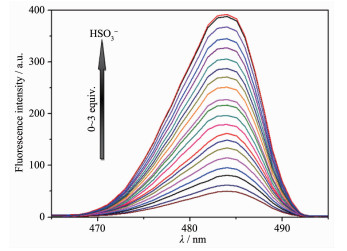
The spectral responses of probe FP1 (10 μmol· L-1) towards HSO3- were measured in EtOH/Tris-HCl buffer solution (1:9, V/V, pH 7.4). Upon addition of HSO3- (0~3 equiv.), the absorption peak of FP1 at 412 nm gradually disappeared, and another absorption peak at 290 nm increased progressively (Fig.S1). It's indicated that a new substance was produced in the reaction of FP1 with HSO3-. The fluorescence emission of FP1 was recorded with excitation at 292 and 415 nm according to the absorption. For the 292 nm excitation, free FP1 (10 μmol·L-1) displayed weak fluorescence, and its fluorescence quantum yield (Φ) was determined to be 0.08. With the addition of varying concentrations of HSO3-, the fluorescence emission intensity of FP1 at 484 nm gradually increased, and achieved saturation(Φ=0.52) after the addition of about 3 equiv. of HSO3-, which could be attributed to the recovered fluorescence of 2-phenyl benzimidazole moiety (Fig. 1). In particular, an excellent linear relationship (linear correlation coefficient R2=0.9892) between the fluorescence intensity of FP1 and the concentration of HSO3- in a range of 1.2~8.1 μmol·L-1 was observed (Fig. 2). The detection limit for HSO3- was estimated to be 0.14 μmol·L-1 according to signal to noise ratio (S/N=3), which is lower than some reported fluorescence probes for HSO3- (Table S1).
λex=292 nm, cHSO3-=0~30 μmol·L-1
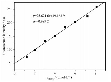
cHSO3-=1.2~8.1 μmol·L-1
As depicted in Fig.S2, when FP1 was excited at 415 nm, a gradual decrease in the maximum emission intensity at 578 nm was observed with the addition of HSO3- (0~3 equiv.), suggesting the π-π conjugation of FP1 was interrupted due to the nucleophilic attack of HSO3-(Fig.S2). However, FP1 did not displayed a ratiometric response to HSO3- under the same excitation wavelength because of the huge fluorescence intensity difference between 484 and 578 nm. The results showed that FP1 was a typical donor-acceptor skeleton with a long wavelength and weak emission properties, which was similar with a semi-cyanine fluorescent probe reported before[25]. So FP1 was more suitable as a turn-on fluorescent probe to detect HSO3- with good sensitivity.
It's significant that a probe is highly selective to a specific analyte. In order to investigate the selectivity of probe FP1 (10 μmol·L-1), its fluorescence responses to 3 equiv. of different analytes (F-, Cl-, Br-, I-, S2-, HS-, SO42-, HSO4-, HSO3-, SO32-, SCN-, CO32-, HCO3-, ClO-, NO3-, NO2-, Ac-, Cys, Hcy, GSH) were measured. As shown in Fig. 3, only addition of HSO3-to the solution of FP1 led to a remarkable fluore-scence enhancement at 484 nm, while other analytes caused negligible effects on the fluorescence intensity. Moreover, a competition experiment was also carried out to confirm the selective recognition of HSO3- by FP1 (Fig. 4). It's found that FP1 (10 μmol·L-1) showed a selective signaling behavior toward 3 equiv. of HSO3- in the presence of 30 equiv. of other competitive species. Obviously, the coexistent interferents had little influence on the detection of HSO3-, which made it feasible for practical applications. These results indicated that FP1 can detect HSO3- with good selectivity and strong anti-interference ability.
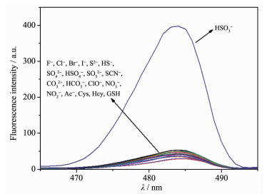
cFP1=10 μmol·L-1, canalyte=30 μmol·L-1
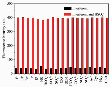
cFP1=10 μmol·L-1, cinterferent=300 μmol·L-1
The time-dependent fluorescent response of FP1 to HSO3-was evaluated at room temperature (Fig. 5). Upon addition of HSO3-, the fluorescence intensity of FP1 enhanced quickly and nearly reached a plateau within 2 min, indicating that FP1 is a fast response probe to HSO3-. As illustrated in the inset photograph shown in Fig. 5, under UV light irradiation at 365 nm, a distinct fluorescence color change (from colorless to blue) can be easily observed after the addition of HSO3- to a solution of FP1. This off-on fluorescence response together with an obvious color change enabled FP1 to visually determine HSO3-.
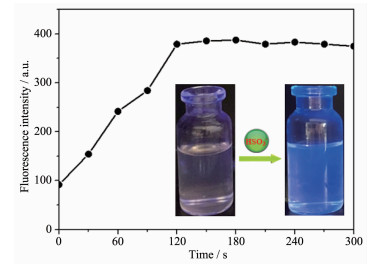
cFP1=10 μmol·L-1, c=30 μmol·L-1; Inset: fluorescence color of FP1 before (left) and after (right) addition of HSO3- under UV light of 365 nm
The influence of pH on probe FP1 was shown in Fig. 6. FP1 itself was stable over a wide pH range of 2~12. When FP1 was incubated with HSO3-, the fluorescence intensity enhanced markedly and remained stable with increasing pH value from 6 to 9. Hence, FP1 can be suitable for detecting HSO3- in the physiological pH range.
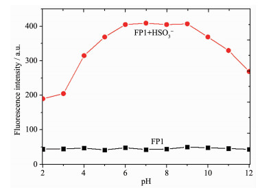
cFP1=10 μmol·L-1
It was reported that HSO3- could add rapidly to α, β-unsaturated compounds[26]. As shown in Fig.S3, after addition of HSO3- into the solution of FP1, the prominent peak at m/z 472.26 corresponding to the addition product[M+SO3]- was clearly observed in ESI-MS spectra. The result suggested that the reaction mechanism was the same as that previously reported (Scheme 2)[27-29]. The Michael addition of HSO3- toward the double bond in probe FP1 would occur owing to the strong nucleophilic ability of HSO3-, which leads to the dramatic fluorescent enhancement.
To investigate the practical utility of probe FP1 to real samples, the levels of HSO3- in granulated sugar, soft sugar, and crystal sugar were determined by using a standard addition method[30]. These sugar samples were directly examined first and further spiked with HSO3- at various concentrations (2.00, 4.00 and 6.00 μmol·L-1), and the fluorescence responses of probe FP1 at 484 nm were measured. As shown in Table 1, the relative standard deviations (RSD) of the test results were all less than 5% for these bisulfite spiked samples, and satisfactory recoveries (95.29%~103.51%) were also observed, which suggested the accuracy and reliability of the proposed method for bisulfite determination. It's indicated that probe FP1 can be applied for the quantitative detection of bisulfite in real samples.
 下载:
导出CSV
下载:
导出CSV
| Sample | HSO3-level/(μmol·L-1) | Added/(μmol·L-1) | Found/(μmol·L-1) | Recovery/% | RSD/% |
| Granulated sugar | 3.12 | 2.00 | 5.02 | 98.05 | 4.23 |
| 4.00 | 7.37 | 103.51 | |||
| 6.00 | 8.69 | 95.29 | |||
| Soft sugar | 2.63 | 2.00 | 4.75 | 102.59 | 3.16 |
| 4.00 | 6.51 | 98.19 | |||
| 6.00 | 8.33 | 96.52 | |||
| Crystal sugar | 1.78 | 2.00 | 3.69 | 97.62 | 1.95 |
| 4.00 | 5.71 | 98.79 | |||
| 6.00 | 7.89 | 101.41 |
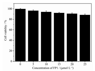
To evaluate the capability of probe FP1 for imaging HSO3- in living cells, the HeLa cells were chosen as a bioassay model. The cytotoxicity of FP1 (0, 5, 10, 15, 20, 25 μmol·L-1) was initially examined by MTT assay (Fig. 7). It's observed that the cell viability was more than 85% even after the concentration of FP1 was up to 25 μmol·L-1 for 24 h, indicating low cytotoxicity and good biocompatibility of FP1. Hence, FP1 could be suitable for the biological applications.
The fluorescence imaging experiments of probe FP1 with and without HSO3- were subsequently performed in living HeLa cells. When HeLa cells were incubated with FP1 (5 μmol·L-1) for 30 min at 37℃, almost no intracellular fluorescence was observed (Fig. 8a). However, HeLa cells exhibited obvious blue fluorescence when the above cells treated with FP1 were further incubated with 20 μmol·L-1 HSO3- for 30 min (Fig. 8c). The significant enhancement observed in intracellular fluorescence was probably a result of the specific response of FP1 to HSO3-. Further bright-field measurements demonstrated that the cells were viable throughout the imaging experiments (Fig. 8b, 8d). Overall, these results showed that FP1 can be applied to imaging HSO3- in living cells.
In summary, we have developed a benzimidazole-derived fluorescent probe FP1 for detection of HSO3-. FP1 has good sensitivity and selectivity to HSO3- with a low detection limit (0.14 μmol·L-1), and it displays a dramatic fluorescent change and a color change on addition of HSO3- within a short response time (2 min). Moreover, FP1 has been successfully applied for the detection of HSO3-in real sugar samples and in living HeLa cells.
Supporting information is available at http://www.wjhxxb.cn
Taylor S L, Higley N A, Bush R K. Adv. Food Res., 1986, 30:1-76 doi: 10.1016/S0065-2628(08)60347-X
Li M X, Feng W Y, Zhang H Y, et al. Sens. Actuators B, 2017, 243:51-58 doi: 10.1016/j.snb.2016.11.132
Giles M A, Danell R. Water Res., 1983, 17:667-676 doi: 10.1016/0043-1354(83)90236-1
Chao J B, Wang X L, Liu Y M, et al. Sens. Actuators B, 2018, 272:195-202 doi: 10.1016/j.snb.2018.05.058
Vally H, Misso N L, Madan V. Clin. Exp. Allergy, 2009, 39:1643-1651 doi: 10.1111/j.1365-2222.2009.03362.x
Sun Y, Zhao D, Fan S W, et al. J. Agric. Food Chem., 2014, 62:3405-3409 doi: 10.1021/jf5004539
Wu W L, Ma H L, Huang M F, et al. Sens. Actuators B, 2017, 241:239-244 doi: 10.1016/j.snb.2016.10.028
陈邦, 王少静, 宋战科, 等.无机化学学报, 2017, 33:1722-1730 doi: 10.11862/CJIC.2017.227CHEN Bang, WANG Shao-Jing, SONG Zhan-Ke, et al. Chinese J. Inorg. Chem., 2017, 33:1722-1730 doi: 10.11862/CJIC.2017.227
Fernandez A, Vendrell M. Chem. Soc. Rev., 2016, 45:1182-1196 doi: 10.1039/C5CS00567A
刘梦琪, 汪欢, 郭昊冉, 等.无机化学学报, 2019, 35:923-929 doi: 10.11862/CJIC.2019.090LIU Qi-Meng, WANG Huan, GUO Hao-Ran, et al. Chinese J. Inorg. Chem., 2019, 35:923-929 doi: 10.11862/CJIC.2019.090
Chen S, Hou P, Wang J, et al. RSC Adv., 2012, 2:10869-10873 doi: 10.1039/c2ra21471g
李东钰, 李照, 杨兴斌, 等.中国科学:化学, 2018, 48:45-57 http://www.cnki.com.cn/Article/CJFDTotal-JBXK201801005.htmLI Dong-Yu, LI Zhao, YANG Xing-Bin, et al. Sci. China Chem., 2018, 48:45-57 http://www.cnki.com.cn/Article/CJFDTotal-JBXK201801005.htm
Song G J, Luo J, Xing X J, et al. New J. Chem., 2018, 42:3063-3068 doi: 10.1039/C7NJ04021K
Wang C, Feng S, Wu L, et al. Sens. Actuators B, 2014, 190:792-799 doi: 10.1016/j.snb.2013.09.045
Sun Y, Li Y, Ma X T, et al. RSC Adv., 2016, 6:79830-79835 doi: 10.1039/C6RA15605C
Luo J, Song G J, Xing X J, et al. New J. Chem., 2017, 41:3986-3990 doi: 10.1039/C7NJ00041C
Li K, Li L L, Zhou Q, et al. Coord. Chem. Rev., 2019, 388:310-333 doi: 10.1016/j.ccr.2019.03.001
Li X, Jin D, Du Y C, et al. Anal. Methods, 2018, 10:4695-4701 doi: 10.1039/C8AY01556B
Xu J C, Yuan H Q, Zeng L T, et al. Chin. Chem. Lett., 2018, 29:1456-1464 doi: 10.1016/j.cclet.2018.08.012
吴彦城, 尤嘉宜, 关丽涛, 等.有机化学, 2015, 35:2465-2486 http://www.cqvip.com/QK/93463X/201512/667668322.htmlWU Yan-Cheng, YOU Jia-Yi, GUAN Li-Tao, et al. Chinese J. Org. Chem., 2015, 35:2465-2486 http://www.cqvip.com/QK/93463X/201512/667668322.html
Dai X, Zhang T, Du Z F, et al. Anal. Chim. Acta, 2015, 888:138-145 doi: 10.1016/j.aca.2015.07.026
Zhang Y, Guan L, Yu H, et al. Anal. Chem., 2016, 88:4426-4431 doi: 10.1021/acs.analchem.6b00061
Wang G, Qi H P, Yang X F. Luminescence, 2013, 28:97-101 doi: 10.1002/bio.2344
Yu L, Wang Q L, Li T T, et al. J. Chem. Res., 2012, 11:632-634
Guo X, Zhu W J, Wei X R, et al. Anal. Methods, 2018, 10:3872-3877 doi: 10.1039/C8AY01543K
Gomez M, Perez E G, Arancibia V, et al. Sens. Actuators B, 2017, 238:578-587 doi: 10.1016/j.snb.2016.07.107
Bi K Y, Tan R, Hao R T, et al. Chin. Chem. Lett., 2019, 30:545-548 doi: 10.1016/j.cclet.2018.11.020
Lan J S, Zeng R F, Ding Y, et al. Sens. Actuators B, 2018, 268:328-337 doi: 10.1016/j.snb.2018.04.047
Zhang D S, Liu A K, Ji R X, et al. Anal. Chim. Acta, 2019, 1055:133-139 doi: 10.1016/j.aca.2018.12.042
Zhang Q, Zhang Y, Ding S S, et al. Sens. Actuators B, 2015, 211:377-384 doi: 10.1016/j.snb.2015.01.122

图 1 Emission intensity of FP1 (10 μmol·L-1) changes with increasing concentrations of HSO3-
λex=292 nm, cHSO3-=0~30 μmol·L-1

图 2 Fluorescence intensity of FP1 as a function of concentration of HSO3-
cHSO3-=1.2~8.1 μmol·L-1

图 3 Fluorescent response of FP1 to different analytes
cFP1=10 μmol·L-1, canalyte=30 μmol·L-1

图 4 Effect of HSO3- on FP1 in the presence of different interferents
cFP1=10 μmol·L-1, cinterferent=300 μmol·L-1

图 5 Time-dependent fluorescence intensity changes of FP1 in the presence of HSO3-
cFP1=10 μmol·L-1, c=30 μmol·L-1; Inset: fluorescence color of FP1 before (left) and after (right) addition of HSO3- under UV light of 365 nm

图 6 Fluorescence intensity of FP1 and FP1+HSO3- over a pH range from 2 to 12 at room temperature
cFP1=10 μmol·L-1

图 8 Fluorescence imaging of HSO3- in HeLa cells by probe FP1: (a) HeLa cells incubated with 5 μmol·L-1 FP1 for 30 min at 37 ℃; (b) Bright-field images of HeLa cells from (a); (c) HeLa cells from (a) after treatment with 20 μmol·L-1 HSO3- for 30 min; (d) Bright-field images of HeLa cells from (c)
表 1 Determination of HSO3- in various sugar samples
| Sample | HSO3-level/(μmol·L-1) | Added/(μmol·L-1) | Found/(μmol·L-1) | Recovery/% | RSD/% |
| Granulated sugar | 3.12 | 2.00 | 5.02 | 98.05 | 4.23 |
| 4.00 | 7.37 | 103.51 | |||
| 6.00 | 8.69 | 95.29 | |||
| Soft sugar | 2.63 | 2.00 | 4.75 | 102.59 | 3.16 |
| 4.00 | 6.51 | 98.19 | |||
| 6.00 | 8.33 | 96.52 | |||
| Crystal sugar | 1.78 | 2.00 | 3.69 | 97.62 | 1.95 |
| 4.00 | 5.71 | 98.79 | |||
| 6.00 | 7.89 | 101.41 |
 下载: 导出CSV

 扫一扫看文章

扫一扫关注我们