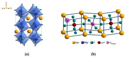
Figure 1.
(a) Crystal structure of orthorhombic-phase NaMgF3, (b) The most possible doping lattice site of Ln3+ ion and magnesium (sodium) vacancies formed in NaMgF3 host lattice to maintain the charge balance
Ultrasmall Lanthanide-doped NaMgF3 Nanocrystals: Controlled Synthesis and Optical Properties
Guo-Wei LI , Yong-Sheng LIU , Fei-Long JIANG , Mao-Chun HONG

Lanthanide (Ln3+)-doped upconversion (UC) nanocrystals (UCNCs) are a unique kind of nanomaterials that can nonlinearly turn low energy irradiation into high energy emission[1-4]. Thanks to their ladder-like 4f-4f transitions[5-7], the doping of Ln3+ ion into the host matrix generally endows these nanomaterials with exceptional optical properties like sharp emission bandwidth, tunable wavelength and long emission lifetime[7-18]. Therefore, Ln3+-doped UCNCs are considered as a promising alternative to the traditional fluorescent probes like quantum dots and organic dyes, and have shown great potential in a wide range of areas including full-color displays, therapeutics, and biological imaging[17, 19-30].
Over the past decade, the selection of host materials to deliver intense upconversion luminescence (UCL) was mainly restricted to the inorganic fluorides, especially in the category of NaREF4 (RE = rare earth)[9, 16, 31-35] due to their perfect chemical stability and low phonon energy less than 500 cm-1[36, 37]. However, all currently available UCNCs may cause unintended acute or chronic toxicity to the living organisms owing to their rich Ln3+ components[38, 39]. Moreover, when compared with green light, red light possesses a better tissue penetration ability (the penetration depth can reach 10~15 nm) that makes it applicable in various areas such as biological imaging, clinical medicine, plant cultivation and cell research[25]. Therefore, it is highly desirable to explore a new class of non-toxic Ln3+-doped UCNCs that can emit large red-to-green (R/G) ratio UC emission for various applications.
Herein, orthorhombic-phase NaMgF3 that displays good biocompatibility[40-44] was chosen as the host material for the synthesis of Ln3+-doped UCNCs. The symmetry of doping site of Ln3+ ion in the NaMgF3 host lattice was demonstrated for the first time by means of the high-resolution photoluminescence spectroscopy of Eu3+ at low temperature (10 K). More importantly, intense UCL emissions of Ln3+-doped NaMgF3 UCNCs ranging from UV to visible and to NIR spectral regions were readily achieved after the doping of typical UCL couples of Yb3+/Er3+, Yb3+/Tm3+ and Yb3+/Ho3+. Among the as-synthesized Ln3+-doped NaMgF3 UCNCs, large UCL R/G ratio of Er3+ was achieved in the NaMgF3: Yb3+/Er3+ UCNCs, thereby enabling them highly promising for various applications such as bioimaging, biodetection, and biotherapy.
Mg(CH3CO2)3·4H2O (99%), Yb(CH3CO2)3·4H2O (99.999%), Er(CH3CO2)3·4H2O (99.99%), Ho(CH3CO2)3·4H2O (99.99%), Tm(CH3CO2)3·4H2O (99.99%), Eu(CH3CO2)3·4H2O (99.99%), oleic acid (OA, technical grade 90%) and trioctylamine (TOA, 98%) were purchased from Sigma-Aldrich (China). NaOH (96%) and NH4F (98%)were purchased from Aladdin (China). Cyclohexane, methanol, and ethanol were purchased from Sinopharm Chemical Reagent Co., China.
The orthorhombic-phase NaMgF3: Ln3+ (Ln3+ = Yb3+, Er3+, Ho3+, Tm3+, and Eu3+) NCs were synthesized via a modified co-precipitation method at high temperature (310 ℃). In a typical procedure for the synthesis of NaMgF3: 4%Yb3+/1%Er3+ NCs, a mixture of 0.475 mmol Mg(CH3CO2)3·4H2O, 0.02 mmol Yb(CH3CO2)3·4H2O, 0.005 mmol Er(CH3CO2)3·4H2O, 5 mL OA and 15 mL TOA was added into a 100 mL three-neck flask. The solution was heated to 155 ℃ under N2 flow with constant stirring for 30 min to form a clear solution, and then cooled down to room temperature. Thereafter, 10 mL of methanol solution containing NH4F (1.5 mmol) and NaOH (1.5 mmol) was added, and the resultant solution was stirred at 75 ℃ for 30 min. After the methanol was evaporated, the solution was heated to 310 ℃ under N2 flow with vigorous stirring for 60 min, and then cooled down to room temperature naturally. The resulting NaMgF3: Yb/Er UCNCs were precipitated by the addition of acetone, collected by centrifugation at 12000 rpm for 5 min, and washed with cyclohexane and ethanol for several times. Finally, the powder samples were made after being dried at 50 ℃ for 8 hours.
The crystallization phase of as-synthesized samples was analyzed by powder X-ray diffraction (XRD) on MiniFlex2 (Rigaku, Japan) from 20° to 80° with Cu-Κα radiation (λ = 0.154187 nm). The sample images of morphology, size and lattice fringes were obtained by transmission electron microscopy (TEM) and high-resolution TEM (HTEM) on Tecnai F20 (USA, 200kV) equipped with energy-dispersive X-ray spectroscopy (EDS). Photoluminescence (PL) emission, excitation spectra and PL decay curve for samples were recorded with UV/V/NIR Fluorescence Spectrometer (FLS980, Edinburgh Instruments) equipped with both continuous (450 W) xenon and pulsed flash lamps. The upconversion luminescence spectra were carried out upon 980 nm excitation provided by a continuous-wave semiconductor laser diode. The effective lifetime (τeff) was determined by:
|
|
where I0 and I(t) represent the maximum luminescence intensity and the luminescence intensity at time t after cutoff of the excitation light, respectively.
Fig. 1a presents the unit cell of orthorhombic-phase NaMgF3 crystal (space group Pbnm (62), a = 5.365, b = 5.492, c = 7.674 Å, Z = 4). The detailed atomic parameters including atom sites and orientations of NaMgF3 are shown in Table 1, where Na+ and Mg2+ ions have only one site, but F- ions have two sites (F1 and F2). According to previous reports in the literature[45], the interatomic distance between Mg–F(1) (1.9779(4) Å) and Mg–F(2) (1.9790(7) and 1.9802(7) Å) are nearly identical in the NaMgF3 lattice, indicating that Mg2+ ions occupy a crystallographic site with symmetry of Oh, the center of octahedra built by six F- ions. Theoretically, Ln3+ ions are easier to replace the Mg2+ ions in the NaMgF3 lattice, and magnesium or sodium vacancies are formed to maintain the charge balance (Fig. 1b)[41, 43].
 DownLoad:
CSV
DownLoad:
CSV
| Phase | Atom | Site | x/a | y/b | z/c |
| Monoclinic | Na | 4c | 0.9896(9) | 0.0452(8) | 0.25 |
| Mg | 4b | 0 | 0.5 | 0 | |
| F1 | 4c | 0.0872(5) | 0.4741(5) | 0.25 | |
| F2 | 8d | 0.7030(3) | 0.2953(3) | 0.0464(3) |
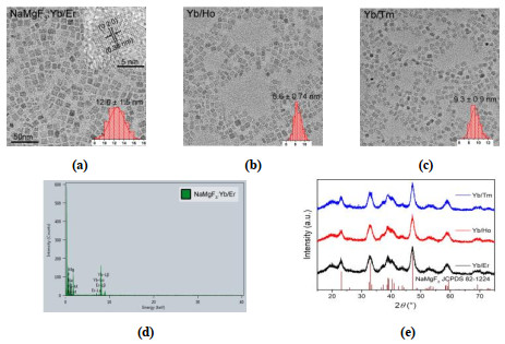
Monodisperse NaMgF3: Ln3+ (Ln3+ = Yb3+/Er3+, Yb3+/Ho3+, Yb3+/Tm3+) UCNCs were synthesized via a modified high temperature co-precipitation method[46, 47] by using oleic acid (OA) and trioctylamine (TOA) as surfactant and solvent. As shown in Fig. 2a~2c, representative transmission elec-tron microscopy (TEM) images for the NaMgF3 UCNCs doped with Yb3+/Er3+ (4/1 mol%), Yb3+/Ho3+ (4.5/0.5 mol%) and Yb3+/Tm3+ (4.8/0.2 mol%) demonstrate their nearly square morphologies with average sizes of 12.6 ± 1.5, 8.6 ± 0.74, and 9.3 ± 0.9 nm, respectively. Clear lattice fringes with a d-spacing of 0.38 nm are observed in the high-resolution TEM (HR-TEM) image of NaMgF3: Yb3+/Er3+ UCNCs, in good agreement with the lattice spacing of (020) plane of orthorhombic-phase NaMgF3 (JCPDS No. 82-1224) (inset of Fig. 2a). Componential analysis by EDX reveals the presence of host elements of Na, Mg and F and the dopants of Yb3+ and Er3+ in the as-synthe- sized Ln3+-doped UCNCs (Fig. 2d). All as-synthesized samples can be indexed in accordance with the standard pattern of orthorhombic-phase NaMgF3 crystal (JCPDS No. 82-1224) (Fig. 2e). These results clearly indicate that the category of Ln3+ dopants has negligibly impact on the size, morphology and crystal phase of the resulting NaMgF3: Ln3+ NCs.
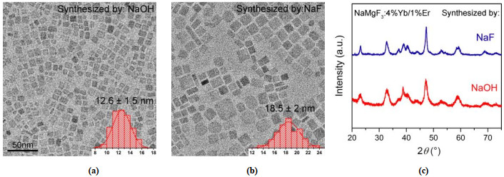
However, it is found that different Na precursors have a great influence on the size of the resulting NaMgF3: Ln3+ NCs. As shown in Fig. 3a and 3b, it is obvious that the NaMgF3: Ln3+ NCs synthesized with NaOH precursor possessed a smaller size with a narrower size distribution than those synthesized with NaF precursor. This can be further substantiated by the XRD analysis. Although both XRD patterns match the standard orthorhombic-phase NaMgF3 crystal in terms of the line positions, the full width at half maximum of the samples synthesized with NaOH precursor is wider than that synthesized with NaF precursor (Fig. 3c). The variation in the nanocrystal size is quite understandable owing to the controlled growth of NCs associated with OA- concentration in the solvents. It is well established that a high concentration of OA- is able to limit the increase of nanocrystal size[13]. In comparison with NaF precursor that hardly reacted with OA, NaOH precursor is easily able to react with OA to release a higher concentration of OA-, thus resulting in a smaller nanocrystal size of NaMgF3: Ln3+ NCs.
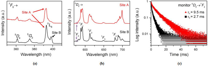
It is well known that the Eu3+ ion was usually employed as a sensitive optical/structural probe to study the crystal structure of host material due to its sensitive response to the crystal-field (CF) environment. By comparing the CF splitting number of the transitions of 5D0 → 7FJ (J = 0, 1, 2, 3, 4, 5 and 6) with theoretical predictions[47, 48], multiple spectroscopic sites of Eu3+ within different CF surroundings can be easily identified in Ln3+-doped NCs, especially for the case of 5D0 → 7F1, 2 transitions. Generally, the 5D0 → 7F1 transition of Eu3+ exhibits a magnetic dipole transition nature that is insensitive to the change of CF surroundings. On the contrary, the 5D0 → 7F2 transition of Eu3+ possessing an electric dipole transition nature is hypersensitive to different local crystal structures. Moreover, according to the electric dipole selection rule, the 5D0 → 7F0 transition of Eu3+ is only allowed in 10 site symmetries including C1, Cs, C2, C2v, C4, C4v, C3, C3v, C6 and C6v (Table S1). On this basis, the high-resolution excitation, emission spectra and decays of NaMgF3: 5%Eu3+ NCs were comprehensively surveyed at low temperature (10 K) to confirm the symmetry of the doping site of Ln3+.
Fig. 4a compares the PL excitation spectra of NaMgF3: Eu3+ NCs by monitoring the 5D0 → 7F2 transition of Eu3+ at 609 and 612 nm, respectively. The characteristic PL bands arising from the 7F0 → 5D4 (361.2 nm), 7F0 → 5G2 (375 nm) and 7F0 → 5L6 (393 nm) transitions were readily detected for both cases. However, a marked difference for these two cases was observed, where an extra peak centered at 398.2 nm assigned to the 7F0 → 5L6 transition appeared for the second case, clearly demonstrating that Eu3+ occupies two different crystalline sites in the NaMgF3 lattice (hereafter defined as sites A and B).
To further determine the specific doping sites of Eu3+ in NaMgF3 NCs, we then mearsured the high-resolution site-selective emission spectra under the excitation at 393 and 398.2 nm originating from the 7F0 → 5L6 transition of Eu3+ at 10 K. As shown in Fig. 4b (upper), five well-resolved emission peaks centered at 578, 591, 612, 648 and 699 nm assigned to the intra-4f transitions from 5D0 to its lower states of 7FJ (J = 0~4) of Eu3+ were observed under the excitation at 393 nm (site A). Theoretically, if Eu3+ occupies the site of ideal Oh symmetry in NaMgF3: Eu3+ NCs, the 5D0 → 7F0, 2, 4 transitions would be strictly forbidden and only magnetic dipole transition of 5D0 → 7F1 can be detected (Table S1). However, the site symmetry of Eu3+ in NaMgF3: Eu3+ NCs would degrade due to the mismatch of valences and ion radiuses between Eu3+ and Mg2+. Specifically, 1, 2, 2 and 4 well-resolved emission lines assigned to the 5D0 → 7F0, 5D0 → 7F1, 5D0 → 7F2 and 5D0 → 7F4 transitions were observed (Fig. 4b, upper), indicative of a C4v site symmetry for site A (Table S1). In contrast, 3 well-resolved emission lines assigned to the 5D0 → 7F1 transition were detected for site B (Fig. 4b, lower), suggesting that this site occupies a lower symmetry than site A. Based on branching rules of the 32 point groups (Fig. S1), C4v can theoratically be degraded to C4, C2v, C2, Cs and C1. Therefore, site B of Eu3+ in NaMgF3: Eu3+ NCs occupies a C2v site symmetry, as a result of 5 emission lines for the 5D0 → 7F4 transition. These results can be further confirmed by their corresponding PL decay lifetime (Fig. 4c). The lifetime of 5D0 → 7F2 transition for site A was determined to be 9.5 ms, much longer than that of site B (2.7 ms). In a word, Eu3+ ions of site A are incorporated in the inner lattice sites of a high symmetry in NaMgF3 NCs, while Eu3+ ions of site B occupy the distorted lattice sites near the surface or surface sites.
After demonstrating the successful hetero-valence doping of Ln3+ ions, we then doped the typical Ln3+ couple of Yb3+/Er3+ into the lattice of NaMgF3 NCs via a modified high temperature co-precipitation method. To determine the optimal doping amount of Ln3+, different amounts of Ln3+ were doped into the NaMgF3 lattice firstly, which was clearly revealed by the XRD analysis (Fig. 5a). Notably, the orthorhombic crystal phase of NaMgF3: Ln3+ NCs kept nearly unchanged as the doping amount of Ln3+ gradually increased (Fig 5a). The magnified XRD pattern shown in Fig. 5a (right) suggested a heterogeneous phase that can be assigned to the α-NaYbF4 crystal (JCPDS No. 77-2043) when a high doping amount of 20.5% Ln3+ (Yb3+/Er3+) was introduced into the NaMgF3 lattice. To avoid the possible impact of impurity phase in NaMgF3: Ln3+ (Ln3+ = Yb3+/Er3+, Yb3+/Ho3+ and Yb3+/Tm3+) UCNCs, we then selected 5% as a total doping concentration for the subsequent UCL measurements.
Under excitation at 980-nm diode laser with a power density of 50 W·cm-2, the as-synthesized NaMgF3: Yb3+/Er3+ UCNCs exhibit intense red and extremely weak green UC emissions, which are assigned to the 4F9/2 → 4I15/2 (654 nm) and 2H11/2/4S3/2 → 4I15/2 (515~565 nm) transitions of Er3+, respectively (Fig. 5b). Moreover, the integrated UC emission intensity was found to increase at first and then decrease as the concentration of Er3+ gradually increased, thus obtaining the optimal Yb3+/Er3+ doping concentration of 4%/1% mol. It should be noted that the NaMgF3: Yb3+/Er3+ UCNCs exhibit high UCL R/G ratios of Er3+ regardless of the doping concentration of Yb3+/Er3+. Such high UCL R/G ratios of Er3+ can be explained when looking at the crystal structure of NaMgF3 lattice that features a high-symmetry substituted site of Mg2+ by Ln3+[2].
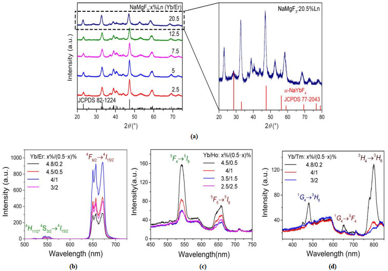
In addition, efficient UCL was also achieved when the typical UCL couples of Yb3+/Ho3+ and Yb3+/Tm3+ were introduced into the NaMgF3 host lattice. As shown in Fig. 5c, the NaMgF3: Yb3+/Ho3+ UCNCs exhibit green and red UC emissions, which are assigned to the 5F4 → 5I8 (542 nm) and 5F5 → 5I8 (655 nm) transitions of Ho3+, respectively. As for the case of Yb3+/Tm3+, the NaMgF3: Yb3+/Tm3+ UCNCs exhibit green, red and NIR UC emissions, which is assigned to 1G4 → 3H6, 1G4 → 3F4, and 3H4 → 3H6 transitions of Tm3+, respectively (Fig. 5d). The optimal doping concentration of Yb3+/Ho3+ and Yb3+/Tm3+ in the NaMgF3 lattice is determined to be 4.5%/0.5% and 4.8%/0.2% mol. Taken together, the as-synthesized ultrasmall NaMgF3: Ln3+ UCNCs are a new class of Ln3+-doped UCNCs that exhibit efficient UCL properties, thereby making them highly promising for various applications.
In this paper, we described a new class of orthorhombic-phase NaMgF3 ultrasmall (~10 nm) NCs doped with different Ln3+ ions that were synthesized via a modified high-temperature co-precipitation method. Monodisperse and uniform Ln3+-doped NaMgF3 NCs with tunable nanocrystal size were obtained by varying the category of Na precursors including NaOH and NaF. By taking advantage of high-resolution photoluminescence spectro-scopy of Eu3+ at low temperature (10 K), the symmetry of doping site of Ln3+ was demonstrated for the first time in NaMgF3 NCs. More importantly, owing to the successful Ln3+ doping, intense UCL in the UV, visible and NIR spectral regions for these Ln3+-doped NaMgF3 NCs were obtained by changing the type of Ln3+ dopant couple of Yb3+/Er3+, Yb3+/Tm3+ and Yb3+/Ho3+. These findings reported here unambiguously demonstrate that Ln3+-doped NaMgF3 NCs without containing toxic elements hold great promise as one of the possible candidates for diverse biological applications both in vitro and in vivo.
Ding, Q.; Zhao, S.; Li, L.; Shen, Y.; Shan, P.; Wu, Z.; Li, X.; Li, Y.; Liu, S.; Luo, J. Abrupt structural transformation in asymmetric ABPO4F (A = K, Rb, Cs). Inorg. Chem. 2019, 58, 1733−1737. doi: 10.1021/acs.inorgchem.8b02754
Fu, H.; Peng, P.; Li, R.; Liu, C.; Liu, Y.; Jiang, F.; Hong, M.; Chen, X. A general strategy for tailoring upconversion luminescence in lanthanide-doped inorganic nanocrystals through local structure engineering. Nanoscale 2018, 10, 9353−9359. doi: 10.1039/C8NR01519H
Li, Y.; Liang, F.; Zhao, S.; Li, L.; Wu, Z.; Ding, Q.; Liu, S.; Lin, Z.; Hong, M.; Luo, J. Two non-π-conjugated deep-UV nonlinear optical sulfates. J. Am. Chem. Soc. 2019, 141, 3833−3837. doi: 10.1021/jacs.9b00138
Hehlen, M. P.; Brik, M. G.; Krämer, K. W. 50th anniversary of the Judd-Ofelt theory: an experimentalist's view of the formalism and its application. J. Lumin. 2013, 136, 221−239. doi: 10.1016/j.jlumin.2012.10.035
Bettinelli, M.; Carlos, L. D.; Liu, X. G. Lanthanide-doped upconversion nanoparticles. Phys. Today 2015, 68, 38−43.
Qin, X.; Liu, X.; Huang, W.; Bettinelli, M.; Liu, X. Lanthanide-activated phosphors based on 4f-5d optical transitions: theoretical and experimental aspects. Chem. Rev. 2017, 117, 4488−4527. doi: 10.1021/acs.chemrev.6b00691
Wang, R.; Guo, Q.; Qian, Y.; Xing, L.; Xu, Y. Upconversion properties of LiNbO3 single crystals co-doped with Ho and Yb. Chin. J. Struct. Chem. 2011, 30, 1597−1603.
Back, M.; Ueda, J.; Ambrosi, E.; Cassandro, L.; Cristofori, D.; Ottini, R.; Riello, P.; Sponchia, G.; Asami, K.; Tanabe, S.; Trave, E. Lanthanide-doped bismuth-based fluoride nanocrystalline particles: formation, spectroscopic investigation, and chemical stability. Chem. Mater. 2019, 31, 8504−8514. doi: 10.1021/acs.chemmater.9b03164
Bian, W.; Lin, Y.; Wang, T.; Yu, X.; Qiu, J.; Zhou, M.; Luo, H.; Yu, S. F.; Xu, X. Direct identification of surface defects and their influence on the optical characteristics of upconversion nanoparticles. ACS Nano 2018, 12, 3623−3628. doi: 10.1021/acsnano.8b00741
Chen, B.; Kong, W.; Liu, Y.; Lu, Y.; Li, M.; Qiao, X.; Fan, X.; Wang, F. Crystalline hollow microrods for site-selective enhancement of nonlinear photoluminescence. Angew. Chem. Int. Ed. 2017, 56, 10383−10387. doi: 10.1002/anie.201703600
Chen, B.; Kong, W.; Wang, N.; Zhu, G.; Wang, F. Oleylamine-mediated synthesis of small NaYbF4 nanoparticles with tunable size. Chem. Mater. 2019, 4779−4786.
Ding, S.; Yang, X. F.; Song, E. H.; Liang, C. L.; Zhou, B.; Wu, M. M.; Zhou, W. Z.; Zhang, Q. Y. An efficient synthetic strategy for uniform perovskite core-shell nanocubes NaMgF3: Mn2+, Yb3+@NaMgF3: Yb3+ with enhanced near infrared upconversion luminescence. J. Mater. Chem. C 2018, 6, 2342−2350. doi: 10.1039/C7TC05416E
Fischer, S.; Swabeck, J. K.; Alivisatos, A. P. Controlled isotropic and anisotropic shell growth in beta-NaLnF4 nanocrystals induced by precursor injection rate. J. Am. Chem. Soc. 2017, 139, 12325−12332. doi: 10.1021/jacs.7b07496
Huang, P.; Zheng, W.; Gong, Z.; You, W.; Wei, J.; Chen, X. Rare earth ion- and transition metal ion-doped inorganic luminescent nanocrystals: from fundamentals to biodetection. Mater. Today Nano 2019, 5, 100031−23. doi: 10.1016/j.mtnano.2019.100031
Liu, M.; Shi, Z.; Wang, X.; Zhang, Y.; Mo, X.; Jiang, R.; Liu, Z.; Fan, L.; Ma, C. G.; Shi, F. Simultaneous enhancement of red upconversion luminescence and CT contrast of NaGdF4: Yb, Er nanoparticles via Lu3+ doping. Nanoscale 2018, 10, 20279−20288. doi: 10.1039/C8NR06968A
Pang, M.; Zhai, X.; Feng, J.; Song, S.; Deng, R.; Wang, Z.; Yao, S.; Ge, X.; Zhang, H. One-step synthesis of water-soluble hexagonal NaScF4: Yb/Er nanocrystals with intense red emission. Dalton Trans. 2014, 43, 10202−10207. doi: 10.1039/c4dt00708e
Zheng, W.; Zhou, S.; Chen, Z.; Hu, P.; Liu, Y.; Tu, D.; Zhu, H.; Li, R.; Huang, M.; Chen, X. Sub-10 nm lanthanide-doped CaF2 nanoprobes for time-resolved luminescent biodetection. Angew. Chem. Int. Ed. 2013, 52, 6671−6676. doi: 10.1002/anie.201302481
Huang, Q.; Yu, H.; Ma, E.; Zhang, X.; Cao, W.; Yang, C.; Yu, J. Upconversion effective enhancement by producing various coordination surroundings of rare-earth ions. Inorg. Chem. 2015, 54, 2643−2651. doi: 10.1021/ic5027976
Gu, Y.; Guo, Z.; Yuan, W.; Kong, M.; Liu, Y.; Liu, Y.; Gao, Y.; Feng, W.; Wang, F.; Zhou, J.; Jin, D.; Li, F. High-sensitivity imaging of time-domain near-infrared light transducer. Nat. Photon. 2019, 13, 525−531. doi: 10.1038/s41566-019-0437-z
Li, L.; Yu, Y.; Zhou, Z.; Li, Q. PEG assisted hydrothermal synthesis of β-NaYF4: Yb3+, Er3+ microrods for upconversion photoluminescence display. Chin. J. Struct. Chem. 2014, 33, 1865−1874.
Liu, Q.; Feng, W.; Yang, T.; Yi, T.; Li, F. Upconversion luminescence imaging of cells and small animals. Nat. Protoc. 2013, 8, 2033−2044. doi: 10.1038/nprot.2013.114
Song, X.; Li, S.; Guo, H.; You, W.; Shang, X.; Li, R.; Tu, D.; Zheng, W.; Chen, Z.; Yang, H.; Chen, X. Graphene-oxide-modified lanthanide nanoprobes for tumor-targeted visible/NIR-II luminescence imaging. Angew. Chem. Int. Ed. 2019, 58, 18981−18986. doi: 10.1002/anie.201909416
Wang, J.; Lin, H.; Cheng, Y.; Cui, X.; Gao, Y.; Ji, Z.; Xu, J.; Wang, Y. A novel high-sensitive upconversion thermometry strategy: utilizing synergistic effect of dual-wavelength lasers excitation to manipulate electron thermal distribution. Sens. Actuators B-Chem. 2019, 278, 165−171. doi: 10.1016/j.snb.2018.09.086
Xu, M.; Zou, X.; Su, Q.; Yuan, W.; Cao, C.; Wang, Q.; Zhu, X.; Feng, W.; Li, F. Ratiometric nanothermometer in vivo based on triplet sensitized upconversion. Nat. Commun. 2018, 9, 2698−7. doi: 10.1038/s41467-018-05160-1
Yu, L.; Liu, Y.; Chen, X. Lanthanide-doped upconversion nano-bioprobes for in-vitro detection of tumor marker. Chin. J. Lumin. 2018, 39, 27−49. doi: 10.3788/fgxb20183901.0027
Zhang, M.; Zhai, X.; Lei, P.; Yao, S.; Xu, X.; Dong, L.; Du, K.; Li, C.; Feng, J.; Zhang, H. Selective enhancement of green upconversion luminescence from NaYF4: Yb, Er microparticles through Ga3+ doping for sensitive temperature sensing. J. Lumin. 2019, 215, 116632−116639. doi: 10.1016/j.jlumin.2019.116632
Zhou, L.; Fan, Y.; Wang, R.; Li, X.; Fan, L.; Zhang, F. High-capacity upconversion wavelength and lifetime binary encoding for multiplexed biodetection. Angew. Chem. Int. Ed. 2018, 57, 12824−12829. doi: 10.1002/anie.201808209
Zhuo, Z.; Liu, Y.; Liu, D.; Huang, P.; Jiang, F.; Chen, X.; Hong, M. Manipulating energy transfer in lanthanide-doped single nanoparticles for highly enhanced upconverting luminescence. Chem. Sci. 2017, 8, 5050−5056. doi: 10.1039/C7SC01393K
Wang, Y.; Wei, T.; Cheng, X.; Ma, H.; Pan, Y.; Xie, J.; Su, H.; Xie, X.; Huang, L.; Huang, W. Insights into Li+-induced morphology evolution and upconversion luminescence enhancement of KSc2F7: Yb/Er nanocrystals. J. Mater. Chem. C 2017, 5, 3503−3508. doi: 10.1039/C7TC00649G
Wu, Y.; Sun, Y.; Zhu, X.; Liu, Q.; Cao, T.; Peng, J.; Yang, Y.; Feng, W.; Li, F. Lanthanide-based nanocrystals as dual-modal probes for SPECT and X-ray CT imaging. Biomaterials 2014, 35, 4699−4705. doi: 10.1016/j.biomaterials.2014.02.034
Dong, H.; Sun, L. D.; Li, L. D.; Si, R.; Liu, R.; Yan, C. H. Selective cation exchange enabled growth of lanthanide core/shell nanoparticles with dissimilar structure. J. Am. Chem. Soc. 2017, 139, 18492−18495. doi: 10.1021/jacs.7b11836
Liu, H.; Xu, H.; Huang, Q.; Cao, W.; Yu, H.; Yu, Y. Upconversion luminescence properties of NaY0.92Yb0.05Er0.03F4 enhanced by Zr4+ codoping. Chin. J. Struct. Chem. 2017, 36, 1743−1751.
Xu, J.; Tu, D.; Zheng, W.; Shang, X.; Huang, P.; Cheng, Y.; Wang, Y.; Chen, X. Interfacial defects dictated in situ fabrication of yolk-shell upconversion nanoparticles by electron-beam irradiation. Adv. Sci. 2018, 5, 1800766−9. doi: 10.1002/advs.201800766
Wang, M.; Tian, Y.; Zhao, F.; Li, R.; You, W.; Fang, Z.; Chen, X.; Huang, W.; Ju, Q. Alleviating the emitter concentration effect on upconversion nanoparticles via an inert shell. J. Mater. Chem. C 2017, 5, 1537−1543. doi: 10.1039/C6TC05289D
Xiang, J.; Ge, F.; Yu, B.; Yan, Q.; Shi, F.; Zhao, Y. Nanocomplexes of photolabile polyelectrolyte and upconversion nanoparticles for near-infrared light-triggered payload release. ACS Appl. Mater. Interfaces 2018, 10, 20790−20800. doi: 10.1021/acsami.8b05063
Ju, D.; Song, F.; Khan, A.; Song, F.; Zhou, A.; Gao, X.; Hu, H.; Sang, X.; Zadkov, V. Simultaneous dual-mode emission and tunable multicolor in the time domain from lanthanide-doped core-shell microcrystals. Nanomaterials 2018, 8, 1023−11. doi: 10.3390/nano8121023
Liu, X.; Wang, Y.; Li, X.; Yi, Z.; Deng, R.; Liang, L.; Xie, X.; Loong, D. T. B.; Song, S.; Fan, D.; All, A. H.; Zhang, H.; Huang, L.; Liu, X. Binary temporal upconversion codes of Mn2+-activated nanoparticles for multilevel anti-counterfeiting. Nat. Commun. 2017, 8, 899−7. doi: 10.1038/s41467-017-00916-7
Lei, P.; An, R.; Yao, S.; Wang, Q.; Dong, L.; Xu, X.; Du, K.; Feng, J.; Zhang, H. Ultrafast synthesis of novel hexagonal phase NaBiF4 upconversion nanoparticles at room temperature. Adv. Mater. 2017, 29, 1700505−4. doi: 10.1002/adma.201700505
Sun, Y.; Feng, W.; Yang, P.; Huang, C.; Li, F. The biosafety of lanthanide upconversion nanomaterials. Chem. Soc. Rev. 2015, 44, 1509−1525. doi: 10.1039/C4CS00175C
Du, Y. P.; Zhang, Y. W.; Yan, Z. G.; Sun, L. D.; Gao, S.; Yan, C. H. Single-crystalline and near-monodispersed NaMF3 (M = Mn, Co, Ni, Mg) and LiMAlF6 (M = Ca, Sr) nanocrystals from cothermolysis of multiple trifluoroacetates in solution. Chem. Asian J. 2007, 2, 965−974. doi: 10.1002/asia.200700054
Wang, J.; Zhang, J.; Xie, J.; Li, Y.; Wang, L.; Zhang, Q. Phase controllable synthesis of NaMgF3: Yb3+, Er3+ nanocrystals with effective red upconversion luminescence. J. Mater. Sci. : Mater. Electron. 2018, 29, 18320−18330. doi: 10.1007/s10854-018-9946-7
Liu, L.; Wang, S.; Zhao, B.; Pei, P.; Fan, Y.; Li, X.; Zhang, F. Er3+ sensitized 1530 to 1180 nm second near-infrared window upconversion nanocrystals for in vivo biosensing. Angew. Chem. Int. Ed. 2018, 57, 7518−7522. doi: 10.1002/anie.201802889
Wang, J.; Wang, F.; Wang, C.; Liu, Z.; Liu, X. Single-band upconversion emission in lanthanide-doped KMnF3 nanocrystals. Angew. Chem. Int. Ed. 2011, 50, 10369−10372. doi: 10.1002/anie.201104192
Zhong, Y.; Ma, Z.; Wang, F.; Wang, X.; Yang, Y.; Liu, Y.; Zhao, X.; Li, J.; Du, H.; Zhang, M.; Cui, Q.; Zhu, S.; Sun, Q.; Wan, H.; Tian, Y.; Liu, Q.; Wang, W.; Garcia, K. C.; Dai, H. In vivo molecular imaging for immunotherapy using ultra-bright near-infrared-IIb rare-earth nanoparticles. Nat. Biotechnol. 2019, 37, 1322−1331. doi: 10.1038/s41587-019-0262-4
Pischedda, V.; Ferraris, G.; Raade, G. Single-crystal X-ray diffraction study on neighborite (NaMgF3) from Gjerdingselva, Norway. N. Jb. Miner. Abh. 2005, 182, 23−29. doi: 10.1127/0077-7757/2005/0028
Fu, H.; Liu, Y.; Jiang, F.; Hong, M. Controlled synthesis and optical properties of lanthanide-doped Na3ZrF7 nanocrystals. Chin. J. Struct. Chem. 2018, 37, 1737−1748.
Yu, L.; Li, G.; Liu, Y.; Jiang, F.; Hong, M. Lanthanide-doped KGd2F7 nanocrystals: controlled synthesis, optical properties, and spectroscopic identification of the optimum core/shell architecture for highly enhanced upconverting luminescence. Cryst. Growth Des. 2019, 19, 2340−2349. doi: 10.1021/acs.cgd.9b00040
Liu, Y.; Tu, D.; Zhu, H.; Chen, X. Lanthanide-doped luminescent nanoprobes: controlled synthesis, optical spectroscopy, and bioapplications. Chem. Soc. Rev. 2013, 42, 6924−6958. doi: 10.1039/c3cs60060b

Figure 1 (a) Crystal structure of orthorhombic-phase NaMgF3, (b) The most possible doping lattice site of Ln3+ ion and magnesium (sodium) vacancies formed in NaMgF3 host lattice to maintain the charge balance

Figure 2 Representative low-resolution TEM images for the as-synthesized NaMgF3: Ln3+ UCNCs doped with (a) Yb3+/Er3+, (b) Yb3+/Ho3+ and (c) Yb3+/Tm3+, respectively, and the inset of (a) shows the high-resolution TEM image of a randomly selected NaMgF3: Yb3+/Er3+ nanocrystal. Note that their corresponding nanocrystal sizes and size distribution histograms are inserted in the bottoms of (a)~(c). (d) EDX pattern for the as-synthesized NaMgF3: Yb3+/Er3+ UCNCs. (e) XRD patterns for the as-synthesized NaMgF3: Ln3+ (Ln3+ = Yb3+/Er3+, Yb3+/Ho3+ and Yb3+/Tm3+) UCNCs

Figure 3 Typical low-resolution TEM image for NaMgF3: Yb3+/Er3+ UCNCs synthesized with (a) NaOH and (b) NaF precursors, respectively. Insets of (a) and (b) show their corresponding nanocrystal sizes and size distribution histograms. (c) XRD patterns for the as-synthesized NaMgF3: Yb3+/Er3+ UCNCs with NaOH and NaF precursors

Figure 4 (a) High-resolution PL excitation spectra for the NaMgF3: 5%Eu3+ NCs by monitoring the 5D0 → 7F2 transition of Eu3+ at 609 (red) and 612 nm (black). (b) Site-selective PL emission spectra for the NaMgF3: 5%Eu3+ NCs under excitation at 393 nm (red) and 398.3 nm (black), respectively. (c) PL decays for the 5D0 → 7F2 transition of Eu3+ in NaMgF3: 5%Eu3+ NCs under site-selective excitation at 393 (red) and 398.2 nm (black) by monitoring the emission at 612 and 609 nm, respectively. Noted that all the PL spectra and decays were measured at low temperature (10 K)

Figure 5 (a) XRD patterns for the as-synthesized NaMgF3: Yb3+/Er3+ UCNCs with different amounts of Yb3+/Er3+ from 2.5% to 20.5%. The specific XRD pattern for NaMgF3: 20.5%Ln3+ UCNCs is shown in the right side. UCL spectra for NaMgF3: Ln3+ UCNCs with different amounts of (b) Yb3+/Er3+, (c) Yb3+/Ho3+, and (d) Yb3+/Tm3+ under excitation of a 980-nm diode laser at a power density of 50 W/cm2
Table 1. Atomic Parameters in the NaMgF3 Crystal
| Phase | Atom | Site | x/a | y/b | z/c |
| Monoclinic | Na | 4c | 0.9896(9) | 0.0452(8) | 0.25 |
| Mg | 4b | 0 | 0.5 | 0 | |
| F1 | 4c | 0.0872(5) | 0.4741(5) | 0.25 | |
| F2 | 8d | 0.7030(3) | 0.2953(3) | 0.0464(3) |
 下载: 导出CSV

 扫一扫看文章

扫一扫关注我们