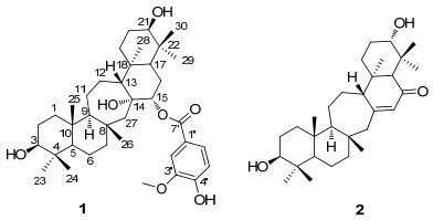
Figure 1.
Structures of compounds 1 and 2
A New Serratane Triterpenoid from Lycopodiella cernua
Yu Jin , Zhengwan Huang , Yingna Huang , Yingting Xu , Jian Yan , Jie Yang , Zhongyu Zhou , Xiaoyi Wei

Lycopodiella cernua (L.) Pic. Serm, belonging to the family Lycopodiaceae, has been used as a traditional Chinese folk medicine to treat rheumatism, quadriplegia and contusion for centuries.[1] Palhinhaea cernua (L.) Franco & Vasc. is a synonym of Lycopodiella cernua. Previous phytochemical study of this plant species has led to the isolation of serratane triterpenoids, [2-6] alkaloids, [7-13] flavonoids, [14] and neolignans.[4] Serratane triterpenoids and lycopodium alkaloids are characteristic components of L. cernua plant. Serratane is a unique class of pentacyclic triterpenoids with a central seven-membered ring C, which is rich in Lycopodiaceae family. Recently, the structural classification, biological activities and hypotheses about biosynthetic pathways of serratane-type triterpenes from Lycopodiaceae family was comprehensively reviewed.[15] As part of our research on novel serratane triterpenoids from L. cernua, [2, 6] two serratane triterpenoids, 3β, 14α, 15α, 21β-tetrahydroxyserrat-15-(3'-methoxyl-4'-hydroxybenzoa-te) (1), and 16-oxoserrat-14-en-3β, 21α-diol (2), were isolated from the whole plant of Lycopodiella cernua (Figure 1). Compound 1 was a new one. Herein, we report the isolation, structural identification and anti-influenza A virus (IAV) activity of these two compounds.
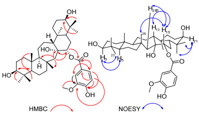
Compound 1 was obtained as a white amorphous powder. The molecular formula of 1 was determined as C38H58O7 by positive HRESIMS (m/z [M+H]+ 627.4247, calcd for C38H59O7: 627.4255) in combination with the 13C and DEPT NMR analysis. The 1H NMR spectrum (Table 1) displayed the signals for seven methyls at δH 0.74 (s, 3H, H-23), 0.92 (s, 3H, H-24), 0.84 (s, 3H, H-25), 1.07 (s, 3H, H-26), 1.02 (s, 3H, H-28), 0.95 (s, 3H, H-29), 0.87 (s, 3H, H-30), an oxygenated methyl at δH 3.92, three methines at δH 3.11 (dd, J=11.5, 4.8 Hz, 1H, H-3), 3.36 (t, J=2.5 Hz, 1H, H-21), and 4.94 (dd, J=11.3, 4.6 Hz, 1H, H-15), and a 1, 3, 4-trisubstituted benzene ring at δH 7.68 (d, J=1.9 Hz, 1H, H-2'), 6.86 (d, J=8.2 Hz, 1H, H-5'), 7.70 (dd, J=8.2, 1.9 Hz, 1H, H-6'). The 13C and DEPT NMR spectra suggested the presence of ten quaternary carbons, ten methines, ten methylenes, and eight methyls. The 13C NMR spectra (DEPT) showed signal at δC 55.8 for a methylene, which was characteristic signal of C-27 in serratane-type triterpenoid.[6] By detailed analysis of the HMBC spectrum of compound 1 and comparison of its NMR data with those of 3β, 14α, 15α, 21α-tetrahydroxyserrat-3-(3′-me- thoxyl-4′-hydroxybenzoate), [2] the serratane triterpenoid skeleton was assigned as 3, 14, 15, 21-tetrahydroxyserratane. Except 30 carbons signals for a serratane triterpenoid, the remaining signals in the 1H NMR and 13C NMR spectra of compound 1 indicated the presence of a 3'-methoxy-4'- hydroxybenzoate group through comparing corresponding NMR data with 3β, 14α, 15α, 21α-tetrahydroxyserrat-3-(3'- methoxyl-4'-hydroxybenzoate).[2] The ester linkage between C-15 of serratane triterpenoid and C-7' of 3'-meth- oxy-4'-hydroxybenzoate was established by heteronuclear multiple bond correlation (HMBC) correlation (Figure 2) from H-15 (δH 4.94) to C-7′ (δC 168.0). The big coupling constant (J=11.5 Hz) of H-3 indicated ax-ax coupling with axial H-2, while the small coupling constant (J=4.8 Hz) of H-3 indicated ax-eq coupling with equatorial H-2. Therefore, H-3 was at axial bond and was α-orientated.[3] In the same way, H-21 appeared to be equatorial (on the α-face) according to its small coupling constants of 2.5 Hz.[3] The nuclear overhauser effect spectroscopy (NOESY) correlation (Figure 2) between H-15 and CH3-26β, confirmed the β configuration of H-15. According to literature, [16] osmolation of lycernuic acid A diacetate gave two isomeric glycols. The minor glycol was proven to be 14α, 15α-diol, while the major one to be 14β, 15β-diol. The chemical shifts of C-17 in 14α, 15α-diol and 14β, 15β-diol were at δC 48.0 and 40.9, respectively. Compound 1 had a chemical shift of C-17 at δC 47.6, which indicated 14α, 15α configurations of two hydroxyl groups in compound 1. Therefore, compound 1 was elucidated as 3β, 14α, 15α, 21β-tetrahydroxyserrat-15-(3′-methoxyl-4′-hydroxybenzo-ate) as shown in Figure 1.
 下载:
导出CSV
下载:
导出CSV
| No. | δH | δC |
| 1 | 1.78~1.80 (m, 1H), 0.99~1.01 (m, 1H) | 39.9 |
| 2 | 1.58~1.60 (m, 2H) | 28.2 |
| 3 | 3.11 (dd, 11.5, 4.8, 1H) | 79.7 |
| 4 | 40.0 | |
| 5 | 0.78 (dd, 9.1, 5.2, 1H) | 57.1 |
| 6 | 1.44~1.46 (m, 2H) | 19.6 |
| 7 | 1.24~1.26 (m, 2H) | 46.7 |
| 8 | 38.6 | |
| 9 | 1.13 (dd, 11.2, 2.2, 1H) | 63.2 |
| 10 | 39.3 | |
| 11 | 1.92~1.94 (m, 1H), 1.22~1.24 (m, 1H) | 27.8 |
| 12 | 1.72~1.74 (m, 1H), 1.47~1.49 (m, 1H) | 24.6 |
| 13 | 1.37~1.39 (m, 1H) | 59.7 |
| 14 | 78.1 | |
| 15 | 4.94 (dd, 11.3, 4.6, 1H) | 81.0 |
| 16 | 1.89~1.91 (m, 1H), 1.68~1.70 (m, 1H) | 25.3 |
| 17 | 1.57~1.59 (m, 1H) | 47.6 |
| 18 | 39.8 | |
| 19 | 1.49~1.51 (m, 1H), 1.40~1.42 (m, 1H) | 33.7 |
| 20 | 2.00~2.02 (m, 1H), 1.56~1.58 (m, 1H) | 26.2 |
| 21 | 3.36 (t, 2.5, 1H) | 76.7 |
| 22 | 38.7 | |
| 23 | 0.92 (s, 3H) | 28.6 |
| 24 | 0.74 (s, 3H) | 16.1 |
| 25 | 0.84 (s, 3H) | 16.6 |
| 26 | 1.07 (s, 3H) | 21.4 |
| 27 | α, 1.36 (d, 15.5, 1H), β, 1.66 (d, 15.5, 1H) | 55.8 |
| 28 | 1.02 (s, 3H) | 16.5 |
| 29 | 0.87 (s, 3H) | 22.6 |
| 30 | 0.95 (s, 3H) | 29.0 |
| 1' | 122.9 | |
| 2' | 7.68 (d, 1.9, 1H) | 114.0 |
| 3' | 148.8 | |
| 4' | 152.9 | |
| 5' | 6.86 (d, 8.2, 1H) | 116.0 |
| 6' | 7.70 (dd, 8.2, 1.9, 1H) | 125.5 |
| 7' | 168.0 | |
| 3'-OCH3 | 3.92 (s, 3H) | 56.6 |
The structure of known compound 2 was identified as 16-oxoserrat-14-en-3β, 21α-diol (2) by interpretation of its spectroscopic data, as well as by comparison with reported values.[17]
Compounds 1 and 2 were evaluated for their anti-IAV activity against A/WSN/33 (H1N1). Preliminary antiviral screening showed that compounds 1 and 2 displayed 9% and 36% inhibition activity at 50 µmol•L-1, respectively.
In conclusion, two serratane triterpenoids, 3β, 14α, 15α, 21β- tetrahydroxyserrat-15-(3'-methoxyl-4'-hydroxybenzoate) (1) and 16-oxoserrat-14-en-3β, 21α-diol (2), were isolated from the whole plant of Lycopodiella cernua. Compound 1 was a new one. These two compounds were isolated from Lycopodiella genus for the first time.
Optical rotations were measured on a Perkin-Elmer Model 341 polarimeter (Perkin-Elmer, Inc., Waltham, MA). UV spectra were recorded on a Perkin-Elmer Lambda 650 UV-Vis spectrophotometer. Nuclear magnetic resonance (NMR) spectra were recorded on a Bruker DRX-400 NMR spectrometer (Bruker Biospin Gmbh, Rheistetten, Germany) and a Bruker AVANCE 600 instrument. ESIMS was collected on an MDS SCIEX API 2000 LC/MS/MS instrument. HRESI-MS was acquired on a time-of-flight mass spectrometer (Bruker maxis 4G). Medium pressure liquid chromatography (MPLC) and preparative HPLC were carried out on a CXTH P3000 instrument (Beijing Chuang Xin Tong Heng Science and Technology Co., Ltd, Beijing, China) equipped with a UV 3000 UV-vis Detector. A Fuji-C18 column (10 µm, 30 mm×250 mm) was used for preparative HPLC. For column chromatography, silica gel (80~100 mesh and 200~300 mesh Qingdao Haiyang Chemical Co., Qingdao, China), and Sephadex LH-20 (Pharmacia Fine Chemical Co., Ltd., Oppsala, Sweden) were performed. Thin-layer chromatography (TLC) was conducted on pre-coated silica gel plates (HSGF254, Yantai Jiang you Silica Gel Development Co., Ltd., Yantai, China) and spot detection was performed by spraying 10% H2SO4 in ethanol, followed by heating.
The information of L. cernua plant material was published in our previous study.[2]
The extraction procedure was previously published, [2] and six serratane triterpenoids were isolated in our previous study.[2] The remaining subfractions of L. cernua extracts were isolated in this study. Subfractions A and B were obtained from EtOAc fraction of L. cernua extracts after silica gel column chromatography and medium pressure liquid chromatography (MPLC). Subfraction A was applied to Sephadex LH-20 column chromatography (CHCl3-MeOH, V:V=1:4), and followed by preparative HPLC (80% aqueous CH3OH, 10 mL/min, Rt=84 min) to afford compound 1 (11 mg). Subfraction B was separated by Sephadex LH-20 column chromatography (CHCl3-MeOH, V:V=1:4) to give subfractions B1 and B2. Compound 2[17] (5 mg) was obtained from subfraction B2 by preparative HPLC (84% aqueous CH3OH, 10 mL/min, Rt=84 min).
3β, 14α, 15α, 21β-tetrahydroxyserrat-15-(3'-methoxyl-4'-hydroxybenzoate) (1): White amorphous powder, m.p. 220~222 ℃;
Two isolated compounds 1 and 2 were evaluated for their anti-IAV activities according to the previously reported 3-(4, 5-dimethylthiazol-2yl)-2, 5-diphenyltetrazolium bromide (MTT) assay, [18] using zanamivir (200 nmol•L-1) as a positive control. Test compounds were preliminary screened at two concentrations of 50 and 10 μmol•L-1, and those which had more than 50% inhibition of influenza virus infection activity were further experimented for cytotoxicity and IC50 evaluation. In detail, Madin Derby canine kidney (MDCK) cells were cultured overnight in Dulbecco's modified Eagle's medium (DMEM) supplemented with 10% fetal bovine serum and 1% penicillin/streptomycin in 96 well plates. A/WSN/33 (H1N1) influenza virus (100 TCID50), which was propagated from 8-day-old chicken embryos, was added and incubated for 1 h. Then the supernatant was removed and washed with phosphate-buffered saline (PBS), and MDCK cells infected with influenza virus were exposed to 200 µL of compounds 1 and 2 at 50 or 10 μmol•L-1. After 48 h of incubation, 100 μL of MTT solution, which was diluted by the medium to 0.5 mg/mL, was added and retained at 37 ℃ for 4 h. Then the supernatant was removed, followed by the addition of 150 μL of dimethyl sulfoxide (DMSO) to dissolve the formazan product. The optical density for each well was measured on the Tecan Genios Pro microplate reader (Bedford, MA, USA) at 570 nm.
Supporting Information 1H NMR, 13C NMR, HSQC, HMBC, and NOESY spectra of compounds 1 and 2. The Supporting Information is available free of charge via the Internet at http://sioc-journal.cn.
肖培根, 新编中药志第三卷, 化学工业出版社, 北京, 2002, p. 135.Xiao, P. G. Modern Chinese Material Medica, Chemical Industry Press, Beijing, 2002, p. 135(in Chinese).
Yan, J.; Zhou, Z.-Y.; Zhang, M.; Wang, J.; Tan, J.-W. Planta Med. 2012, 78, 1387. doi: 10.1055/s-0032-1314999
Wei, J. J.; Wang, W. Q.; Song, W. B.; Xuan, L. J. Fitoterapia 2018, 127, 151. doi: 10.1016/j.fitote.2018.02.011
Li, J.; Xu, P. S.; Tan, L. H.; Zou, Z. X.; Wang, Y. K.; Long, H. P.; Zhou, G.; Li, G.; Xu, K. P.; Tan, G. S. Fitoterapia 2017, 119, 45. doi: 10.1016/j.fitote.2017.04.005
Liang, L. F.; Chen, Q. H.; Xu, J. H.; Liu, T.; Song, X. N.; Chen, H. Y.; Chen, H. Chem. Nat. Compd. 2019, 55, 759. doi: 10.1007/s10600-019-02803-7
Yan, J.; Sun, L.-R.; Zhang, X.-M.; Li, Z.-R.; Zhou, L.; Qiu, M.-H. Chem. Pharm. Bull. 2009, 57, 1381. doi: 10.1248/cpb.57.1381
Zhao, F. W.; Sun, Q. Y.; Yang, F. M.; Hu, G. W.; Luo, J. F.; Tang, G. H.; Wang, Y. H.; Long, C. L. Org. Lett. 2010, 12, 3922. doi: 10.1021/ol101602n
Zhao, F. W.; Sun, Q. Y.; Yang, F. M.; Luo, J. F.; Hu, G. W.; Liu, F.; Wang, Y. H.; Long, C. L. J. Brazil. Chem. Soc. 2012, 23, 349. doi: 10.1590/S0103-50532012000200023
Zhang, D. B.; Chen, J. J.; Zhang, L.; Song, Q. Y.; Gao, K. Phytochem. Lett. 2014, 10, 76. doi: 10.1016/j.phytol.2014.08.008
Tang, Y.; Xiong, J.; Zou, Y. K.; Zhang, H. Y.; Hu, J. F. Phytochemistry 2016, 131, 130. doi: 10.1016/j.phytochem.2016.08.010
Tang, Y.; Xiong, J.; Zou, Y.; Nay, B.; Wang, L. J.; Hu, J. F. Chin. Chem. Lett. 2016, 27, 969. doi: 10.1016/j.cclet.2016.02.014
Dong, L. B.; Yang, J.; He, J.; Luo, H. R.; Wu, X. D.; Deng, X.; Peng, L. Y.; Cheng, X.; Zhao, Q. S. Chem. Commun. 2012, 48, 9038. doi: 10.1039/c2cc34676a
Dong, L. B.; Gao, X.; Liu, F.; He, J.; Wu, X. D.; Li, Y.; Zhao, Q. S. Org. Lett. 2013, 15, 3570. doi: 10.1021/ol401411m
Jiao, R. H.; Ge, H. M.; Shi, D. H.; Tan, R. X. J. Nat. Prod. 2006, 69, 1089. doi: 10.1021/np060038a
Boonya-udtayan, S.; Thasana, N.; Jarussophon, N.; Ruchirawat, S. Fitoterapia 2019, 136, 104181. doi: 10.1016/j.fitote.2019.104181
Zhang, Z. Z.; ElSohly, H. N.; Jacob, M. R.; Pasco, D. S.; Walker, L. A.; Clark, A. M. J. Nat. Prod. 2002, 65, 979. doi: 10.1021/np0200616
杨国勋, 藏毅, 胡长玲, 熊娟, 胡金锋, 中草药, 2014, 45, 3524. doi: 10.7501/j.issn.0253-2670.2014.24.004Yang, G.; Zang, Y.; Hu, C.; Xiong, J.; Hu, J. Chin. Trad. Herbal Drugs 2014, 45, 3524(in Chinese). doi: 10.7501/j.issn.0253-2670.2014.24.004
Luo, X.; Yang, J.; Chen, F.; Lin, X.; Chen, C.; Zhou, X.; Liu, S.; Liu, Y. Front. Chem. 2018, 6, 282 doi: 10.3389/fchem.2018.00282
Table 1. 1H NMR and 13C NMR (600 and 150 MHz, resp.) data for 1 in CD3OD
| No. | δH | δC |
| 1 | 1.78~1.80 (m, 1H), 0.99~1.01 (m, 1H) | 39.9 |
| 2 | 1.58~1.60 (m, 2H) | 28.2 |
| 3 | 3.11 (dd, 11.5, 4.8, 1H) | 79.7 |
| 4 | 40.0 | |
| 5 | 0.78 (dd, 9.1, 5.2, 1H) | 57.1 |
| 6 | 1.44~1.46 (m, 2H) | 19.6 |
| 7 | 1.24~1.26 (m, 2H) | 46.7 |
| 8 | 38.6 | |
| 9 | 1.13 (dd, 11.2, 2.2, 1H) | 63.2 |
| 10 | 39.3 | |
| 11 | 1.92~1.94 (m, 1H), 1.22~1.24 (m, 1H) | 27.8 |
| 12 | 1.72~1.74 (m, 1H), 1.47~1.49 (m, 1H) | 24.6 |
| 13 | 1.37~1.39 (m, 1H) | 59.7 |
| 14 | 78.1 | |
| 15 | 4.94 (dd, 11.3, 4.6, 1H) | 81.0 |
| 16 | 1.89~1.91 (m, 1H), 1.68~1.70 (m, 1H) | 25.3 |
| 17 | 1.57~1.59 (m, 1H) | 47.6 |
| 18 | 39.8 | |
| 19 | 1.49~1.51 (m, 1H), 1.40~1.42 (m, 1H) | 33.7 |
| 20 | 2.00~2.02 (m, 1H), 1.56~1.58 (m, 1H) | 26.2 |
| 21 | 3.36 (t, 2.5, 1H) | 76.7 |
| 22 | 38.7 | |
| 23 | 0.92 (s, 3H) | 28.6 |
| 24 | 0.74 (s, 3H) | 16.1 |
| 25 | 0.84 (s, 3H) | 16.6 |
| 26 | 1.07 (s, 3H) | 21.4 |
| 27 | α, 1.36 (d, 15.5, 1H), β, 1.66 (d, 15.5, 1H) | 55.8 |
| 28 | 1.02 (s, 3H) | 16.5 |
| 29 | 0.87 (s, 3H) | 22.6 |
| 30 | 0.95 (s, 3H) | 29.0 |
| 1' | 122.9 | |
| 2' | 7.68 (d, 1.9, 1H) | 114.0 |
| 3' | 148.8 | |
| 4' | 152.9 | |
| 5' | 6.86 (d, 8.2, 1H) | 116.0 |
| 6' | 7.70 (dd, 8.2, 1.9, 1H) | 125.5 |
| 7' | 168.0 | |
| 3'-OCH3 | 3.92 (s, 3H) | 56.6 |
 下载: 导出CSV

 扫一扫看文章

扫一扫关注我们