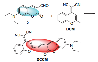
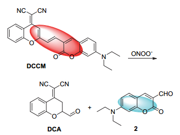
Scheme 1.
Synthesis of probe DCCM

Peroxynitrite (ONOO-) is an important reactive oxygen species (ROS) in living organisms.[1, 2] In living systems, peroxynitrite is produced by the diffusion-controlled reaction of nitric oxide (NO) and superoxide radicals (O2•-).[3] ONOO- can react with a variety of biologically active species, such as genetic substances, lipids, proteins, iron-sulfur clusters and thiols because of its strong oxidability and reactive activity, thereby leading to cell necrosis and apoptosis.[4~6] The imbalance of ONOO- is also associated with many diseases, such as cardiovascular disease, neurodegenerative disorders, Alzheimer's disease, diabetes, circulatory shock, inflammatory diseases and cancers.[7, 8] However, ONOO- also plays a positive role, assisting the immune system to combat pathogens.[9, 10] Therefore, it is important to develop a method for rapid and efficient detection of ONOO- in living organisms. Because peroxynitrite has a very short half-life (<10 ms), it is difficult to detect it in living cells by traditional analytical methods.[11, 12] Fluorescence imaging, which has a high sensitivity and strong selectivity, is an excellent method for real-time detection and monitoring of ONOO- in living cells.[13, 14] Fluorescent probes for ONOO- have been designed[15], however they are mainly intensity-type probes. Until now, few ratiometric fluorescent ONOO- probes have been reported.[16] Ratiometric fluorescent probes, which detect the analytes using two different fluorescence signals, can eliminate most of the environmental fluorescence background, enabling more accurate tests in complex biological environments.[17~22] Therefore, there is a need for development of a ratiometric fluorescent probe which can be used to detect ONOO-, and provide information on ONOO- inside cells through fluorescence imaging. In this study, we have designed and synthesized a new near-infrared ratiometric fluorescent probe, DCCM (Scheme 1), based on dicyanomethylene-4H-pyran (DCM) and 7-(diethylamino)coumarin-3-formaldehyde, for the accurate detection of ONOO-. The fluorophore, DCM, has a large Stokes shift, strong photostability and high fluorescent quantum yield, and is commonly used in near-infrared fluorescent probe.[23~27] The introduction of DCM leads to the formation of a strong push-pull electron system with the excellent electron donor 7-diethylamine coumarin, ensuring the maximum emission wavelength of the DCCM probe reaches 697 nm, in the near-infrared region. As a probe, DCCM exhibits a good response towards ONOO,- with a significant color change from purple to light pink, along with fluorescence color changes from heliotrope to blue. DCCM also has good selectivity and sensitivity for the detection of ONOO-, and has been successfully applied to biological imaging in HeLa cells.
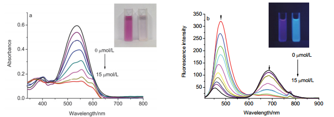
First, the fluorescence spectrum of DCCM in different proportions N, N-dimethylformamide/4-(2-hydroxyethyl)- 1-piperazineethanesulfonic acid (DMF/HEPES) buffer solution (0.01 mol•L-1, pH=7.4) from 9:1 to 1:9 (V:V) was investigated. It was found that the DMF/HEPES buffer solution (V:V=9:1, 0.01 mol•L-1, pH=7.4) is most suitable for fluorescence detection because of its high fluorescence emission intensity at 697 nm. As shown in Figure 1, with increasing ONOO- concentration, the intensity of the absorption peak at 535 nm gradually decreased, while an absorption peak at 390 nm progressively increased, accompanied by a color change of the solution, from purple to light pink (Figure 1a). As shown in Figure 1b, with higher concentration of ONOO-, the fluorescence emission peak at 697 nm gradually decreases, with the appearance of a new emission peak at 480 nm. The fluorescent color of the solution changed from heliotrope to blue simultaneously. Notably, the fluorescence intensity response of the DCCM probe for ONOO- has a strong linear correlation (R2=0.99) in the range of 0~5.6 μmol•L-1. Moreover, the detection limit for ONO- O- by DCCM was calculated to be 6.0×10⁻⁷ mol•L-1. This data proves that DCCM probe has the same order of sensitivity compared with 224 nmol•L-1 of a recently reported ratiomatric fluorescent probe for ONOO- detection.[28]
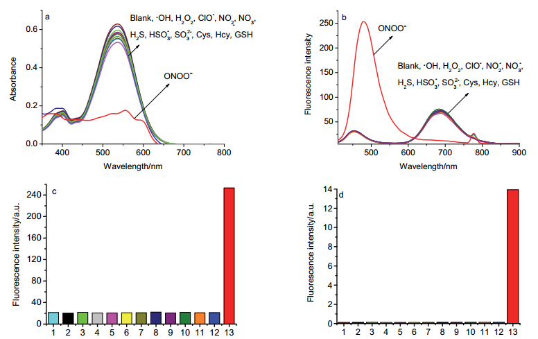
An investigation of the UV-vis absorption and fluores-cence selectivity of DCCM for ONOO- was carried out in the same DMF/HEPES buffer solution. The selectivity of DCCM probe was determined by comparing its response in the presence of various analytes including reactive oxygen species, reactive nitrogen species, reactive sulfur species, and metal ions, as well as amino acids. These experiments indicated that in the presence of other analytes, there was no change in the absorption or the fluorescent emission of DCCM, and the color of the DCCM solution was unaltered. Only with the addition of ONOO-, the absorption peak was quenched and shifted from 535 nm to 390 nm (Figure 2a), and the fluorescent emission peak was shifted from 697 nm to 480 nm, with the emission color changing from heliotrope to blue (Figure 2b). Therefore, these results indicate that DCCM is highly selective and can detect ONOO- without interference from other analytes.
Based on a previous report, [3] a mechanism of interaction between DCCM and ONOO- has been proposed (Scheme 2).
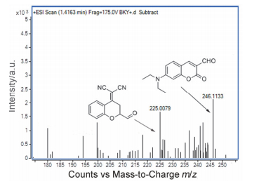
DCCM itself has a strong red fluorescence, but its double bond is broken by interacting with ONOO-. This shifts the maximum fluorescence emission peak to 480 nm. It was preliminarily determined that the probe DCCM interacted with ONOO- to generate a coumarin compound.Mass spectrometry was used to verify this reaction mechanism. First, DCCM itself was analyzed. The theoretical relative molecular mass of DCCM is [M+H]+m/z 436.1583, while the measured relative molecular mass was [M+H]+ m/z 436.1657. Then, in a DMF/HEPES buffer solution (V:V=9:1, 0.01 mol•L-1, pH=7.4), ONOO- (15 μmol•L-1) was added to a DCCM (10 μmol•L-1) solution. After reacting for a while, mass spectrometry was performed on the products. As shown in Figure 3b, peaks at [M+H]+m/z 225.0079 and 246.1133 were observed after the reaction, indicating that DCCM reacts with ONOO- to form a coumarin compound. The conversion rate of DCCM to coumarin 2 is relatively higher (88.9%) by comparing the DCCM (10 μmol/L) fluorescence emission intensity at 480 nm after the addition of ONOO- (15 μmol•L-1) with coumarin 2 (10 μmol•L-1) in DMF/ HEPES buffer solution (V:V=9:1, 0.01 mol•L-1, pH=7.4).
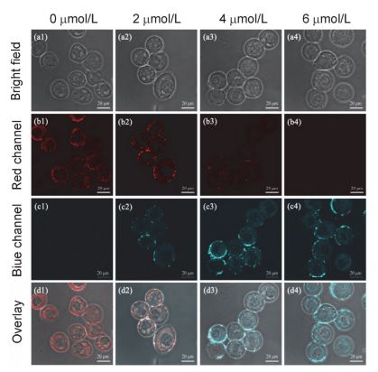
Prior to being applied to cell imaging, MTS assay was performed to assess the biocompatibility of DCCM. After addition of DCCM (40 μmol•L-1), which was much higher than the concentration (6 μmol•L-1) for cell imaging, the cell survival rate was 97.83% after 48 h. This result indicated that DCCM had low cytotoxicity to HeLa cells. To research the application of DCCM as an imaging probe for ONOO- in living cells, then DCCM was usedfor imaging ONOO- in HeLa cells. The HeLa cells were incubated with 10 μmol•L-1 DCCM for 120 min, and then were exposed to ONOO- (0, 2, 4 or 6 μmol•L-1) for another 60 min. As shown in Figure 4, the HeLa cells exhibited bright red fluorescence after incubation with DCCM for 120 min, but no blue fluorescence was observed in the blue channel. After the DCCM-loaded HeLa cells were treated with ONOO- (2, 4, 6 μmol•L-1) for 60 min, the fluorescence in the red channel decreased gradually, and increased gradually in the blue channel. These results indicate that DCCM could be used to perform fluorescence imaging of ONOO- in HeLa cells.
A near-infrared fluorescence probe for ONOO- detection, DCCM, was synthesized using a simple aldol condensation reaction between DCM and 7-(diethylamino)- coumarin-3-formaldehyde. As a ONOO- probe, DCCM displayed a remarkable colorimetric (from purple to light pink) and ratiometric fluorescence response (from heliotrope to blue) in the presence of ONOO-. The mechanism of the reaction was shown to be the oxidation and breaking of a carbon-carbon double bond by ONOO-. DCCM exhibited high selectivity and sensitivity for ONOO-, as well as low limit of detection (6.0×10⁻⁷ mol•L-1). Also, DCCM was successfully used to measure the presence of peroxynitrite in HeLa cells. Based these results, we believe that this research provides a promising means for investigating the effect of ONOO- in various physiological and pathological conditions.
Unless otherwise indicated, all chemicals were purchased from Aladdin, Macklin and “9 Ding chem”, and used without further purification. The crude product was separated and purified in silica gel (300~400 mesh) columns to obtain probe DCCM. A stock solution of DCCM was made by dissolving an appropriate amount in 5 mL of DMF. UV-vis absorption spectra were measured at 25 ℃ using a UV-240ZPC spectrophotometer. Fluorescence emission spectra were measured at 25 ℃ with an F-4600 fluorescence spectrum analyzer spectrometer (slit: 5 nm). 1H NMR (400 MHz) and 13C NMR spectra (101 MHz) were obtained using a Bruker Avance spectrometer. ESI mass spectra were obtained using a Xevo TQ-S and Q- TOF mass spectrometer. Confocal imaging of ONOO- in HeLa cells was carried out on a F100 fluorescence spectroscopy confocal microscope.
Compounds 3, 2 and DCM were synthesized according to literature with commercially available starting materials.[29, 30]
DCM (378 mg, 1.8 mmol) and compound 2 (414 mg, 1.6 mmol) were dissolved in 40 mL of toluene, then piperidine (0.6 mL) and acetic acid (0.6 mL) were added to the solution. The resulting mixture was refluxed for 4 h under nitrogen. After that, the mixture was cooled to room temperature and concentrated under vacuum, and the crude product was purified using silica gel column chromatography to isolate DCCM as a brown solid(330 mg, 46% yield). 1H NMR (500 MHz, CDCl3)δ: 8.94 (d, J=8.2 Hz, 1H), 7.83 (s, 1H), 7.75 (t, J=7.4 Hz, 1H), 7.60~7.53 (m, 2H), 7.47 (dd, J=15.0, 7.1 Hz, 1H), 7.41~7.36 (m, 2H), 6.90 (s, 1H), 6.68 (d, J=8.2 Hz, 1H), 6.55 (s, 1H), 3.49 (d, J=7.1 Hz, 4H), 1.28 (t, J=6.9 Hz, 6H). HRMS calcd for C27H22N3O3[M+H]+ 436.1661, found 436.1657. HRMS: calcd for C27H21N3O3Na[M+Na]+ 458.1481, found 458.1469. Fusing point is 167.7 ℃.
HeLa cells were cultured in modified Eagle's medium (DMEM, Invitrogen) supplemented with 10% fetal bovine serum (FBS) at 37 ℃, in a 5% CO2 incubator. Two days before use, roughly 1.0×10⁵ cells were inoculated onto a culture dish (37 ℃, 5% CO2). First, the dishes were rinsed several times with 4-(2-hydroxyethyl)-1-piperazineethane- sulfonic acid (HEPES) buffer. The cells were then incubated and probe DCCM (10 μmol•L-1) for 120 min in HEPES buffer solution, then washed with HEPES buffer and treated with ONOO- (either 0, 2, 4, or 6 μmol•L-1) for 60 min. Fluorescence images were then collected using a confocal microscope with a 40× objective.
Supporting Information Synthetic methods of probe DCCM.1H NMR and HR-MS spectra for compounds. The Supporting Information is available free of charge via the Internet at http://sioc-journal.cn/.
Wu L. L., Wang Y., Weber, Maria.; Liu L. Y., Sedgwick A., Bull S., Huang C., James T.-D...Chem. Commun, 2019, 55:3674. doi: 10.1039/C9CC90119A
Li Y., Zhao Z. W., Xiao Y. S., Wang X., Jiao X. Y., Xie X. L., Zhang J., Tang B...Anal. Chem, 2019, 91:6097. doi: 10.1021/acs.analchem.9b00636
Xie X. L., Tang F. Y., Liu G. G., Li Y., Su, X X.; Jiao X. Y., Wang X., Tang B...Anal. Chem, 2018, 90:11629. doi: 10.1021/acs.analchem.8b03207
Lee D. Y., Lim C. S., Ko G. Y. J., Kim D. Y., Cho M. Y. K., Nam S. J., Kim H. M., Yoon J...J. Anal. Chem, 2018, 90:9347. doi: 10.1021/acs.analchem.8b01960
Zhu M. Y., Zhou H., Ji D. D., Li G., Wang F., Song D. Y., Deng B., Li C., Qiao R. Z...Dyes Pigm, 2019, 168:77. doi: 10.1016/j.dyepig.2019.04.046
Li J. B., Chen L. L., Wang Q. Q., Liu H. W., Hu X. X., Yuan L., Zhang X. B...Anal. Chem, 2018, 90:4167. doi: 10.1021/acs.analchem.8b00198
Li Z. L., Liu C. Y., Yu C., Chen Y. N., Jia P., Zhu H. C., Zhang X., Yu Y., Zhu B., Sheng W.. Analyst 2019, 144:3442. doi: 10.1039/C9AN00347A
Qu W. B., Niu C. H., Zhang X. Y., Chen W., Yu F. B., Liu H., Zhang X. H., Wang S. F.. Talanta 2019, 197:431. doi: 10.1016/j.talanta.2019.01.065
Li H. Y., Li X. H., Wu X. F., Shi W., Ma H. M...Anal. Chem, 2017, 89:5519. doi: 10.1021/acs.analchem.7b00503
Cheng D., Pan Y., Wang L., Zeng Z. B., Yuan L., Zhang X. B., Chang Y.-T...J. Am. Chem. Soc, 2017, 139:285. doi: 10.1021/jacs.6b10508
Cheng D., Xu W., Yuan L., Zhang X. B...Anal. Chem, 2017, 89:7693. doi: 10.1021/acs.analchem.7b01671
田庆, 陈双虎, 陈景龙, 刘蕊, 汪雨诗, 杨晓朋, 叶勇..有机化学, 2019, 39:1. doi: 10.6023/cjoc201808030Tian Q., Chen S. H., Chen J. L., Liu L., Wang Y. S., Yang X. P., Ye Y...Chin. J. Org. Chem, 2019, 39:1 (in Chinese). doi: 10.6023/cjoc201808030
刘瑞姣, 曾竟..有机化学, 2017, 37:3274. doi: 10.6023/cjoc201706037Liu R. J., Zeng J...Chin. J. Org. Chem, 2017, 37:3274 (in Chinese). doi: 10.6023/cjoc201706037
(a) Zhou D. Y., Ou-Yang J., Li Y. F., Jiang W. L., Tian Y., Yi Z. M., Li C. Y.. Dyes Pigm. 2019, 161: 288. (b) Bi K. Y., Tan R., Hao R. T., Miao L. X., He Y. Q., Wu X. H., Zhang J. F., Xu R.. Chin. Chem. Lett. 2019, 30: 545. (c) Yu L., Qiao Y. M., Miao L. X., He Y. Q., Zhou Y.. Chin. Chem. Lett. 2018, 29: 1545.
Cheng D., Peng J. J., Lv Y., Su D. D., Liu D. J., Chen M., Yuan L., Zhang X. B...J. Am. Chem. Soc, 2019, 141:6352. doi: 10.1021/jacs.9b01374
Zhou D. Y., Li, Y. f.; Jiang W. L., Tian Y., Fei J. J., Li C. Y...Chem. Commun, 2018, 54:11590. doi: 10.1039/C8CC07389A
Zhu D. J., Yan X. W., Ren A., Xie W., Duan Z. Z.. Anal. Chim. Acta 2019, 1058:136. doi: 10.1016/j.aca.2019.01.013
Xiong H. Q., He L., Zhang Y., Wang J. P., Song X. Z., Yang Z. G...Chin. Chem. Lett, 2019, 30:1075. doi: 10.1016/j.cclet.2019.02.008
Tang X., Zhu Z., Liu R. J., Tang Y.. Spectrochim. Acta, A 2019, 219:576. doi: 10.1016/j.saa.2019.04.042
Chen H., Sun T., Qiao X. G., Tang Q. O., Zhao S. C., Zhou Z.. Spectrochim. Acta, A 2018, 204:196. doi: 10.1016/j.saa.2018.06.037
Feng S., Fang Y., Feng W. Y., Xia Q. F., Feng G. Q...Dyes. Pigm, 2017, 146:103. doi: 10.1016/j.dyepig.2017.07.002
Hu Q., Li W., Qin C. Q., Zeng L. T., Hou J. T.. Spectroscopy 2018, 204:196.
Deng B. B., Ren M. G., Kong X. Q., Zhou K., Lin W. Y.. Sensors. Actuators, B 2018, 255:963. doi: 10.1016/j.snb.2017.08.146
Li Q., Yan C. X., Zhang J., Guo Z. Q., Zhu W. H...Dyes. Pigm, 2019, 162:802. doi: 10.1016/j.dyepig.2018.11.019
Li H. D., Li Y. Q., Yao Q. C., Fan J. L., Sun W., Long S., Shao K., Du J. J., Wang J. Y., Peng X...J. Chem. Sci, 2019, 10:1619. doi: 10.1039/C8SC04685A
Duan C., Zhang J. F., Hu Y. B., Zeng L. T., Su D. D., Bao G. M...Dyes. Pigm, 2019, 162:459. doi: 10.1016/j.dyepig.2018.10.057
Yao Y. X., Gui L. J., Gao B. K., Yuan Z. W., Chen Y. S., Wei C., He Q., Wang F., Xu M. J., Chen H. Y...New J. Chem, 2019, 43:1785. doi: 10.1039/C8NJ04255A
Shu W., Wu Y. L., Shen T. J., Cui J., Kang H., Jing J., Zhang X. L...Dyes Pigm, 2019, 170:107609. doi: 10.1016/j.dyepig.2019.107609
Wu J. S., Liu W. M., Zhuang X. Q., Wang F., Wang P. F., Tao S. L., Zhang X. H., Wu S. K., Lee S. T...Org. Lett, 2007, 9:33. doi: 10.1021/ol062518z
Qi Y., Huang Y., Li B., Zeng F., Wu S. Z...Anal. Chem, 2018, 90:1014. doi: 10.1021/acs.analchem.7b04407

Figure 1 (a) UV-vis absorption spectra changes and (b) fluorescence spectra changes of probe DCCM (10 μmol•L-1) in DMF/HEPES buffer solution (V:V=9:1, 0.01 mol•L-1, pH=7.4) upon addition of increasing amount of ONOO- (0~15 μmol•L-1) (λex=390 nm, slits 5 nm×5 nm

Figure 2 (a) UV-vis absorption spectrum, (b) fluorescence emission spectrum, (c) fluorescence intensity (F480) and (d) fluorescence intensity ratios (F480/F697) of probe DCCM (10 μmol•L-1) in the presence of various analytes in DMF/HEPES buffer solution (V:V=9:1, 0.01 mol•L-1, pH=7.4)1: Blank, 2: •OH, 3: H2O2, 4: ClO-, 5:NO2- , 6:NO3- , 7: H2S, 8: HSO3-, 9: SO32-, 10: Cys, 11: Hcy, 12: GSH, 13: ONOO-. 15 μmol•L-1. λex=390 nm, slits 5 nm×5 nm

Figure 4 Confocal fluorescence images of probe DCCM in HeLa cells incubated with different concentrations of ONOO-The HeLa cells were incubated with probe DCCM (10 μmol•L-1) for 120 min, and then were fed with ONOO- (0, 2, 4, 6 μmol•L-1) for another 60 min. (a1~a4) bright field map; (b1~b4) red light channel map; (c1~c4) blue light channel map; (d1~d4) superposition map. Images were received by collecting the emissions at 450~500 nm and the emissions at 650~700 nm upon excitation at 390 nm. Scale bar: 20 μm

 扫一扫看文章

扫一扫关注我们
 下载:
下载:

 下载:
下载:

